/
1 Pathogens 1 Pathogens

1 Pathogens - PowerPoint Presentation

pamella-moone
pamella-moone . @pamella-moone
Follow
407 views
Uploaded On 2015-12-01

1 Pathogens - PPT Presentation

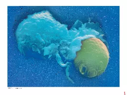
such as bacteria fungi and viruses INNATE IMMUNITY all animals Rapid response Recognition of traits shared by broad ranges of pathogens using a small set of receptors Recognition of traits ID: 210758

antigen cell receptor cells cell antigen cells receptor antibody pathogen response receptors mhc fragment molecule pore recognition class intron

Share:

Link:

Embed:

Download Presentation from below link

Download Presentation The PPT/PDF document "1 Pathogens" is the property of its rightful owner. Permission is granted to download and print the materials on this web site for personal, non-commercial use only, and to display it on your personal computer provided you do not modify the materials and that you retain all copyright notices contained in the materials. By downloading content from our website, you accept the terms of this agreement.


Presentation Transcript

Slide1

1Slide2

Pathogens

(such as bacteria,

fungi, and viruses)

INNATE IMMUNITY

(all animals)

• Rapid response

Recognition of traits shared

by broad ranges ofpathogens, using a smallset of receptors

Recognition of traits

specific to particularpathogens, using a vastarray of receptors

• Slower response

Barrier defenses:

SkinMucous membranesSecretions

Internal defenses:

Phagocytic cellsNatural killer cellsAntimicrobial proteinsInflammatory response

Humoral response:

Antibodies defend againstinfection in body fluids.

Cell-mediated response:

Cytotoxic cells defendagainst infection in body cells.

ADAPTIVE IMMUNITY(vertebrates only)

2Slide3

Pathogen

PHAGOCYTIC

CELL

Vacuole

Lysosome

containing

enzymes

3Slide4

Thymus

Peyer’s

patches

(small

intestine)

Appendix(cecum)

Adenoid

Tonsils

Lymphatic

vessels

Spleen

Lymphnodes

Lymphnode

Bloodcapillary

Interstitialfluid

Tissuecells

Lymphatic vessel

Lymphatic vessel

Masses ofdefensive cells

4Slide5

Pathogen

Splinter

Mast

cell

Macro-

phage

Capillary

Red

blood cells

Neutrophil

Signaling

molecules

Movementof fluid

Phagocytosis

5Slide6

Antigen

receptors

Mature B cell

Mature T cell

6Slide7

Cytoplasm of B cell

Antigen-

binding

site

B cell

antigen

receptor

B cell

Lightchain

Disulfide

bridgeAntigen-binding site

Variable

regions

Constant regions

Transmembraneregion

Heavy chains

Plasmamembrane

C

C

C

C

VV

V

V

7Slide8

Antibody

Antigen

receptor

B cell

Antigen

Epitope

Pathogen

(a) B cell antigen receptors and antibodies

Antibody C

Antibody B

Antibody A

Antigen

(b) Antigen receptor specificity

8Slide9

T cell

antigen

receptor

T cell

Cytoplasm of T cell

Plasma

membrane

chain

chainDisulfide

bridge

Antigen-bindingsite

Variable

regions

Constantregions

Transmembraneregion

V

V

C

C

9Slide10

Displayed

antigen

fragment

MHC

molecule

Antigenfragment

Pathogen

Host cell

T cell

T cell antigen

receptor(a) Antigen recognition by a T cell

(b) A closer look at antigen presentation

Antigen

fragmentMHC

moleculeHost cell

Top view

10Slide11

DNA of

undifferentiated

B cell

DNA of

differentiatedB cell

Recombination deletes DNA between

randomly selected V segment and

J segmentFunctional gene

Transcription

RNA processing

Translation

pre-mRNA

mRNA

Light-chain polypeptide

Antigen receptorB cell

Variableregion

Constantregion

2

3

4

1

Poly-A tail

Cap

V

39

J

5

J

5

V

39

V

37

V

38

V

39

V

37

V

38

V

39

V

40

J

5

J

5

J

4

J

3

J

2

J

1

Intron

Intron

V

C

C

C

C

C

C

C

C

C

V

V

V

V

Intron

11Slide12

Antigen

Antigen

receptor

Antibody

Plasma cells

Memory cells

B cells that

differ in

antigenspecificity

12Slide13

Antigen-

presenting

cell

Pathogen

Antigen fragment

Class II MHC molecule

Accessory protein

Antigen receptor

Helper T cell

Cytokines

Humoral

immunity

Cell-

mediatedimmunity

B cell

Cytotoxic T cell

3

2

1

13Slide14

Cytotoxic T cell

3

1

2

Accessory

protein

Class I MHC

molecule

Infected

cell

Antigen

receptor

Antigen

fragment

Perforin

Pore

Gran-

zymes

Released

cytotoxic

T cellDying

infected cell

14Slide15

Pathogen

3

1

2

Antigen-presenting

cell

Antigen

fragment

Class II

MHC

molecule

Antigen

receptor

Accessory

protein

Helper T cell

B cell

Cytokines

Activated

helper T cell

Memory B cells

Plasma cells

Secreted

antibodies

15Slide16

Opsonization

Neutralization

Antibody

Virus

Bacterium

Macrophage

Activation of complement system and pore

formation

Complement proteins

Formation of membrane

attack complex

Flow of water

and ions

Pore

Antigen

Foreign

cell

16Slide17

IgE

Allergen

Histamine

Granule

Mast cell

17