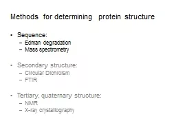
Sequence Edman degradation Mass spectrometry Secondary structure Circular Dichroism FTIR Tertiary quaternary structure NMR Xray crystallography Protein sequencing approaches depend on what is known and what is the goal ID: 599867
Download Presentation The PPT/PDF document "Methods for determining protein structur..." is the property of its rightful owner. Permission is granted to download and print the materials on this web site for personal, non-commercial use only, and to display it on your personal computer provided you do not modify the materials and that you retain all copyright notices contained in the materials. By downloading content from our website, you accept the terms of this agreement.
Slide1
Methods for determining protein structure
Sequence:
Edman degradation
Mass spectrometry
Secondary structure:
Circular Dichroism
FTIR
Tertiary, quaternary structure:
NMR
X-ray crystallographySlide2
Protein sequencing approaches depend on what is known and what is the goal
Protein is unknown, from organism with no DNA sequence information – starting from scratch
Purify protein & separate chains (if multimer)
Fragment and sequence each chain
Fragment differently and sequence
Reassemble sequence based on overlapping fragments
Protein is unknown or known, and comes from an organism with known DNA sequence
Purify protein (& separate chains)
Fragment chain(s) and sequence or measure mass
Use sequence database to reassemble sequenceSlide3
Protein sequencing from scratch
Step 0: Purify the protein
Step 1: Separate the chains (if
multimeric
)
If needed, reduce disulfides and block free thiolsSlide4
Protein sequencing from scratch
Step 0: Purify the protein
Step 1: Separate the chains (if
multimeric
)
Step 2: Fragment each polypeptideEnzymatically, with endopeptidase
, chemically (e.g. with cyanogen bromide), or physically (e.g. through collision in MS)Slide5
Step 2: Fragment each polypeptide
Cyanogen bromide (CNBr): R
n-1
= MetSlide6
Protein sequencing from scratch
Step 0: Purify the protein
Step 1: Separate the chains (if
multimeric
)
Step 2: Fragment each polypeptideStep 3: Sequence the fragmentsVia, e.g.,
Edman
degradation or Mass spectrometrySlide7
Sequence peptides with mass spectrometry
(MS/MS)Slide8
MS cleavage occurs mainly at peptide bonds, and charge is retained in one productSlide9
Protein sequencing from scratch
Step 0: Purify the protein
Step 1: Separate the
chains (if
multimeric
)Step 2: Fragment each polypeptideStep 3: Sequence the fragments
Step 4: Reconstruct the sequenceSlide10
Protein sequencing approaches depend on what is known and what is the goal
Protein is unknown, from organism with no DNA sequence information – starting from scratch
Purify protein & separate chains (if multimer)
Fragment and sequence each chain
Fragment differently and sequence
Reassemble sequence based on overlapping fragments
Protein is unknown or known, and comes from an organism with known DNA sequence
Purify protein (& separate chains)
Fragment chain(s) and sequence or measure mass
Use sequence database to reassemble sequenceSlide11
There are different approaches for using mass spectrometry to sequence a protein
Bottom-Up Proteomics
Fragment protein (e.g. enzymatically) and separate fragments
Ionize fragments, trap in the spectrometer, and measure m/z
Select one m/z peak and fragment (e.g. by collision)
Measure m/z of the smaller fragments and use a database to match the peaks to known sequencesSlide12
There are different approaches for using mass spectrometry to sequence a protein
Top-Down Proteomics
Ionize
whole
protein(s), trap in the spectrometer, and measure m/z
Use the instrument to select one m/z peak and fragment the protein (e.g. by collision)
Measure m/z ratios of the fragments and use a database to match the peaks to known sequences
OR Select a peak and fragment again, then match to sequence (Selection and fragmentation may be repeated over and over)Slide13
In shotgun proteomics, mass spec. is used to sequence mixtures of proteins
Mixture of many proteins
Mixture of peptides from different proteins
Enzymatic digest
Separation of peptides,
Ionization in MS, Fragmentation
Matched sequences
Submit peaks to databaseSlide14
Methods for determining protein structure
Sequence:
Edman degradation
Mass spectrometry
Secondary structure:
Circular Dichroism
FTIR
Tertiary, quaternary structure:
NMR
X-ray crystallographySlide15
Circular
dichroism
(CD) measures amide absorption of circularly polarized UV light
Ellipticity
(
De
) is
the difference in absorption of left-handed and right-handed circularly polarized light
Different secondary structures show different patterns of
ellipticity
Protein’s CD spectrum is ‘
deconvoluted
’ to estimate fractional contribution of helix, sheet, turn, and
coilSlide16
Proteins with different compositions of 2
structure give different CD spectraSlide17
Fourier transform infrared (FTIR) spectra show amide absorption of infrared light
Peak frequencies show bond stretching and bending, which vary with protein conformation
C=O stretching frequency of amide I band correlates with secondary structure
Protein’s FTIR spectrum is ‘
deconvoluted
’ to estimate fractional contribution of helix, sheet, and coilSlide18
Methods for determining protein structure
Sequence:
Edman degradation
Mass spectrometry
Secondary structure:
Circular Dichroism
FTIR
Tertiary, quaternary structure:
NMR
X-ray crystallographySlide19
Proteins have too many protons to be resolved by one-dimensional NMRSlide20
2D NMR
separates proton peaks and can
reveal approximate distances between nearby atoms
a
b
c
d
Cross-peaks indicate protons are within 5Å of each otherSlide21
NMR-derived distance constraints are used to calculate likely protein conformationsSlide22
X-ray crystallography reveals the layout of repeating electron density
X-rays
Protein crystal
Diffraction pattern
Data processing
Diffracted
X-rays
Electron density mapSlide23
Electron density map allows for positioning of protein atoms, revealing structure