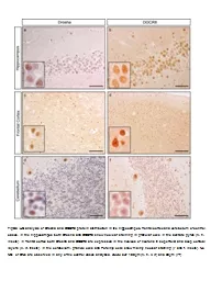
IHC analysis of Drosha and DGCR8 protein distribution in the hippocampus frontal cortex and cerebellum of control cases In the hippocampus both Drosha and DGCR8 show nuclear staining in granular cells In the dentate ID: 595994
Download Presentation The PPT/PDF document "FigS3" is the property of its rightful owner. Permission is granted to download and print the materials on this web site for personal, non-commercial use only, and to display it on your personal computer provided you do not modify the materials and that you retain all copyright notices contained in the materials. By downloading content from our website, you accept the terms of this agreement.
Slide1
FigS3
IHC analysis of Drosha and DGCR8 protein distribution in the hippocampus, frontal cortex and cerebellum of control cases. In the hippocampus both Drosha and DGCR8 show nuclear staining in granular cells In the dentate
gyrus
(
a
,
b
, insets). In frontal cortex both Drosha and DGCR8 are expressed in the nucleus of neurons in superficial and deep cortical layers (
c
,
d
, insets). In the cerebellum, granule cells and Purkinje cells show mainly nuclear staining (
e
and
f
, insets). No NCI or DNs are observed in any of the control cases analyzed
.
Scale bar
; 100μm (
a
,
b
,
c
,
d
) and 50μm (
e
-
f
)