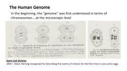
Germ Cell Division 1876 Oskar Hertwig recognized for describing the events of meiosis for the first time in sea urchin eggs Germ Cell Division 1890 August Weismann determined the significance of meiosis for reproduction and inheritance and that ID: 759229
Download Presentation The PPT/PDF document "The Human Genome In the beginning, the ..." is the property of its rightful owner. Permission is granted to download and print the materials on this web site for personal, non-commercial use only, and to display it on your personal computer provided you do not modify the materials and that you retain all copyright notices contained in the materials. By downloading content from our website, you accept the terms of this agreement.
Slide1
The Human Genome
In the beginning, the “genome” was first understood in terms of chromosomes….at the microscopic level
Germ Cell Division
1876 – Oskar
Hertwig
recognized for describing the events of meiosis for the first time in sea urchin eggs
Slide2Germ Cell Division
1890 – August Weismann determined the significance of meiosis for reproduction and inheritance and that
two cell divisions are necessary to go from the diploid to the haploid number of chromosomes
Slide3It was not until 1956 that the correct number of human chromosomes, 46, was determined by Indonesia-born cytogeneticist Joe
Hin
Tijo.
Genome…. Chromosomes….. Chromatin Nucleosome
Slide4Do It……………………………..
…how big is the human genome?…how do you begin to explore the human genome?
Writing the genome exercise…..
Slide5How big is the human genome?
Virus (SV40) 5,243
4 1,311 (5 x 10³ )
Bacteria (E. coli) 5,400,000 5,400 1,000 (5 x 10⁶ )
Eukaryote (Human) 3,200,000,000 25,000 128,000 (3 x 10⁹ )
Base pairs
per genome
Number of genes
Base pairs per gene
Why is the human genome so big?
Slide6Slide7Imagine that,….you were an aspiring graduate student in biochemistry in the early 1970’s…with a growing interest in genomes. You knew that your fellow graduate students in molecular biology (the grasshoppers) were all excited about small viral genomes (~5,000 base pairs) as they started to cut these small genomes into discrete fragments with restriction endonucleases. But you (the ant) had bigger aspirations. You wanted to learn something about the human genome….
What tool did biochemists have that allowed them to learn something surprising about the human genome?
SPECTROPHOTOMETRY
Slide8Why does DNA absorb light at 260 nm?
Slide9Slide10Hyperchromicity
Some smart people noticed that the
rate
at which the single stranded DNA “reannealed”
was related to the size of the genome from which the DNA was isolated.
Slide11Slide12In the 1970’s….before we began sequencing DNA, Cot curves confused many young graduate students … and revealed something interesting about the human genome.
Co = DNA concentration
t = time to “re-anneal”
Draw a curve on the chart to the left representing the reannealing of human DNA.
Human
Slide13In the 1970’s….before we began sequencing DNA, Cot curves confused many young graduate students … and revealed something interesting about the human genome.
Co = DNA concentration
t = time to “re-anneal”
The human genome is composed of both repeated DNA sequences and unique DNA sequences.
Human
Only ~ 1.5% of the human genome encodes proteins.
Slide14(1974)
Slide15Slide16CORE DNA
CORE PARTICLE
contains 8 histones and 145 base pairs of DNA
LINKER DNA
20-120 base pairs
First-ever illustration of a nucleosome….drawn by Sally L., an undergraduate “dishwasher” in Dr. George Beaudreau’s lab, Oregon State University, ~ 1975.
Chromatin,….as “Beads – On – A – String”
HISTONE CORE
(2H2a, 2H2b, 2H3, 2H4)
Slide17Crystal structure of the nucleosome core particle at 2.8 A
˚ resolutionKarolin Luger, Armin W. Ma¨ der, Robin K. Richmond, David F. Sargent & Timothy J. RichmondInstitut fu¨r Molekularbiologie und Biophysik ETHZ, ETH-Ho¨nggerberg, CH-8093 Zu¨rich, Switzerland. ............ ............ ............ ........... ............ ............ ............ ........... ............ ............ ........ .... ........... ............ ............ ............ ........... ............ ............ ............ ............ ...........
NATURE |VOL 389 | 18 SEPTEMBER 1997
Notice how difficult it is to communicate detailed structural information in a flat 2-D image
Slide18The N-terminal tails of the histones….
protrude to the outside surface of the nucleosome…through “aligned minor grooves”.
Slide19Do It…………………………………………
…. Explore nucleosome structure.
…. Explore nucleosome model kit / epigenetics kit
Slide20Slide21Slide22Slide23Slide24Comprehensive Mapping of Long-Range Interactions Reveals Folding Principles of the Human Genome
Erez Lieberman-Aiden et.al., Science Vol 326 9 October 2009
What is the 3D structure of chromatin…in the nucleus?
…the concept of a “
fractal globule
”
Slide25Overview of Hi-C ….
a Chromosome Conformation Capture technology
Slide26http://www.bioedonline.org/library/media
/photos-and-video/a-3d-map-of-the-human-genome/
Researchers at Baylor College of Medicine, Rice University, the Broad Institute of MIT and Harvard, and Harvard University describe the results of a five-year effort to map, in unprecedented detail, how the 2-meter long human genome folds inside the nucleus of a cell. Their results show that the cell — like a microscopic
origamist
— modulates its function by folding the genome into an almost limitless variety of shapes.
Slide27Comprehensive Mapping of Long-Range Interactions Reveals Folding Principles
of the Human Genome
Erez
Lieberman-Aiden et.al., Science Vol 326 9 October 2009