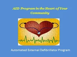
/
JCM OSCE PWH AED 5 /10/2022 JCM OSCE PWH AED 5 /10/2022

JCM OSCE PWH AED 5 /10/2022 - PowerPoint Presentation

phoebe
phoebe . @phoebe
Follow
65 views
Uploaded On 2023-11-22

JCM OSCE PWH AED 5 /10/2022 - PPT Presentation

Question 1 57M GPH Sudden onset severe back pain when lifting water bottle Bilateral lower limb pain numbness and weakness GCS 15 BP17281 P98 SpO2 99 RA Bil UL power 55 bil LL power 15 ID: 1034436

diagnosis question important cardiac question diagnosis cardiac important arrest pain anterior patient complications dislocation injury ecg joint posterior clinical

Share:

Link:

Embed:

Download Presentation from below link

Download Presentation The PPT/PDF document "JCM OSCE PWH AED 5 /10/2022" is the property of its rightful owner. Permission is granted to download and print the materials on this web site for personal, non-commercial use only, and to display it on your personal computer provided you do not modify the materials and that you retain all copyright notices contained in the materials. By downloading content from our website, you accept the terms of this agreement.


Presentation Transcript

1. JCM OSCEPWH AED5/10/2022

2. Question 157/MGPHSudden onset severe back pain when lifting water bottleBilateral lower limb pain, numbness and weaknessGCS 15, BP172/81, P98, SpO2 99% RABil UL power 5/5, bil LL power 1/5

3. Question 1aState 3 other important physical signs

4. Question 1aState 3 other important physical signsNeurological: sensation, tone, reflexes, sensory level, anal tone, distended bladderLL vascular status: peripheral pulses, mottling/ pallor, temperature, ankle-brachial pressure indexAortic dissection: RR delay, bilateral arm BP discrepancy

5. Question 1bState 2 possible differential diagnoses

6. Question 1bState 2 possible differential diagnosesSpinal cord compressionSpinal cord infarctionAortic dissectionAcute bilateral lower limb ischemiaCVA

7. Question 1cState 3 ED investigations

8. Question 1cState 3 ED investigationsPOCTEcho USG doppler for LL pulsesImagingCXRCTBX-ray LS spineContrast CT aortogram

9. Question 1dContrast CT was done. Please describe the important radiological findings.What is the diagnosis?

10. Question 1

11.

12.

13. Question 1dDescribe the important radiological findingsInfra-renal abdominal aorta completely non-opacified all the way down to proximal bilateral common iliac arteries Atherosclerotic wall changes of aortaWhat is the diagnosis?Acute aortic occlusion

14. Acute Aortic OcclusionUncommon vascular emergency with high mortality (20-50%) and amputation rateThrombosis most common cause due to aging population (embolism now less common due to anticoagulants)Infra-renal most common location

15. Acute Aortic OcclusionLL acute pain, paralysis, mottlingAbdominal painAcute hypertensive crisis in renal artery involvement

16. Question 1eState 4 ED management for this patient

17. Question 1eState 4 ED management for this patientSupportivePain controlIVF hydrationHeparinisationDefinitiveConsult vascular surgery for thromboembolectomy +/- revascularisation with bypass

18. Question 1fState 4 possible complications for this condition

19. Question 1fState 4 possible complications for this conditionAcute renal failureLower limb ischemiaCompartment syndromeMesenteric ischemiaRhabdomyolysisAMIARDS

20. Question 1gState 3 risk factors for this condition

21. Question 1gState 3 risk factors for this conditionThrombosisSmokingDMBlunt abdominal traumaEmbolicValvular heart diseaseLow flow stateMajor surgeryHypercoagulable state e.g. protein C or S deficiency

22. Back to our patientEmergency endarterectomy of aorta Intra-op finding: aortic occlusion by firm gelatinous thrombusComplicated with rhabdomyolysis/ reperfusion syndrome

23. Back to our patientCT also showed a LA myxoma with thrombus over LA and LVLA myxoma excisionRecovery uneventful

24. Question 2A 30 year old man crushed his bike. He complained of right shoulder pain and right chest wall pain.BP135/72. P78. SpO2 100% on RA. RR28/min.

25. Question 2aDescribe the clinical photo

26. Question 2aDescribe the clinical photoSwelling over the right sternoclavicular joint

27. Question 2bDescribe the X-rays

28.

29.

30.

31. Question 2bDescribe the X-ray(Frontal) Joint space widening and asymmetry over right sternoclavicular joint. Rib fractures.(Oblique) Widening and asymmetry of the right sternoclavicular joint with abnormal position of the clavicle in relation to the manubrium.

32. Question 2cWhat other X-ray view can you order? What additional advantage does it have?

33. Question 2cWhat other X-ray view can you order? What additional advantage does it have?Serendipity (Rockwood) view. It can differentiate anterior and posterior sternoclavicular joint dislocation.

34. Question 2dWhat further investigation will you do?

35. Question 2dWhat further investigation will you do?CT scan

36. Question 2eWhat is the likely diagnosis? Name one important differential diagnosis if the patient is younger.

37. Question 2eWhat is the likely diagnosis?Traumatic anterior dislocation of right sternoclavicular joint.Name one important differential diagnosis if the patient is younger.Medial clavicle fracture-dislocation (salter I or II physeal fractures)

38. Question 2fWhat is the usual mechanism for this injury?

39. Question 2fWhat is the usual mechanism for this injuryHigh energy trauma e.g. contact sports, MVCForces to the posterior shoulder or to the lateral shoulder if held posteriorlyRarely non-traumatic subluxation, infection, rheumatologic diseases, hypermobility

40. Question 2gName the 2 main types of this injury. What are the complications?

41. Question 2gName the 2 main types of this injury.Anterior and posterior.What are the complications?Tracheal injuryPneumothoraxSubclavian vessels damageMediastinal compressionOesophageal ruptureBrachial plexopathyThoracic outlet syndrome

42. Question 2hWhat is your management for this patient?

43. Question 2hWhat is your management for this patient?Pain controlTemporary immobilisation with arm slingMonitor airway and breathingMonitor right upper limb neurovascular statusConsult orthopedics for reduction of his right SCJ dislocation

44. ManagementConservativeChronic anterior dislocation with minimal symptoms (>3 weeks old)Closed reductionAcute anterior and posterior dislocationOpen reductionAcute posterior dislocation failed closed reductionChronic anterior or posterior dislocation with persistent symptoms

45. Question 3A 69 year old woman complained of chest discomfort and dizziness for 3 days. Presented today with LOC 1min.GCS 15. BP131/90. P134. RR14. SpO2 99% RA.

46. Question 3aName 2 clinical prediction rules for syncope

47. Question 3aName 2 clinical prediction rules for syncopeSan Francisco Syncope RuleOESIL Score for SyncopeEGSYS Score Canadian Syncope Risk Score

48. Question 3bDescribe the ECG findings

49. Question 3bDescribe the ECG findingsAtrial Fibrillation with ventricular rate 154bpm

50. Question 3cDescribe the cardiac monitor stripe

51. Question 3cDescribe the cardiac monitor stripeAlternating narrow complex tachycardia and long sinus pauses (up to 6 seconds)

52. Question 3dWhat is your diagnosis?

53. Question 3dWhat is your diagnosis?Tachy-brady syndrome

54. Question 3eWhat is the definitive treatment?Name 3 other indications for this treatment.

55. Question 3eWhat is the definitive treatment?Permanent pacemaker insertionName 3 other indications for this treatmentSinus node dysfunction with symptomatic bradycardiaSymptomatic chronotropic incompetence3rd degree AV blockSymptomatic advanced 2nd degree AV block Trifascicular blockBifascicular block with syncope

56. Question 4A 20 year old man fainted while hiking in the rain. GCS 15, BP 150/90, P100, RR 14, SpO2 99% RA

57. Question 4aDescribe the clinical photos. Name the physical sign. What is the diagnosis?

58.

59. Question 4aDescribe the clinical photosErythematous marks in fern-like pattern over neck, chest and backName the physical signLichtenberg figures.What is the diagnosis?Lightening injury.

60. Question 4bName the mechanisms of injury. Which one has the worst prognosis?

61. Question 4bName the mechanisms of injury. Which one has the worst prognosis?Direct strike (worst prognosis)Side flashContactGround currentUpward streamerBall lighteningBlunt injuries (muscular contractions, explosion)

62. Question 4cState 3 possible neurological complications

63. Question 4cState 3 possible neurological complicationsConfusionAmnesiaKeraunoparalysisHypoxic encephalopathyIntracranial hemorrhageParaplegia, hemiplegiaMotor neuron diseaseAmyotrophic lateral sclerosisParkinsonismSpinal cord injury

64. Question 4dState 3 possible complications in other systems

65. Question 4dState 3 possible complications in other systemsBurnsRhabdomyolysisTransient blindnessTympanic membrane perforationAutonomic dysfunctionBone fractures and dislocationsPsychiatric e.g. sleeping disorder

66. Question 4eHis companion was found to be in cardiac arrest. Name 2 mechanisms for immediate cardiac arrest.

67. Question 4eHis companion was found to be in cardiac arrest. Name 2 mechanisms for immediate cardiac arrest.Respiratory arrest as a result of CNS medulla respiratory center paralysisAsystole from depolarization of myocardium

68. Question 4fThere are 4 other companions on the hill who suffered from minor falls but could still walk. If you were the on-scene medical team, would you save the patient in cardiac arrest first or the 4 ambulatory companions and why?

69. Question 4fThe patient in cardiac arrest should receive medical attention first.Reversed triage should not be adopted in lightening mass casualties as anyone showing signs of life at scene has high chance of survival. So those in cardiac or respiratory arrest should receive the greatest attention.

70. Question 5A 60 year old man complained of chest pain for 3 days.

71. Question 5aState the important ECG findings

72. Question 5aState the important ECG findingsBiphasic T waves over V2 - 4

73. Question 5bWhat is the diagnosis?

74. Question 5bWhat is the diagnosis?Wellens syndrome

75. Question 5cWhat is the underlying pathology?

76. Question 5cWhat is the underlying pathology?Critical occlusion of proximal LAD

77. Question 5dWhat is the definitive treatment?

78. Question 5dWhat is the definitive treatment?Early PCI

79. Question 5eSudden increased chest pain. ECG was repeated. Describe the important ECG findings.

80. Question 5e

81. Question 5eDescribe the important ECG findings Tall peaked T waves with minimal ST elevation over V3-4 (Hyperacute T waves)

82. Question 5fWhat is the diagnosis? What is the underlying pathology?

83. Question 5fWhat is the diagnosis? What is the underlying pathology?Anterior MIAcute occlusion of proximal LAD

84. Question 5gState 3 ED management

85. Question 5gState 3 ED managementAspirin and antiplateletsNitratesPain controlConsult cardiac for immediate cardiac reperfusion therapy (PPCI/ TNK)