/
Toxicity Test With MEK inhibitor in ( Toxicity Test With MEK inhibitor in (

Toxicity Test With MEK inhibitor in ( - PowerPoint Presentation

riley
riley . @riley
Follow
66 views
Uploaded On 2023-05-20

Toxicity Test With MEK inhibitor in ( - PPT Presentation

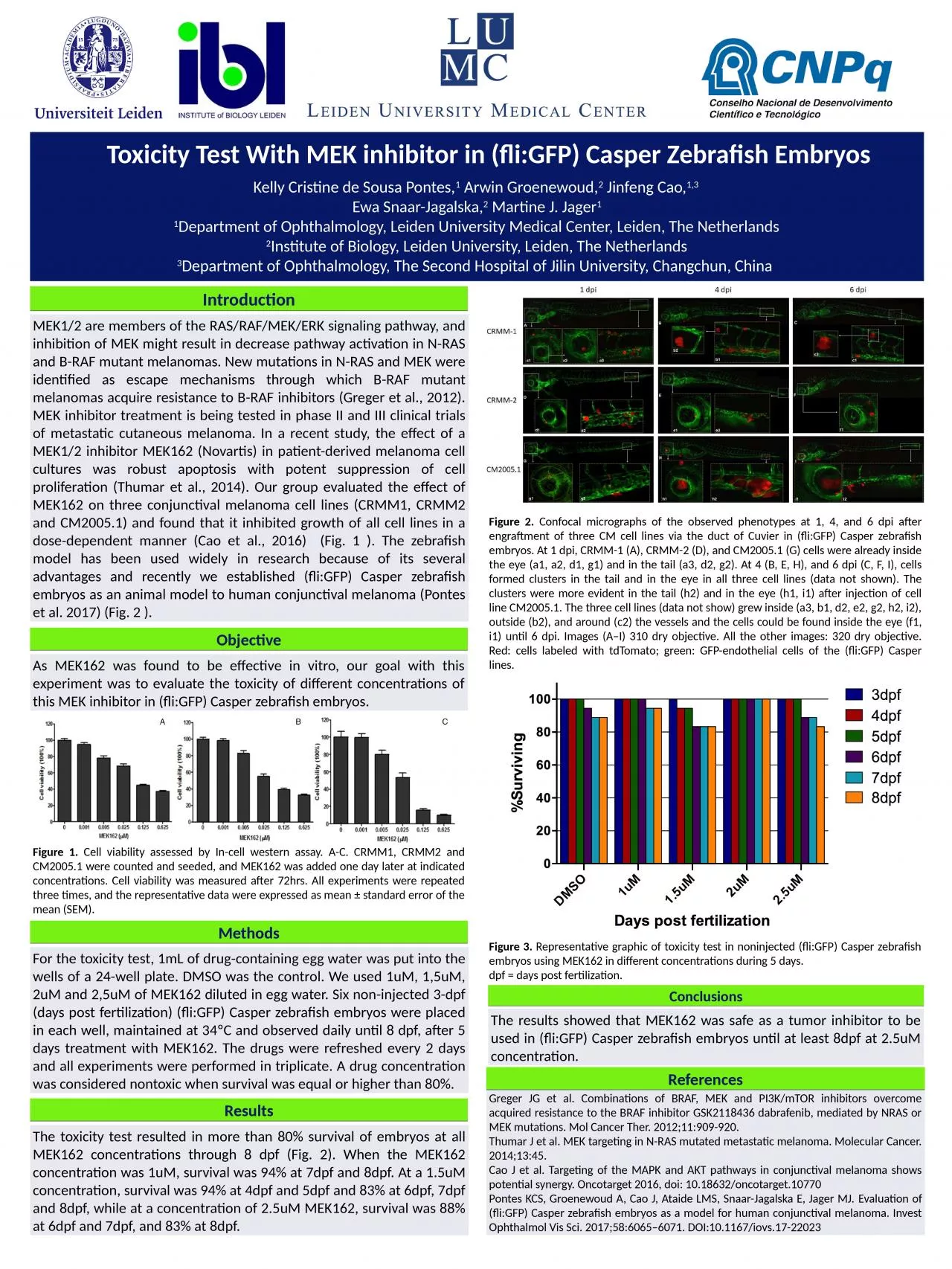
fliGFP Casper Zebrafish Embryos Kelly Cristine de Sousa Pontes 1 Arwin Groenewoud 2 Jinfeng Cao 13 Ewa SnaarJagalska 2 Martine J Jager 1 1 Department of Ophthalmology Leiden University Medical Center Leiden The Netherlands ID: 998561

mek162 cell mek gfp cell mek162 gfp mek fli casper zebrafish embryos melanoma inhibitor lines 5um survival concentration days

Share:

Link:

Embed:

Download Presentation from below link

Download Presentation The PPT/PDF document "Toxicity Test With MEK inhibitor in (" is the property of its rightful owner. Permission is granted to download and print the materials on this web site for personal, non-commercial use only, and to display it on your personal computer provided you do not modify the materials and that you retain all copyright notices contained in the materials. By downloading content from our website, you accept the terms of this agreement.


Presentation Transcript

1. Toxicity Test With MEK inhibitor in (fli:GFP) Casper Zebrafish Embryos Kelly Cristine de Sousa Pontes,1 Arwin Groenewoud,2 Jinfeng Cao,1,3 Ewa Snaar-Jagalska,2 Martine J. Jager11Department of Ophthalmology, Leiden University Medical Center, Leiden, The Netherlands2Institute of Biology, Leiden University, Leiden, The Netherlands3Department of Ophthalmology, The Second Hospital of Jilin University, Changchun, China IntroductionMEK1/2 are members of the RAS/RAF/MEK/ERK signaling pathway, and inhibition of MEK might result in decrease pathway activation in N-RAS and B-RAF mutant melanomas. New mutations in N-RAS and MEK were identified as escape mechanisms through which B-RAF mutant melanomas acquire resistance to B-RAF inhibitors (Greger et al., 2012). MEK inhibitor treatment is being tested in phase II and III clinical trials of metastatic cutaneous melanoma. In a recent study, the effect of a MEK1/2 inhibitor MEK162 (Novartis) in patient-derived melanoma cell cultures was robust apoptosis with potent suppression of cell proliferation (Thumar et al., 2014). Our group evaluated the effect of MEK162 on three conjunctival melanoma cell lines (CRMM1, CRMM2 and CM2005.1) and found that it inhibited growth of all cell lines in a dose-dependent manner (Cao et al., 2016) (Fig. 1 ). The zebrafish model has been used widely in research because of its several advantages and recently we established (fli:GFP) Casper zebrafish embryos as an animal model to human conjunctival melanoma (Pontes et al. 2017) (Fig. 2 ). MethodsFor the toxicity test, 1mL of drug-containing egg water was put into the wells of a 24-well plate. DMSO was the control. We used 1uM, 1,5uM, 2uM and 2,5uM of MEK162 diluted in egg water. Six non-injected 3-dpf (days post fertilization) (fli:GFP) Casper zebrafish embryos were placed in each well, maintained at 34ºC and observed daily until 8 dpf, after 5 days treatment with MEK162. The drugs were refreshed every 2 days and all experiments were performed in triplicate. A drug concentration was considered nontoxic when survival was equal or higher than 80%. ResultsThe toxicity test resulted in more than 80% survival of embryos at all MEK162 concentrations through 8 dpf (Fig. 2). When the MEK162 concentration was 1uM, survival was 94% at 7dpf and 8dpf. At a 1.5uM concentration, survival was 94% at 4dpf and 5dpf and 83% at 6dpf, 7dpf and 8dpf, while at a concentration of 2.5uM MEK162, survival was 88% at 6dpf and 7dpf, and 83% at 8dpf.ReferencesGreger JG et al. Combinations of BRAF, MEK and PI3K/mTOR inhibitors overcome acquired resistance to the BRAF inhibitor GSK2118436 dabrafenib, mediated by NRAS or MEK mutations. Mol Cancer Ther. 2012;11:909-920.Thumar J et al. MEK targeting in N-RAS mutated metastatic melanoma. Molecular Cancer. 2014;13:45.Cao J et al. Targeting of the MAPK and AKT pathways in conjunctival melanoma shows potential synergy. Oncotarget 2016, doi: 10.18632/oncotarget.10770Pontes KCS, Groenewoud A, Cao J, Ataide LMS, Snaar-Jagalska E, Jager MJ. Evaluation of (fli:GFP) Casper zebrafish embryos as a model for human conjunctival melanoma. Invest Ophthalmol Vis Sci. 2017;58:6065–6071. DOI:10.1167/iovs.17-22023ConclusionsThe results showed that MEK162 was safe as a tumor inhibitor to be used in (fli:GFP) Casper zebrafish embryos until at least 8dpf at 2.5uM concentration.Figure 1. Cell viability assessed by In-cell western assay. A-C. CRMM1, CRMM2 and CM2005.1 were counted and seeded, and MEK162 was added one day later at indicated concentrations. Cell viability was measured after 72hrs. All experiments were repeated three times, and the representative data were expressed as mean ± standard error of the mean (SEM).As MEK162 was found to be effective in vitro, our goal with this experiment was to evaluate the toxicity of different concentrations of this MEK inhibitor in (fli:GFP) Casper zebrafish embryos. ObjectiveFigure 2. Confocal micrographs of the observed phenotypes at 1, 4, and 6 dpi after engraftment of three CM cell lines via the duct of Cuvier in (fli:GFP) Casper zebrafish embryos. At 1 dpi, CRMM-1 (A), CRMM-2 (D), and CM2005.1 (G) cells were already inside the eye (a1, a2, d1, g1) and in the tail (a3, d2, g2). At 4 (B, E, H), and 6 dpi (C, F, I), cells formed clusters in the tail and in the eye in all three cell lines (data not shown). The clusters were more evident in the tail (h2) and in the eye (h1, i1) after injection of cell line CM2005.1. The three cell lines (data not show) grew inside (a3, b1, d2, e2, g2, h2, i2), outside (b2), and around (c2) the vessels and the cells could be found inside the eye (f1, i1) until 6 dpi. Images (A–I) 310 dry objective. All the other images: 320 dry objective. Red: cells labeled with tdTomato; green: GFP-endothelial cells of the (fli:GFP) Casper lines.Figure 3. Representative graphic of toxicity test in noninjected (fli:GFP) Casper zebrafish embryos using MEK162 in different concentrations during 5 days. dpf = days post fertilization.