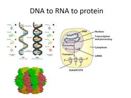
/
Cell Unit III: Cell Division, Cell Cycle, Transcription and Translation Cell Unit III: Cell Division, Cell Cycle, Transcription and Translation

Cell Unit III: Cell Division, Cell Cycle, Transcription and Translation - PowerPoint Presentation

roberts
roberts . @roberts
Follow
347 views
Uploaded On 2022-06-20

Cell Unit III: Cell Division, Cell Cycle, Transcription and Translation - PPT Presentation

Chapters 12 13 16 17 Limits to Cell Growth The larger a cell becomes the more demands a cell places on its DNA If extra copies of DNA are not made an information crisis would occur The cell also has more trouble moving nutrients and wastes across the cell membrane ID: 921133

cell dna cells amp dna cell amp cells chromosomes amino called division protein rna cycle meiosis mrna nucleotides genetic

Share:

Link:

Embed:

Download Presentation from below link

Download Presentation The PPT/PDF document "Cell Unit III: Cell Division, Cell Cycle..." is the property of its rightful owner. Permission is granted to download and print the materials on this web site for personal, non-commercial use only, and to display it on your personal computer provided you do not modify the materials and that you retain all copyright notices contained in the materials. By downloading content from our website, you accept the terms of this agreement.


Presentation Transcript

Slide1

Cell Unit III: Cell Division, Cell Cycle, Transcription and Translation

Chapters 12, 13, 16, 17

Slide2

Limits to Cell Growth

The larger a cell becomes, the more demands a cell places on its DNA

If extra copies of DNA are not made, an “information crisis” would occur

The cell also has more trouble moving nutrients and wastes across the cell membrane

Food, oxygen, water, and wastes move through the cell membrane

The rate at which the exchange takes place depends on the surface area of the cell

The rate at which food and oxygen are used up and wastes produced depends on the cell’s volume

Slide3

Ratio of Surface Area to Volume

Volume increases much more rapidly than surface area causing the ratio of surface area to volume to decrease

This decrease creates serious problems for the cell such as:

Inability to remove wastes from the cell

Lack of sufficient oxygen and food entering through the cell membrane

Slide4

Division of the Cell

The process by which a cell divides into two new daughter cells is called cell division

Before cell division occurs, the cell replicates, or copies, all of its DNA

Each daughter cell gets one complete set of genetic information

Each daughter cell also has an increased ratio of surface area to volume

Slide5

Slide6

Cell Division

Each cell has only one set of genetic information

must be copied before cell division begins

The first stage, division of the cell nucleus, is called mitosis

The second stage, division of the cytoplasm, is called cytokinesis

Reproduction by mitosis is classified as asexual

Mitosis is the source of new cells when a multicellular organism grows and develops

Slide7

Slide8

Chromosomes

Chromosomes are made of DNA (genetic information) and proteins (histones)

The cells of every organism have a specific number of chromosomes

Fruit flies = 8, human = 46, carrots = 18

Chromosomes are not visible in most cells except during cell division

Each chromosome consists of two identical sister chromatids which separate during cell division

Each pair of chromatids is attached in an area called the centromere

Slide9

Slide10

The Cell Cycle

Interphase is the period in between periods of cell division

The cell cycle is the series of events that cells go through as they grow and divide

During the cell cycle, a cell grows, prepares for division, and divides to form two daughter cells, each of which then begins the cycle again

The cell cycle consists of four phases

M, S, G

1

, and G

2

Slide11

The Cell Cycle

Slide12

The Cell Cycle

Slide13

Events of the Cell Cycle

During the normal cell cycle, interphase can be quite long, whereas the process of cell division takes place quickly

The G

1

phase is a period in which cells do most of their growing

In the S phase, chromosomes are replicated and the synthesis of DNA molecules takes place

During the G

2

phase, many of the organelles and molecules required for cell division are produced

Slide14

Slide15

Mitosis

Prophase:

Chromosomes become visible, centrioles begin to organize the spindle and move to opposite ends of the cell, fibers attach to centromeres, nucleolus and nuclear envelope disappear

Metaphase:

Chromosomes line up across the center of the cell

Anaphase:

Centromeres split and individual

chromatids

are separated into two groups near the poles

Telophase:

Chromosomes disperse, nuclear envelope and nucleolus re-form, spindle breaks apart

Slide16

Mitosis

Slide17

Cytokinesis

Cytokinesis

is the division of the cytoplasm itself and usually occurs at the same time as

telophase

In most animal cells, the cytoplasm is drawn inward until the cytoplasm is pinched into two nearly equal parts. This is called a cleavage furrow.

In plants, a structure known as the cell plate forms midway between the divided nuclei

Slide18

Slide19

Cytokinesis in Animal Cells

Slide20

Controls on Cell Division

When placed on a petri dish with a thin layer of nutrient solution, cells will grow until they form a thin layer on the bottom of the dish

When cells come into contact with other cells, they respond by not growing

If cells are removed from the center of the dish, the cells bordering the open space will divide until they have filled the space

Controls on cell growth and division can be turned off and on

Slide21

Cell Cycle Regulators

Several scientists discovered that cells in mitosis contained a protein that when injected into a

nondividing

cell, would cause a mitotic spindle to form

They called this protein

cyclin

because it seemed to regulate the cell cycle

Cyclins

regulate the timing of the cell cycle in eukaryotic cells

Proteins that respond to events inside the cell are called internal regulators

External regulators respond to events outside of the cell

Slide22

Cell Cycle Regulators

Slide23

Uncontrolled Cell Growth

Cancer is a disorder in which some of the body’s own cells lose the ability to control growth

Cancer cells do not respond to the signals that regulate the growth of most cells

They divide uncontrollable and form masses of cells called tumors that can damage the surrounding tissues

Causes include smoking, radiation, and viral infections

Damaged or defective p53 genes cause the cells to lose the information needed to respond to signals that would normally control their growth

Slide24

p53

is a protein that functions to block the cell cycle if the DNA is damaged. If the damage is

severe,

this protein can cause apoptosis (cell death).

p53 levels are increased in damaged cells. This allows time to repair DNA by blocking the cell cycle.

A p53 mutation is the most frequent mutation leading to cancer.

p27

is a protein that binds to

cyclin

and

cdk

blocking entry into S phase. Recent research (

Nature Medicine

3, 152 (1997)) suggests that breast cancer prognosis is determined by p27 levels. Reduced levels of p27 predict a poor outcome for breast cancer patients.

Slide25

Slide26

Uncontrolled Cell Growth

Slide27

CHAPTER 13: MEIOSIS AND SEXUAL CYCLES

Meiosis

- cell division that reduces the diploid # to the haploid # in the formation of sex cells (gametes).

Example (

Humans)

- 46 chromosomes is reduced to 23.

MOST IMPORTANT

- the cells produced at the end of meiosis contain one chromosome of each

homologous

(matching) pair.

Slide28

GENE

-

HEREDITARY INFORMATION, IN A SECTION OF A DNA

MOLECULE ON A CHROMOSOME.

LOCUS (LOCI)

- A GENE’S SPECIFIC LOCATION ON A CHROMOSOME.

TERMS:

CLONE

- A GROUP OF GENETICALLY IDENTICAL INDIVIDUALS

(

WHAT MITOSIS PRODUCES)

ASEXUAL REPRODUCTION

- REPRODUCTION

W/O

SEX (NO MALE/FEMALE;

1

PARENT; OFFSPRING

IS A CLONE OF PARENT.

HOMOLOGOUS CHROMOSOMES

- A MATCHING PAIR ALWAYS ONE FROM EACH PARENT.(one paternal/ one maternal.)

Slide29

AUTOSOMES

- CHROMOSOMES NOT DIRECTLY INVOLVED IN DETERMINING SEX. (IN

HUMANS: 22

HOMOLOGOUS PAIR).

SEX CHROMOSOMES

- THE CHROMOSOMES DIRECTLY INVOLVED IN DETERMINING SEX (IN HUMANS THE LAST HOMOLOGOUS PAIR).

(a) CALLED (X) & (Y) CHROMOSOMES.

(b) XX = FEMALE & XY = MALE.

FERTILIZATION

(or SYNGAMY)

- UNION OF GAMETES.

KARYOTYPE:

DISPLAY

OF AN INDIVIDUAL’S CHROMOSOMES. CHROMOSOMES ARE COLLECTED DURING

METAPHASE

.

THIS IS DONE BY NUMBER, SIZE & TYPE CHROMOSOME.

(c) In other organisms:

(1) Insects (Grasshoppers, Roaches): X-O sex chromosomes. O represents no sex chromosome = Male

(2) Birds, Butterflies and some fish: Z-W sex chromosomes. Female gamete determines sex. Males are ZZ, Females are ZW

(3) Parthenogenesis – wasps, bees and ants. If the egg is fertilized it becomes a female and is diploid. If the egg is unfertilized it is male and haploid.

Slide30

THE HUMAN LIFE CYCLE:

(

characteristic of most animals)

Gametes are the only haploid cells.

The diploid zygote divides by mitosis producing a diploid organism.

Slide31

MEIOSIS STEPS

:

(

FIG. 13.5.)

(a) Each chromosome replicates. (This shows 1 homologous pair). Remember - sister chromatids & centromere.

(b)

Meiosis I

segregates the homologous pair into 2 different cells (each new daughter cell is in

HAPLOID

).

(c )

Meiosis II

separates sister chromatids into chromosomes. No chromosome duplication)

Slide32

MEIOSIS TERMS:

Synapsis

- ( in

prophase I

) - the duplicated chromosomes pair with their Homologues). This is a

PROCESS.

Homologous chromosomes made of two sister chromatids come together as pairs.

Homologue

- one of a homologous pair.

Tetrad

- the four closely associated chromatids of a homologous pair together. This happens during synapsis.

Crossing over

- (

a process

) reciprocal exchange of genetic material between

nonsister

chromatids

.

Slide33

COMPARING

MITOSIS & MEIOSIS.

MEIOSIS

- Prophase I with -(a) Tetrad & synapsis making a synaptonemal complex (b) Crossing over with the chiasma.

MITOSIS

- No tetrads,

synapsis

, or

crossing over.

DAUGHTER CELL DIFFERENCE -

Mitosis has produced 2 identical cells. Meiosis produced daughter cells with one of each homologous pair.

Slide34

SUMMARY

:

differences

between Mitosis & Meiosis.

Slide35

FIG. 13.8

-

This shows the most

important concept of meiosis (how it produces

genetic variation

in organisms).

INDEPENDENT ASSORTMENT:

At the end of meiosis chromosome pairs distribute themselves independently of one another. This causes 4 different combinations of chromosomes with 2 homologous pair.

Slide36

1st MEIOTIC DIVISION

RESULTS IN INDEPENDENT ASSORTMENT OF MATERNAL & PATERNAL CHROMOSOMES IN DAUGHTER CELLS.

FORMULA:

The number of combinations possible when chromosomes assort independently into gametes during meiosis is 2

n

, where (n) is the haploid # in the organism.

EXAMPLE

-

Human haploid (n) is 23. 2

23

is over 8 million. A male can produce 8 million genetically different combinations of sperm & a female 8 million combinations of eggs.

RANDOM FERTILIZATION

then would produce 8 million x 8 million(over 64 Trillion) possibly different genetic combinations in the offspring.

Slide37

Crossing

Over -

produces individual chromosomes that combine genes inherited from our two parents.

Independent Assortment, Random Fertilization, & Crossing Over

- result ways that genetic variation can be produced.

Slide38

SUMMARY:

Prophase I & Anaphase I

produce the most variation in the 4 new daughter cells.

If clones were genetically different, this would be due to mutation (change in the code of DNA).

Remember these !!!!

Which might be a daughter cell of meiosis I ?

Which might be a daughter cell of meiosis II?

Slide39

CHAPTER

16 - THE

MOLECULAR BASIS OF INHERITANCE

DNA

- most celebrated molecule of all time. It is made of nucleic acids that have the unique ability to direct their own replication.

PROBLEM

: Since a chromosome is made of protein & DNA which one is carrying the genetic material

?

There

can be an infinite # of proteins so it would be a prime candidate to carry genetic material.

Slide40

JAMES WATSON

– CO-FOUNDER OF THE STRUCTURE OF DNA

Watson & Crick working on the DNA structure model. (April 1953)

Slide41

Transformation

of Bacteria - Frederick Griffin.(1928)

The

captions under the picture is all that is needed to explain this experiment.

TRANSFORMATION

- the change in genotype & phenotype due to the assimilation of external DNA by a cell.

Slide42

EVIDENCE THAT VIRAL DNA CAN PROGRAM CELLS (FIG. 16.2)

Virus is made of a protein coat & DNA core. Virus injects DNA into a

Bacteriophage

. DNA coat has radioactive protein coat (S

35)

while DNA is radiated with (P

32

).

HERSHEY-CHASE EXPERIMENT

That

the bacteria are called T2 Phages.

Slide43

ADDITIONAL EVIDENCE THAT DNA IS THE GENETIC MATERIAL OF CELLS

Erwin Chargaff

- Said that the bases of DNA (A, T, C, G) vary from one species to another.

He

also found a regular ratio of bases. (A approximately = T; and G approx. = C). This was known as

Chargaff’s

Rules

.

NOTE:

All these discoveries were before Watson & Crick discovered the double helix structure of DNA.

Slide44

Structure

of a DNA strand

DNA is composed of nucleotides ( 5 carbon sugar, phosphate & a nitrogenous base (A,T,C,G). Phosphate of one nucleotide is attached to the sugar of the next nucleotide.

Slide45

Fig. 16.5 (a) The Double Helix Structure of DNA.

Adenine

(A) is always paired with Thymine (T) & Guanine (G) is always paired with Cytosine (C).

The

nitrogenous bases are held together with Hydrogen bonds (weak).

We

even know the distances between steps of the DNA rungs

. What’s a nm?

Slide46

Notice

the strands are oriented in opposite directions.

This entire structure was worked out by Watson & Crick in 1953 with help from Rosalind Franklin’s x- ray diffraction photo of

DNA

Slide47

Base

Pairing in DNA

.

A

& G are double ring compounds called

Purines

.

T

& C are single ring compounds called

Pyrimidines

.

Each

rung of DNA is made of a

Purine

attached to a

Pyrimidine

. Held together by H bonds.

Slide48

The

SEMICONSERVATIVE

MODEL

- DNA replication model

Slide49

Meselson

& Stahl tested the three hypothesis's on DNA

replication

Slide50

B

eginnings

of how DNA Replicates.

Elongation of DNA at a replication fork is catalyzed by a enzyme called

DNA polymerase.

Rate of elongation in humans is approx.50/sec.

Slide51

Adding a Nucleotide:

A

similar molecule to ATP (NTP) is used to link the new nucleotide to the proper position.

The enzyme that catalyzes the reaction is

DNA POLYMERASE.

Slide52

THE

TWO STRAND OF DNA ARE

ANTIPARALLEL

Know:

Where

the 5’ & 3’ end are.

PROBLEM:

DNA polymerase can ONLY add nucleotides to the free 3’ end of a growing DNA strand.

So

..A new DNA strand can only elongate in the 5’ to 3’ direction.

Slide53

SYNTHESIS

OF LEADING & LAGGING STRANDS DURING DNA REPLICATION.

DNA

polymerase is adding new DNA fragments in a 5’ to 3’ direction continuously along a replication fork, adding to the 3’ end.

Lagging

strand is synthesized in segments called Okazaki fragments. DNA

ligase

joins the fragments into a single DNA strand.

Okazaki fragments are about 100 -200 nucleotides long in eukaryotes.

Slide54

PRIMING

DNA SYNTHESIS

DNA polymerase cannot initiate a polynucleotide strand; it can only add to the 3’ end of an already-started strand.

The

primer is a short segment of RNA synthesized by the enzyme

primase

.

Each primer is eventually replaced by DNA.

Slide55

Slide56

FIG. 16.15 - THE MAIN PROTEINS OF REPLICATION & THEIR FUNCTIONS.

DNA must also be able to form complementary base pairs with both DNA & RNA nucleotides. The sequence of nucleotides will be decoded into a sequence to make amino acids into proteins

.

Replication -> Transcription -> Translation

Slide57

Enzymes

must proofread

DNA during its Replication and repair damage in existing DNA.

Mismatch Repair

fixes mistakes made in DNA. DNA polymerase itself carries out the mismatch repair.

Telomeres

- special sequences of DNA nucleotides found at the end of the DNA molecule. They do not contain genes. They protect the organism’s genes from being eroded through successive rounds of DNA replication

.

Secret to

aging?

http://www.youtube.com/watch?v=J9QApCHsrJk&feature=related

Slide58

Image of Telomere

squeneces

(yellow) on chromosomes

Slide59

Chapter 17 – From Gene to Protein

Transcription

- the synthesis of mRNA (messenger RNA) under the direction of DNA. This is a code to make a polypeptide (protein). This is also the synthesis of any RNA from DNA.

Translation

- the actual synthesis of a polypeptide (which occurs at the ribosomes.)

The difference

in Eukaryotic & Prokaryotic cells.

Gene to RNA to Protein.

Slide60

Basics of the Genetic Code:

1. There is a total of 20 amino acids possible in any protein.

2. 3 Nucleotides on mRNA code for an amino acid. This is called the

triplet

code.

3. Only one strand of DNA is transcribed into mRNA. This strand is called the TEMPLATE strand. The other strand is called the complementary strand.

4. All Translation & Transcription occur in a 3’ to 5’ direction.

5. The mRNA is in triplet bases called CODONS.

Slide61

mRNA

is only a single helix & that

Uracil

(U) is a substitute for Thymine (T).

The

number of nucleotides making up a genetic message must be 3 times the number of amino acids making up the protein.

EXAMPLE

- 4 amino acids

= 12

nucleotides

.

Amino Acids are connected by polypeptide bonds.

Slide62

Learn to read this!!!

AUG

codon

is a start

codon

& the amino acid

Methionine

(Met).

Start

Codon

begins the sentence &

UAA,UAG & UGA = no amino acid but stops the amino acid chain (read in a 5’ to 3’

direction)= STOP CODON, like the period at the end of a sentence

Slide63

Fig. 17.6 The Stages of Transcription

1. RNA binds to the promoter region of DNA (several dozen nucleotides “upstream” from the transcription startpoint).

2. RNA moves “downstream” from promoter, unwinding DNA & elongating RNA at the 3” end (5’ to 3’ direction).

Slide64

3. RNA polymerase transcribes a terminator (this sequence of nucleotides along DNA signals the end of transcription unit.)

4. Eventually RNA is released & the polymerase moves from DNA.

5.

Prokaryotes -

RNA transcript immediately used to make protein.

6.

Eukaryotes

- mRNA will undergo additional processing.

Progresses at about 60 nucleotides/sec in Eukaryotes.

Slide65

RNA

Processing 1st step:

Enzymes modify 2 ends of a eukaryote pre-mRNA molecule.

Cap made of modified guanosine triphosphate added to the 5’ end of RNA.

A Poly(A) tail consiting of 200 adenine nucleotides attached to 3’ end

.

( may helps export mRNA from the nucleus.)

***Role of Cap and Tail - protect

RNA from

degradation****

The leader,

trailer & termination signal.

Leader

& trailer are not translated.

Slide66

RNA

processing (splicing).

Pre-mRNA -

Exons

(Expressed sequence) are keep & the

Introns

(Intervening sequence) are removed (both by enzymes).

Exons

are then spliced together. We now have the processed RNA ready to leave the nucleus & go to the ribosome for translation

.

Slide67

Translation - Basic Concept:

1)

tRNA

picks up amino acids & transport them to the

ribosome

2) Each

tRNA

has an

anticodon

(3 letters) that pick up one of the twenty amino acids.

3) When

the

tRNA’s

deliver their amino acid, they add them to a growing polypeptide chain.

tRNA’s

are now available to pick up another amino acid to repeat the process.

4) New Polypeptide

chain added in the 5’ to 3’ direction.

Slide68

The

Anatomy of a

Ribosome:

Ribosomes

are made of 2 subunits each made of many molecules or

rRNA

(ribosomal

RNA) and proteins.

The

sites on the

ribosome:

(1)

P site

- holds the

tRNA

attached to the growing

polypeptide

(2)

A site

- holds the

tRNA

carrying the amino acid to be added to the polypeptide

chain

(3

) Discharged

tRNA

leaves via the

E site.

Peptide bonding between amino acids maintains the shape of

tRNA

.

Slide69

Fig. 17.15 Initiation of Translation

1. Small ribosomal subunit binds to molecule of mRNA.

2. Initiator tRNA with anticodon UAC base-pairs with the start codon, AUG carrying the amino acid Met.

3. A large ribosomal unit arrives & completes the initiation complex.

4. Initiator tRNA is in the P site. A site is available to tRNA carrying the next amino acid.

5.

Proteins

called initiation factors bring translation components together. GTP provides the energy for all this.

Slide70

GTP

& proteins called elongation factors needed to drive this process.

Slide71

Termination

of Translation

1. When ribosome reaches a termination codon on mRNA, the (A) site of ribosome accepts a protein called a release factor instead of tRNA.

2. Release factor hydrolyzes the bond between tRNA in the P site & the last amino acid of the chain. This frees the polypeptide from the ribosome.

3.

The

2 ribosomal subunits dissociate

Slide72

Fig. 17.18 Polyribosomes

A. An mRNA molecule is generally translated together with several ribosomes in clusters called

polyribosomes.

B. This enables a single mRNA to make many copies of a polypeptide simultaneously.

Slide73

Proteins

can be chemically modified by attachment of sugars, lipids, phosphate groups etc.

Example:

Enzymes may remove leading amino acids from a chain. Sometimes several proteins will join together to allow them to function or one protein may split into several proteins.

Proteins

formed here are only the primary structure & must develop a secondary, tertiary, or even Quaternary structure.

Slide74

Transcription

& Translation in

Bacteria:

Bacteria

(

Prokaroytes

) have no nucleus, so mRNA does not need to move through the membrane to the ribosome.

Streamlined

operations here - Transcription & Translation can be occurring at the same time.

3. There

is not RNA processing in bacteria. (All

exons

).

Slide75

MUTATIONS:

Changes in the genetic code of DNA.

Point Mutations:

Chemical changes in just one or a few base pairs in a single gene.

If a point mutation occurs in a

gamete

or cells giving rise to

them,

it could be transmitted to offspring & future generations.

TYPES OF MUTATIONS:

1.

Base-pair substitution

- replacement of one nucleotide & its partner in the complementary DNA strand with another pair of nucleotides.

Some substitutions are

silent mutations

since genetic code is redundant, there may be no change in the amino acid coded for.

EXAMPLE:

CCG mutated to CCA would make mRNA GGC

become

GGU which is still

glycine

.

Slide76

2.

Missense

Mutation

-

altered

codon

still codes for an amino acid & makes sense although not necessarily the RIGHT sense

. (Make a protein, just not the correct one)

3.

Nonsense mutation -

Alterations that change an amino acid code to a stop

codon

. Almost always leads to a nonfunctional protein.

Slide77

4.

Insertions & deletions

are additions or losses of one or more nucleotide pairs in a gene.

a.

Note this can cause

missense

or nonsense.

Where

the

amino

acid is incorrect in a chain can be important or not.

Frameshift

mutation

- alters “reading frame” of message (# of nucleotides inserted or deleted is not a multiple of 3.

a. (

the big cat) remove the h =

teb

igc

at_.) This will make

all

amino acids downstream from this incorrect.

What can cause Mutations?:

Mutagens

- Physical & chemical agents that cause

mutations or increase the mutation rate.

Examples - X-rays, Radiation, UV light, chemicals (pesticides, radon), Viruses & Bacteria