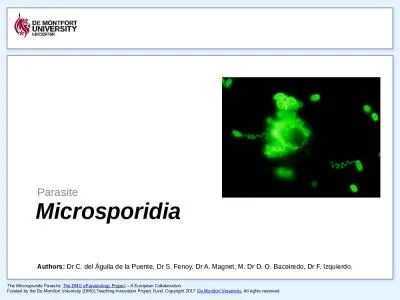
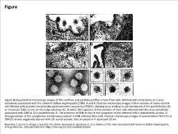
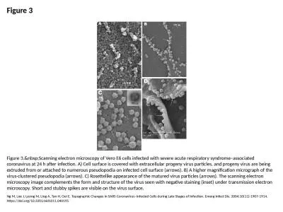
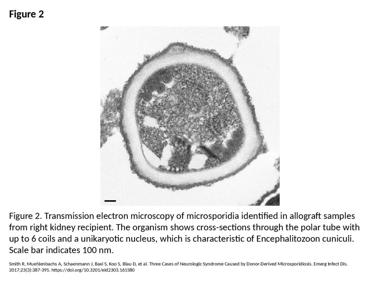
PPT-Figure 2 Figure 2. Transmission electron microscopy of microsporidia identified in allograft
Author : rodriguez | Published Date : 2023-07-19
Smith R Muehlenbachs A Schaenmann J Baxi S Koo S Blau D et al Three Cases of Neurologic Syndrome Caused by DonorDerived Microsporidiosis Emerg Infect Dis 2017233387395
Presentation Embed Code
Download Presentation
Download Presentation The PPT/PDF document "Figure 2 Figure 2. Transmission electron..." is the property of its rightful owner. Permission is granted to download and print the materials on this website for personal, non-commercial use only, and to display it on your personal computer provided you do not modify the materials and that you retain all copyright notices contained in the materials. By downloading content from our website, you accept the terms of this agreement.
Figure 2 Figure 2. Transmission electron microscopy of microsporidia identified in allograft: Transcript
Smith R Muehlenbachs A Schaenmann J Baxi S Koo S Blau D et al Three Cases of Neurologic Syndrome Caused by DonorDerived Microsporidiosis Emerg Infect Dis 2017233387395 httpsdoiorg103201eid2303161580. Fire Protection Laboratory Methods Day. June 25, 2014. Paul M. Anderson. Graduate Research Assistant. University of Maryland. Department of Fire Protection Engineering. Transmission Electron Microscopy. Replica of van 1670 Moody Use the information in this tutorial to supplement the visuals in lab and the Chapters 1 8 and 9 in your lab manual Replica of Culpepper tripod microscope built c 1725 by Co . . del . Águila . de la Puente, . Dr. S. . . Fenoy. , . Dr. A. . . Magnet. , . M. . Dr. D. . O. . Baceiredo. , . Dr. F. . Izquierdo.. 1. Infectious Agent and Morphology. Microsporidia are obligate intracellular parasites with a unique mode of entering host cells via a polar tube. They are true eukaryotes with a nucleus, chromosome separation and an intracytoplasmic membrane system. However, they lack a classical stacked Golgi apparatus, centrioles, peroxisomes and the mitochondria have been substituted by a mitochondria-like organelle called . Peter Harris. Transmission . electron . microscopy. ► What . is transmission electron microscopy?. . ► TEM in biological science . ► CfAM’s TEMs. ► TEM in physical . science . ► Recent developments in TEM. Outline. Introduction. Optical Microscopy. Types of Optical Microscopes. Confocal Microscopy. Laser Scanning Confocal Microscopy. Examples. Optical Microscopy. Optical microscopy is the oldest type of microscope and uses visible light and a system of lenses to create an image of the sample. parasites . Cyclospora. (. Cyclosporosis. ). The first human cases of . Cyclospora. . . cayetanenis. were reported in 1979.. It was originally referred to as . . cyanobacteria. -like bodies or coccidian-. Scanning electron microscope (SEM. ). 2- Transmission Electron Microscope. New Techniques in Microscopy:. Confocal Microscopy (Confocal Scanning Laser Microscope). Light Microscope. Electron Microscope. . Electron microscopy (EM) . is an electron beam which is focused into a small probe across the surface of a specimen . . The first electromagnetic lens was developed in 1926 by . Hans Busch. .. . AIM : T THEORY: Microscope is the most commonly used piece of apparatus in the laboratory. It produces greatly enlarged images of minute objects. LIGHT MICROSCOPE A Light Microscope can be simple or Riquelme C, Escors D, Ortego J, Sanchez CM, Uzelac-Keserovic B, Apostolov K, et al. Nature of the Virus Associated with Endemic Balkan Nephropathy. Emerg Infect Dis. 2002;8(8):869-870. https://doi.org/10.3201/eid0808.020042. O'Rourke LG, Pitulle C, Hegarty BC, Kraycirik S, Killary KA, Grosenstein P, et al. Bartonella quintana in Cynomolgus Monkey (Macaca fascicularis). Emerg Infect Dis. 2005;11(12):1931-1934. https://doi.org/10.3201/eid1112.030045. Ng M, Lee J, Leong M, Ling A, Tan H, Ooi E. Topographic Changes in SARS Coronavirus–infected Cells during Late Stages of Infection. Emerg Infect Dis. 2004;10(11):1907-1914. https://doi.org/10.3201/eid1011.040195. The aims of this course are:. ● . to introduce the principles of scanning electron microscopy. ● . to describe the components of the microscope and explain how they work. ● . to highlight some of the problems which can arise during imaging. Peter Aspinall. http://www.zyvexlabs.com/EIPBNuG/EIPBN%20images/05Ion.jpg. Intro to Electron Microscopy. Similar to optical microscopy except with electrons rather than photons. Used to image samples with a resolution of 10 Å.
Download Document
Here is the link to download the presentation.
"Figure 2 Figure 2. Transmission electron microscopy of microsporidia identified in allograft"The content belongs to its owner. You may download and print it for personal use, without modification, and keep all copyright notices. By downloading, you agree to these terms.
Related Documents