Explore
Featured
Recent
Articles
Topics
Login
Upload
Featured
Recent
Articles
Topics
Login
Upload
Search Results for 'figure patient'
figure patient published presentations and documents on DocSlides.
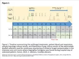
Figure 1 Figure 1. Timeline summarizing the antifungal treatments, patient blood and respiratory sa
by emmy
Lemaire B, Normand A, Forel J, Cassir N, Piarroux ...
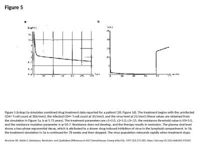
Figure 5 Figure 5. 5a simulates combined drug treatment data reported for a patient (18; F
by ximena
Kirschner DE, Webb G. Resistance, Remission, and Q...
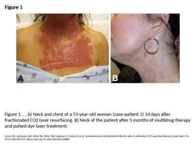
Figure 1 Figure 1. . . A) Neck and chest of a 53-year-old woman (case-patient 1) 14 days after frac
by kaizen
Culton DA, Lachiewicz AM, Miller BA, Miller MB, Ma...
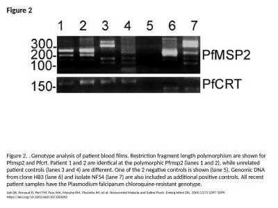
Figure 2 Figure 2. . Genotype analysis of patient blood films. Restriction fragment length polymorp
by jainy
Jain SK, Persaud D, Perl TM, Pass MA, Murphy KM, P...
Figure 1 Location of the
by anderson
colon in the body. Ulcerative Colitis: Introd...
Figure 1Cholangiogram with Large Filling Defects (Stones)
by cleverfan
Endoscopic View of CRE Direct Visualization System...
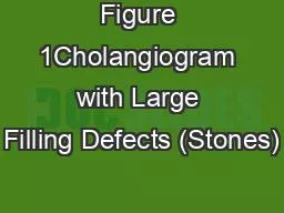
Figure 1 Figure 1. Histologic patterns of cutaneous Kaposi sarcoma (KS) associated with a
by reuben975
Cassar O, Blondot M, Mohanna S, Jouvion G, Bravo F...
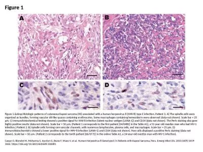
Figure 3 Figure 3. Immunostaining of severe acute respiratory syndrome coronavirus 2 in pulmonary t
by alan
Martines RB, Ritter JM, Matkovic E, Gary J, Bollwe...
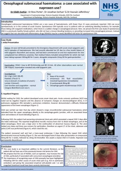
Oesophageal submucosal haematoma: a case associated with naproxen use?
by jovita
Dr Eilidh Duthie. 1. , Dr Ross Porter. 1. , Dr Jon...
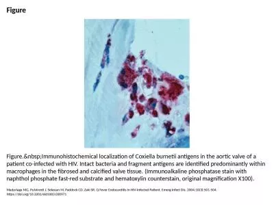
Figure Figure. Immunohistochemical localization of Coxiella burnetii antigens in the aorti
by gelbero
Madariaga MG, Pulvirenti J, Sekosan M, Paddock CD,...
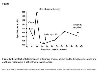
Figure Figure. Effect of tularemia and anticancer chemotherapy on the lymphocyte counts an
by arya
Han XY, Ho LX, Safdar A. Francisella tularensis Pe...
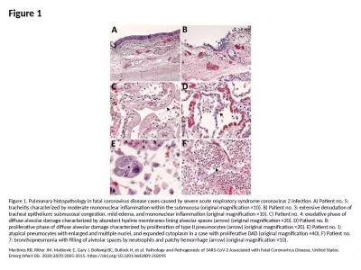
Figure 1 Figure 1. Pulmonary histopathology in fatal coronavirus disease cases caused by severe acu
by piper
Martines RB, Ritter JM, Matkovic E, Gary J, Bollwe...
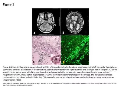
Figure 1 Figure 1. A) Magnetic resonance imaging (MRI) of the patient's brain showing a la
by angelina
Shirwadkar CG, Samant R, Sankhe M, Deshpande R, Ya...
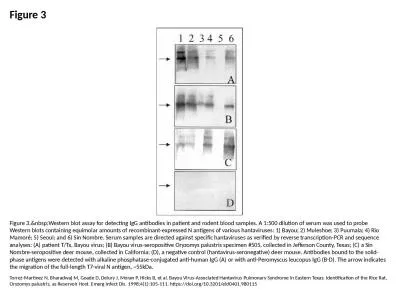
Figure 3 Figure 3. Western blot assay for detecting IgG antibodies in patient and rodent b
by naomi
Torrez-Martinez N, Bharadwaj M, Goade D, Delury J,...
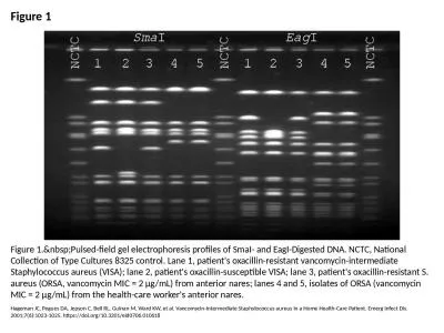
Figure 1 Figure 1. Pulsed-field gel electrophoresis profiles of SmaI- and EagI-Digested DN
by caitlin
Hageman JC, Pegues DA, Jepson C, Bell RL, Guinan M...
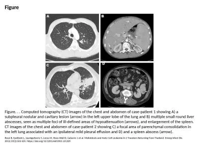
Figure Figure. . . Computed tomography (CT) images of the chest and abdomen of case-patient 1 showi
by belinda
Rossi B, Epelboin L, Jauréguiberry S, Lecso M, Ro...
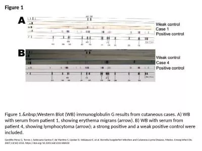
Figure 1 Figure 1. Western Blot (WB) immunoglobulin G results from cutaneous cases. A) WB
by ida
Gordillo-Pérez G, Torres J, Solórzano-Santos F, ...
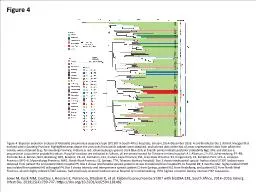
Figure 4 Figure 4. Bayesian evolution analysis of Klebsiella pneumoniae sequence type (ST) 307 in S
by jalin
Lowe M, Kock MM, Coetzee J, Hoosien E, Peirano G, ...
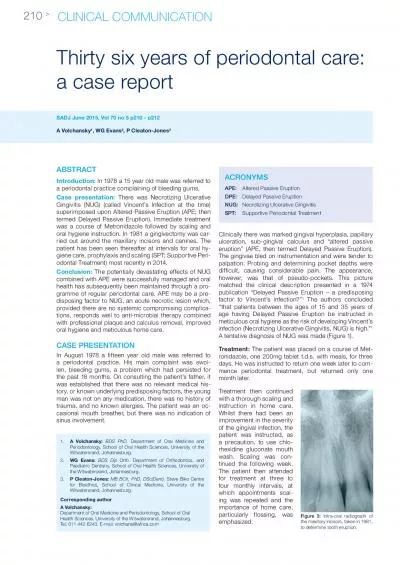
ABSTRntroduction In 1978 a 15 year old male was referred to a periodo
by molly
Thirty six years of periodontal care: a case repor...
CEDIRECTED READING
by brown
July/August 2007 Vol 78/No 6 RADIOLOGIC TECHNOLOGY...
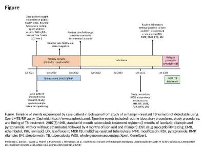
Figure Figure. Timeline of events experienced by case-patient in Botswana from study of a rifampin-
by brodie
Modongo C, Barilar I, Wang Q, Molefi T, Makhondo T...
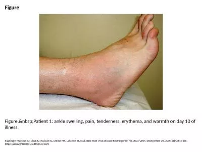
Figure Figure. Patient 1: ankle swelling, pain, tenderness, erythema, and warmth on day 10
by yahya
Klapsing P, MacLean JD, Glaze S, McClean KL, Drebo...
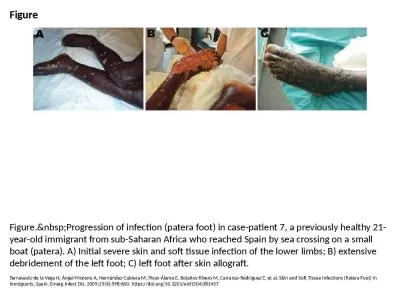
Figure Figure. Progression of infection (patera foot) in case-patient 7, a previously heal
by victor
Ternavasio-de la Vega H, Ángel-Moreno A, Hernánd...
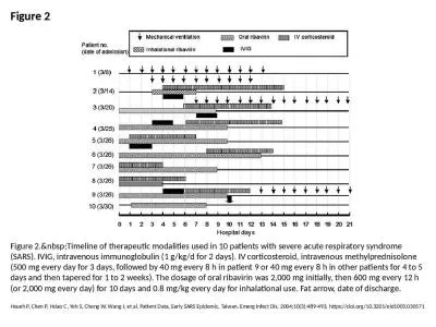
Figure 2 Figure 2. Timeline of therapeutic modalities used in 10 patients with severe acut
by erick524
Hsueh P, Chen P, Hsiao C, Yeh S, Cheng W, Wang J, ...
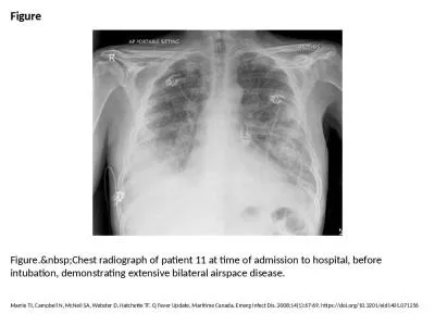
Figure Figure. Chest radiograph of patient 11 at time of admission to hospital, before int
by bryson173
Marrie TJ, Campbell N, McNeil SA, Webster D, Hatch...
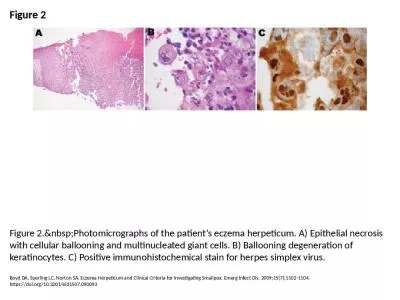
Figure 2 Figure 2. Photomicrographs of the patient’s eczema herpeticum. A) Epithelial ne
by ulises
Boyd DA, Sperling LC, Norton SA. Eczema Herpeticum...
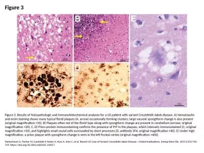
Figure 3 Figure 3. Results of histopathologic and immunohistochemical analyses for a US patient wit
by badra
Maheshwari A, Fischer M, Gambetti P, Parker A, Ram...
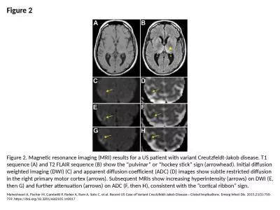
Figure 2 Figure 2. Magnetic resonance imaging (MRI) results for a US patient with variant Creutzfel
by carny
Maheshwari A, Fischer M, Gambetti P, Parker A, Ram...
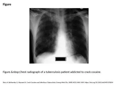
Figure Figure. Chest radiograph of a tuberculosis patient addicted to crack cocaine.
by mia
Story A, Bothamley G, Hayward A. Crack Cocaine and...
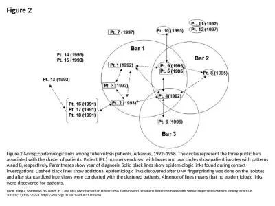
Figure 2 Figure 2. Epidemologic links among tuberculosis patients, Arkansas, 1992–1998.
by lauren
Ijaz K, Yang Z, Matthews HS, Bates JH, Cave MD. My...
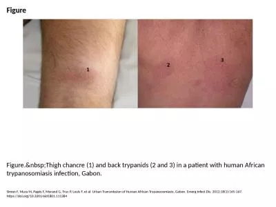
Figure Figure. Thigh chancre (1) and back trypanids (2 and 3) in a patient with human Afri
by martin
Simon F, Mura M, Pagès F, Morand G, Truc P, Louis...
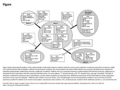
Figure Figure. Schematic illustration of the epidemiologic relationships between patients
by oconnor
Chiu RW, Chim SS, Tong Y, Fung KS, Chan P, Zhao G,...
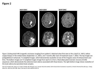
Figure 2 Figure 2. Axial MRI (magnetic resonance imaging) from patient 2 obtained when fir
by cora
Sohn AH, Probert WS, Glaser CA, Gupta N, Bollen AW...
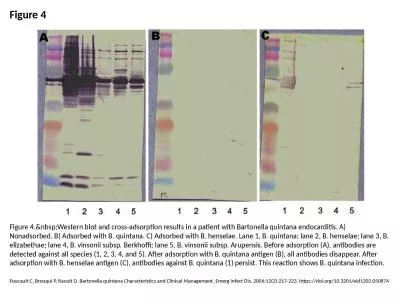
Figure 4 Figure 4. Western blot and cross-adsorption results in a patient with Bartonella
by scarlett
Foucault C, Brouqui P, Raoult D. Bartonella quinta...
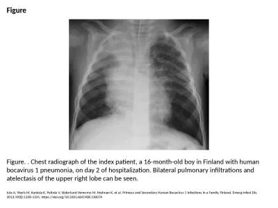
Figure Figure. . Chest radiograph of the index patient, a 16-month-old boy in Finland with human bo
by morgan
Jula A, Waris M, Kantola K, Peltola V, Söderlund-...
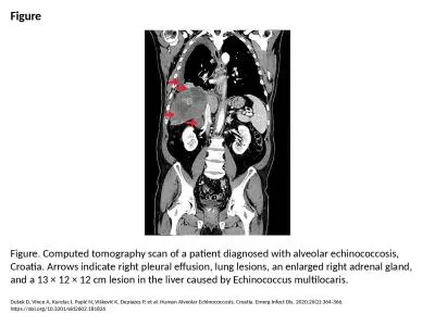
Figure Figure. Computed tomography scan of a patient diagnosed with alveolar echinococcosis, Croati
by freya
Dušek D, Vince A, Kurelac I, Papić N, Višković...
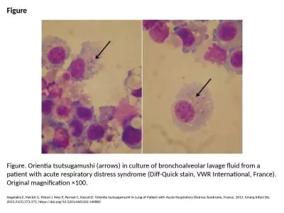
Figure Figure. Orientia tsutsugamushi (arrows) in culture of bronchoalveolar lavage fluid from a pa
by angelina
Angelakis E, Patrick G, Peloni J, Wey P, Perreal C...
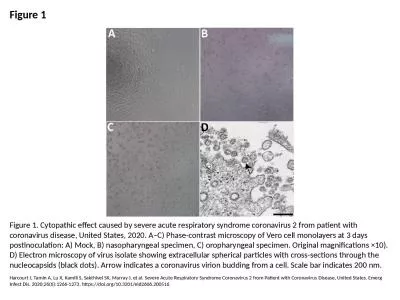
Figure 1 Figure 1. Cytopathic effect caused by severe acute respiratory syndrome coronavirus 2 from
by lam
Harcourt J, Tamin A, Lu X, Kamili S, Sakthivel SK,...
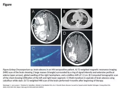
Figure Figure. Chrysosporium sp. brain abscess in an HIV-seropositive patient. A) T2-weigh
by davies
Steininger C, van Lunzen J, Tintelnot K, Sobottka ...
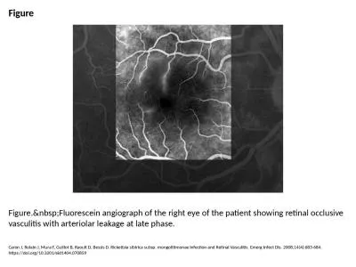
Figure Figure. Fluorescein angiograph of the right eye of the patient showing retinal occl
by berey
Caron J, Rolain J, Mura F, Guillot B, Raoult D, Be...
Load More...