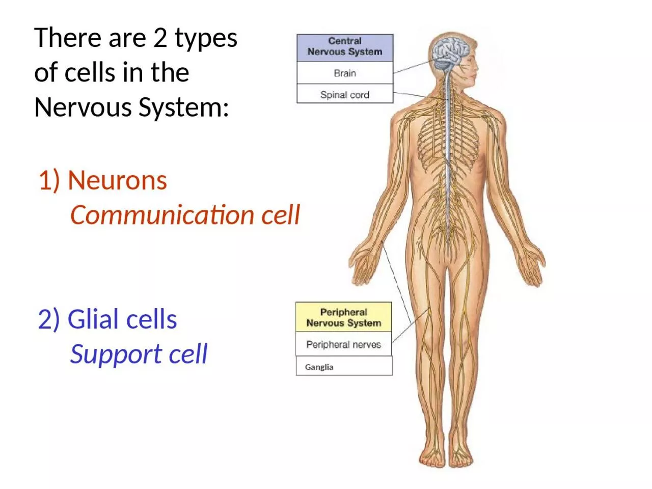
Nervous System 1 Neurons Communication cell 2 Glial cells Support cell Ganglia Incoming Info Processing Info Outgoming Info Typical Motor Neuron Receives incoming info ID: 1044095
Download Presentation The PPT/PDF document "There are 2 types of cells in the" is the property of its rightful owner. Permission is granted to download and print the materials on this web site for personal, non-commercial use only, and to display it on your personal computer provided you do not modify the materials and that you retain all copyright notices contained in the materials. By downloading content from our website, you accept the terms of this agreement.
1. There are 2 typesof cells in the Nervous System:1) Neurons Communication cell2) Glial cells Support cell Ganglia
2. IncomingInfo.ProcessingInfo.OutgomingInfo.Typical MotorNeuronReceives incoming infoReleases the signalProcesses information
3. Sensory PathwayMotor PathwayInterneuron(Integration)(calculate what to do)CNSPNSReceptors(detect changes)EffectorTissue(respond to change)1. Sensory Neuron3. Motor Neuron2. InterneuronThe Feedback Loop
4. TelodendriaThe Motor/Multipolar NeuronHistology of a Neuron
5. AnaxonicBipolarUnipolarMultipolarImportant Anatomical Neurons
6. The 3 major types of Communications coming from Neurons
7. Axon Myelination
8. Glial CellsThe Glial Cells
9. Nervous System TerminologyGray Matter – mostly nerve cell bodies.White Matter – mostly myelinated axons.Nerve fiber – a single axon of a neuron. Tract – a bundle of axons in the CNS.Ganglion – a cluster of nerve cell bodies in PNS.Nucleus – gray matter in CNS with discrete boundaries and common function.Nerve – a bundle of axons in the PNS. Columns – are groups of tracts.
10. Anatomy of a NerveNeurolemmaAxolemma
11. HistologyofNerves
12. What Nerves look like on models
13. What Nerves look like on cadavers
14. In the Spinal cord: White matter = columnsGray matter = hornsThere are 31 pairsof Spinal Nerves 8 Cervical (C1-C8) 12 Thoracic (T1-T12) 5 Lumbar (L1-L5) 5 Sacral (S1-S5) 1 Coccygeal (Co1)
15. Anatomical Features of the Spinal CordShallow posterior median sulcusDeep anterior median fissureCervical and lumbar enlargementsTypically ~18 inches in lengthConus medullaris (= end of cord)
16. Cauda Equina - extension of ventral and dorsal roots (horses tail!).Filum Terminale extends to sacral vertebrae, joins coccygeal ligament, for length-wise stabilization.Denticulate Ligament (extension of dura to pia mater) for lateral (side to side) stabilization.
17. Spinal Plexus:(in the Nervous System)Cervical (C1-C5)Brachial (C5-T1)Lumbar (T12-L4 )Sacral (L4-S4)is a re-arrangement of spinal nerves into functional groups.
18. Cervical (C1-C5)Brachial (C5-T1)e.g. Phrenic n. (C3-C5)e.g. Radial n. (C5-8-T1)e.g. Ulnar n. (C8-T1)e.g. Musculocutaneous n. (C5-C7)e.g. Median n. (C5-8-T1)
19. Lumbar (T12-L4 )Sacral (L4-S4)e.g. Femoral n. (L2-L4)e.g. Obturator n. (L2-L4)e.g. Sciatic n. (L4-S3)e.g. Tibial n. (L4-S3)
20. Spinal Meninges - Dura Mater- Arachnoid- Pia MaterCollagen and elastic fibersDense irregular CT +Simple squamous epitheliumSimple squamous epithelium
21. Lumbar Puncture
22. Cross Section of Spinal Cord HistologyCentral Canal filled with CSF lined with ependymal cellsNow draw the x.s. of spinal cord
23. Spinal Cord Anatomy (x.s.)
24.
25. The Spinal Cord Nuclei
26. Posterior gray horns:Somatic and Visceral Sensory nuclei.Anterior gray horns:Somatic Motor control.Lateral gray horns:Visceral Motor neurons.Gray commissuresAxons of interneurons crossing and unmyelinated axons.The Spinal Cord Nuclei
27. The Spinal Cord Nuclei
28. Spinal PathwaysSpinal Reflexes
29. Lou GehrigAmyotropic Lateral Sclerosis (ALS)