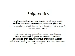
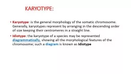
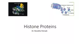
Chromatin is made of repeating units of nucleosomes which consist of 146 base pairs of DNA wrapped around an octamer of four core histone proteins H3 H4 H2A and H2B Introduction Histones are a special group of proteins found in the nuclei of eukaryotic cells responsible for DNA folding and ID: 784965
Download The PPT/PDF document " Histone Proteins Dr. Nivedita Patnaik" is the property of its rightful owner. Permission is granted to download and print the materials on this web site for personal, non-commercial use only, and to display it on your personal computer provided you do not modify the materials and that you retain all copyright notices contained in the materials. By downloading content from our website, you accept the terms of this agreement.
Slide1
Histone Proteins
Dr. Nivedita Patnaik
Slide2Chromatin is made of repeating units of nucleosomes, which consist of 146 base pairs of DNA wrapped around an octamer of four core
histone proteins (H3, H4, H2A and H2B)
Slide3Introduction
Histones are a special group of proteins found in the nuclei of eukaryotic cells responsible for DNA folding and chromatin formation.
Slide4Chemically they are-
highly alkaline basic proteins Histones are positively charged
abundance of positive amino-acids, arginine and lysine
Slide55
Slide6Classes of Histones
There are two main classes of Histones: Core Histones
Linker Histones Core Histones: In core histones following families are included H2A H2B contain more lysine H3
H4 contain more arginine
Two of each of these core histone proteins assembles to form one octameric nucleosome core particle, and 147 base pairs of DNA wrap around this core particle.
Slide7Linker Histones
Linker histone included: H1 H5 highest lysine/arginine ratio
The linker histone protein H1 binds the nucleosome at the starting and ending sites of the DNA, thus locking the DNA into place and help in the formation of higher order structure. H5 histones are individual proteins involve in the packaging of specific region of DNA.
Slide8Function of the histone protein in a chromosome
The DNA is housed in chromosomes in the form of nucleosomes It is basic unit of chromosome or chromatin fiber. It is DNA duplex coiled around a core of eight histone proteins
Positively charged histones are linked with negative charged phosphate groups of DNA
Slide9Some histone proteins function as spools for the thread-like DNA to wrap around
looks like beads on a string
Slide10The nucleosomes + H1 histones = 30 nm spiral Solenoid
It maintains the chromosomal structure
Slide11histone modifications
Histone proteins contain a globular C-terminal domain and an N-terminal tail
The N-terminal tails of histones can undergo a variety of posttranslational covalent modifications including methylation, acetylation, ubiquitylation,
sumoylation
and phosphorylation on specific residues
regulate key cellular processes such as transcription, replication and repair
Slide12Post-translational
histone modifications
12
A
= acetylation
M
= methylation
P
= phosphorylation
U
= ubiquitination
Unlike DNA methylation, histone modifications can lead to either activation or repression depending upon which residues are modified and the type of modifications present
Slide13Histone Acetylation & Deacetylation
Histone acetylation – Histone acetyl transferases (HATs) Adds acetyl groups to histone tails
Reduces positive charge and weakens interaction of histones with DNA Facilitates transcription by making DNA more accessible to RNA polymerase II Histone deacetylation – Histone deacetylases (HDACs) Removes acetyl groups from histone tails
Increases interaction of DNA and histones
Represses transcription
Slide14Acetylation
It is the introduction of an Acetyl functional group to the Lysine amino acid of the histone tail. These reactions are
catalyzed by enzymes with "histone acetyltransferase" (HAT) or "histone deacetylase" (HDAC) activity.
Slide15Effects of Acetylation
-ve charge on histone. reduces affinity of tail for adjacent nucleosomes
creating a transcription permissive environment increase the access of transcription factors
Slide16Methylation
It is the introduction of an Methyl functional group to Lysine or Arginine of the histone tail.
These reactions are catalysed by enzymes with "histone methyltransferase” ‘Arg’ can be methylated once or twice, and ‘Lys’ once, twice or thrice.
Slide17Histone Methylation
Histone methyl transferases (HMTs) Histone lysine methyl transferases(HKMTs) methylate
lys (K) residues Protein argenin methyl transferase (PRMTs) Methylate arge
(R) residues
Methylation can result in activation or repression of expression
trimethylation of histone H3 at lysine 4 (H3K4) is an active mark for transcription
dimethylation
of histone H3 at lysine 9 (H3K9), a signal for transcriptional silencing
Slide18Effects of methylation
Methylation does not neutralize charge but recruit silencing or regulatory proteins that bind methylated histones. Chromodomain containing proteins interact with methylated histone tails.
transcription repression
Slide19Histone phosphorylation (H3)
Histones are phosphorylated during mitosis.Histones are also phosphorylated by signal transduction pathways like the ERK pathway in response to external signals. It is not known how (and if) this phosphorylation contributes to gene expression.
Slide20Histone ubiquitylation
Addition and removal of Ub (a LARGE moiety) to histone tails –
Functions largely unknown in vertebratesubiquitylationH2A K119: repressionH2B K120: activation
H3 and H4: DNA repair (CUL4)
de-ubiquitylation –
Recrutiment
of other proteins in yeast
H2A Dub (PCAF)
H2B Ubp8 (SAGA)
Functions
: transcription
elongation
,
polycomb
repression
Slide21Study of histone modifications
Histone modifications are studied using the chromatin immunoprecipitation (ChIP) assay.
ChIP on chip is the high throughput form of the ChIP assay wherein the immunoprecipitated DNA, instead of being subject to the usual PCR, is hybridized to a microarray chip with printed oligonucleotides corresponding to various regions of the genome.
This helps to study the localization of a specific histone modification to various parts of the genome.
Slide22Slide23Slide24Epigenetic therapy
DNMT INHIBITORS
1.Nucleoside analogue inhibitors
2.Non nucleoside analogue inhibitors
3.Antisense oligonucleotides
HDAC INHIBITORS
1.Hydroxamates
2.Cyclic tetrapeptides
3.Aliphatic acids
4.Benzamides
Slide25DNMTi
Nucleoside analogs
5 aza cytidine,
5 aza 2' deoxy cytidine (
decitabine)
Leads to DNA demethylation
Reactivate epigenetically silenced genes
Used in MDS.
Zebularine, oral, less toxic, under trial
25
Slide26Epigenetic therapy
HDACi
SAHA (
Vorinostat
)
,
Depsipeptide
Leads to
hyperacetylation
Activation of silenced pro-apoptotic genes
Pro-apoptotic genes are
upregulated
Lead to apoptosis in cancer cells.
Cutaneous
T cell lymphoma
EZH2i
Deazaneplanocin
Activate apoptotic cell death in breast cancer
26
Slide27Slide28Slide29Slide30Primary
nonsmall cell lung cancer (NSCLC) is the main cause of malignancy-related mortality in Asian and Western populations.positive correlation between lower levels of H3K9ac, H3K9me3 and H4K16ac and
tumor recurrence. However when patients were further clustered according to histone modification patterns (i.e.
acetylation
dominant,
methylation dominant, co-dominant and modification negative), the acetylation-dominant group exhibited
better survival prognosis, but methylation dominant and
modification negative status was associated with poor prognosis
Slide31THANK YOU