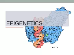
/
Epigenetics Originally defined as “ the branch of biology which studies the causal interactions Epigenetics Originally defined as “ the branch of biology which studies the causal interactions

Epigenetics Originally defined as “ the branch of biology which studies the causal interactions - PowerPoint Presentation

kittie-lecroy
kittie-lecroy . @kittie-lecroy
Follow
350 views
Uploaded On 2018-11-07

Epigenetics Originally defined as “ the branch of biology which studies the causal interactions - PPT Presentation

Waddington 1942 The study of any potentially stable and ideally heritable change in gene expression or cellular phenotype that occurs without changes in WatsonCrick basepairing of DNA ID: 719468

histone rna gene chromatin rna histone chromatin gene dna methylation cell silencing genes biology figure expression modifications mirnas sirnas

Share:

Link:

Embed:

Download Presentation from below link

Download Presentation The PPT/PDF document "Epigenetics Originally defined as “ th..." is the property of its rightful owner. Permission is granted to download and print the materials on this web site for personal, non-commercial use only, and to display it on your personal computer provided you do not modify the materials and that you retain all copyright notices contained in the materials. By downloading content from our website, you accept the terms of this agreement.


Presentation Transcript

Slide1

Epigenetics

Originally defined as “ the branch of biology which studies the causal interactions between genes and their products, which brings the phenotype into being”

Waddington, 1942

“The study of any potentially stable, and ideally, heritable change in gene expression or cellular phenotype that occurs without changes in Watson-Crick base-pairing of

DNA”

Goldberg , Allis and Bernstein. Cell, 2007.Slide2

Three main players

Li and Zhao, Stem Cells Dev., 2008Slide3

ChromatinSlide4

Histone modifications on the nucleosome core particle

Adapted from Turner, B. M., Cell 111 (2002): 285-291.Slide5

Histone modifications determine functionSlide6

Examples of the “histone code” for H3/H4

Ac = acetyl (lysine) Me = methyl (lysine), P = phosphoryl (Ser or Thr)

See table 7-7

Modifications include acetylation, methylation, phosphorylation, and ubiquitylation. Modification patterns comprise the “histone code”.

Principle: “Epigenetic “ marks alter gene functionSlide7

Histone tail modifications alter chromatin accessiblitySlide8

Histone

modifying complexes and

nucleosome

remodeling complexes recognize the modified

histones

through specific domains

Kouzarides

, Cell, 2007Slide9

Is there a histone code?

In

Drosophila

:

During metaphase HP1 is temporarily lost from mitotic chromosomes

This is associated with

phosphorylation

of the neighboring serine residue, mediated by cell cycle

kinase

Aurora BSlide10

Chromatin-remodeling and histone-modifying complexes work together to alter chromatin structureSlide11

Figure 4-43

Molecular Biology of the Cell

(© Garland Science 2008)Slide12

Figure 4-45

Molecular Biology of the Cell

(© Garland Science 2008)

Figure 4-46a

Molecular Biology of the Cell

(© Garland Science 2008)

Spreading of an epigenetic mark and remodellingSlide13

Are the histone

modifications really epigenetic and thus heritable?

Histone

demethylases

,

deacetylases

,

dephosphorylases

Is DNA

methylation

or an RNA

speices

involved

?

Can this mark be propagated during replication?Slide14

Inheritance of histones after DNA replication

Differential labeling of old and new

histones

Results indicate:

Old

histones

are present on both of the daughter chromosomes

Mixing is not entirely random: H3.H4 tetramers and H2A.H2B

dimers

are composed of all new or all old

histones

H3.H4 tetramers remain bound to the DNA

H2A.H2B

dimers

are released and enter the local pool available fir new

nucleosome

assemblySlide15

Inheritance of parental H3:H4 tetramers

could potentially facilitate the

inheritance of chromatin statesSlide16

DNA Methylation

Methylation

to the 5’ position of

cytosines

just before

guanosines

in

CpG

nucleotides

CpG

nucleotides are uncommon in mammalian DNA (about 1%) and are present 10-20 times their average density in selected regions called CG islands (1000-2000 nucleotides long)Slide17

Epigenetic gene regulation: Patterns of DNA methylation can be maintained through cell division

Figure

17-28 Molecular Biology of the GeneSlide18

Role of methylation

in silencing

Figure

17-25, Molecular Biology of the GeneSlide19

DNA methylation

patterns differ between stem cells and differentiated cells

Filled circles:

methylated

cytosines

; Unfilled circle:

unmethylated

cytosines

.

H stands for A, C or T;

N stands for any nucleotide.

Dirk

Schübeler

, Nature. 2009. 462: 296-297.Slide20

DNA

methylation

lies at the heart of Imprinting

Figure 17-26Slide21

Recent study by Ciccone

et al suggests that Imprinting involves

histone

demethylation

and DNA

methylationSlide22

RNA meets Chromatin

Dosage compensation: a strong link between RNA and chromatin

RNA interference can direct Heterochromatin Formation

si

/

miRNAs

can also affect specific gene expression either

post-

transcriptionally

or

post-

translationallySlide23

Short RNAs (21-23

nt

) direct silencing of genes in three different ways

http://www.nature.com/focus/rnai/animations/animation/animation.htm

-RNA interference (RNAi): Several types of short RNAs repress, or silence expression of genes with homology to those short RNAs:

microRNAs (miRNAs) are derived from precursor RNAs encoded within genes

small interfering RNAs (siRNAs) are produced from dsRNAs

This can happen by two mechansims:

post-transcriptional gene silencing (PTGS) and transcriptional gene silencing (TGS).

PTGS can, in turn, be divided into two main mechanisms:

direct sequence-specific cleavage,

and translational repression and RNA degradation. Slide24

Overview

Incorporation of a guide strand RNA into RISC makes the mature complex that is ready to silence gene expression; RISC: RNA-induced silencing complexSlide25

Core features of miRNA and siRNA silencing

Carthew and Sontheimer. 2009 Cell 136, 642–655

In general:

-

miRNAs

- regulators of endogenous genes

-

siRNAs

- defenders of genome integrity in response to foreign or invasive nucleic acids such as viruses,

transposons

, and

transgenes

.

Single-stranded forms of both

miRNAs

and

siRNAs

are found to associate with

effector

assemblies known as RNA-induced silencing complexes (RISCs).Slide26

Machinery includes

:

-Dicer (

Rnase

-III-like enzyme) that generates the

siRNAs

and

miRNAs

-RNA-induced silencing complex (RISC): contains various proteins including a member of the

Argonaute

family (Slicer) and the

siRNA

/

miRNA

which is denatured to a guide RNA. Some RISC complexes can be directed to the nucleus to recruit chromatin modifying complexes….silencing of

transcription

-RNA –dependent RNA polymerase (

RdRP

).Slide27

siRNAs

can

transcriptionally

silence genes by directing chromatin modification ( in fission yeast)

Figure

18-13, Molecular Biology of the geneSlide28

Epigenetic changes may affect miRNA

expression and

vice versaSlide29

Synopsis

Ultimately, expression of genes requires that RNA

Pol

and transcription factors have access to promoter and enhancer sequences to initiate transcription

Chromatin structure around genes is dictated by:

modifications to

histones

and the resulting interactions of

histone

modifiers and chromatin remodelers

DNA

methylation

, which can recruit

histone

modifiers/remodelers

siRNAs

, again ultimately directing chromatin modification and remodeling

si

/

miRNAs

can also affect specific gene expression either post-

transcriptionally

or post-

translationally