/
 © 2014 Pearson Education, Inc.  © 2014 Pearson Education, Inc.

© 2014 Pearson Education, Inc. - PowerPoint Presentation

stefany-barnette
stefany-barnette . @stefany-barnette
Follow
345 views
Uploaded On 2020-04-03

© 2014 Pearson Education, Inc. - PPT Presentation

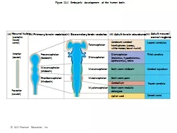
Figure 121 Embryonic development of the human brain Neural tube contains neural canal Primary brain vesicles Secondary brain vesicles Adult brain structures Adult neural ID: 775207

nerve cerebral ventricle mater nerve cerebral ventricle mater pearson education 2014 figure csf lateral spinal brain arachnoid lobe cord

Share:

Link:

Embed:

Download Presentation from below link

Download Presentation The PPT/PDF document " © 2014 Pearson Education, Inc." is the property of its rightful owner. Permission is granted to download and print the materials on this web site for personal, non-commercial use only, and to display it on your personal computer provided you do not modify the materials and that you retain all copyright notices contained in the materials. By downloading content from our website, you accept the terms of this agreement.


Presentation Transcript

Slide1

© 2014 Pearson Education, Inc.

Figure 12.1 Embryonic development of the human brain.

Neural tube

(contains neural canal)

Primary brain vesicles

Secondary brain vesicles

Adult brain structures

Adult neural canal regions

Anterior(rostral)

Prosencephalon(forebrain)

Mesencephalon(midbrain)

Rhombencephalon(hindbrain)

Posterior(caudal)

Telencephalon

Diencephalon

Mesencephalon

Metencephalon

Myelencephalon

Cerebrum: cerebralhemispheres (cortex,white matter, basal nuclei)

Diencephalon(thalamus, hypothalamus,epithalamus), retina

Brain stem: midbrain

Brain stem: pons

Cerebellum

Brain stem: medullaoblongata

Spinal cord

Lateral ventricles

Third ventricle

Cerebral aqueduct

Fourth ventricle

Central canal

Slide2

© 2014 Pearson Education, Inc.

Chapter Opener 12

Slide3

© 2014 Pearson Education, Inc.

Figure 12.2c Brain development.

Cerebral

hemisphere

Diencephalon

Cerebellum

Brain stem

Midbrain

Pons

Medulla oblongata

Birth: Shows adult pattern of structures and convolutions.

Slide4

© 2014 Pearson Education, Inc.

Figure 12.4a Lobes, sulci, and fissures of the cerebral hemispheres.

Anterior

Longitudinal

fissure

Frontal lobe

Cerebral veins

and arteries

covered by

arachnoid

mater

Left cerebral

hemisphere

Parietal lobe

Right cerebral

hemisphere

Occipital

lobe

Superior view

Posterior

Slide5

© 2014 Pearson Education, Inc.

Figure 12.4c Lobes, sulci, and fissures of the cerebral hemispheres.

Frontal lobe

Postcentral

gyrus

Parietal lobe

Central

sulcus

Precentral

gyrus

Parieto-occipital sulcus

(on medial surface

of hemisphere)

Lateral sulcus

Temporal lobe

Occipital lobe

Transverse

cerebral fissure

Pons

Spinal cord

Fissure

(a deep

sulcus)

Gyrus

Cortex (gray matter)

Sulcus

White matter

Lobes and sulci of the cerebrum

Medulla oblongata

Cerebellum

Slide6

© 2014 Pearson Education, Inc.

Figure 12.4a Lobes, sulci, and fissures of the cerebral hemispheres.

Anterior

Longitudinal

fissure

Frontal lobe

Cerebral veins

and arteries

covered by

arachnoid

mater

Left cerebral

hemisphere

Parietal lobe

Right cerebral

hemisphere

Occipital

lobe

Superior view

Posterior

Slide7

© 2014 Pearson Education, Inc.

Figure 12.4c Lobes, sulci, and fissures of the cerebral hemispheres.

Frontal lobe

Postcentral

gyrus

Parietal lobe

Central

sulcus

Precentral

gyrus

Parieto-occipital sulcus

(on medial surface

of hemisphere)

Lateral sulcus

Temporal lobe

Occipital lobe

Transverse

cerebral fissure

Pons

Spinal cord

Fissure

(a deep

sulcus)

Gyrus

Cortex (gray matter)

Sulcus

White matter

Lobes and sulci of the cerebrum

Medulla oblongata

Cerebellum

Slide8

© 2014 Pearson Education, Inc.

Figure 12.9b Basal nuclei. (2 of 2)

Cerebral cortex

Cerebral white matter

Corpus callosum

Anterior horn

of lateral ventricle

Putamen

Globus pallidus

Thalamus

Third ventricle

Inferior hornof lateral ventricle

Head of caudate nucleus

Slide9

Parkinson’s Disease

-Degeneration of dopamine-releasing neurons

-What is dopamine?

-Without dopamine basal nuclei become overactive-tremor

Slide10

Slide11

Huntingtin’s Disease

-The

Huntingtin

gene provides the genetic information for a protein that is also called "

huntingtin

"

-Fatal hereditary disorder

-Huntington protein accumulates in basal nuclei

-Autosomal

dominant mutation

-Any

child of an affected person typically has a 50% chance of inheriting the disease

Slide12

© 2014 Pearson Education, Inc.

Figure 12.10a Midsagittal section of the brain.

Cerebral hemisphere

Septum pellucidum

Interthalamic

adhesion

(intermediate

mass of thalamus)

Interventricular

foramen

Anterior

commissure

Hypothalamus

Optic chiasma

Pituitary gland

Mammillary

body

Pons

Medulla

oblongata

Spinal cord

Corpus callosum

Fornix

Choroid plexus

Thalamus

(encloses third ventricle)

Posterior

commissure

Pineal gland

Epithalamus

Corpora

quadrigemina

Cerebral

aqueduct

Midbrain

Arbor vitae (of cerebellum)

Fourth ventricle

Choroid plexus

Cerebellum

Slide13

© 2014 Pearson Education, Inc.

Thalamus

Hypothalamus

Midbrain

Pons

Medulla

oblongata

Diencephalon

Brain stem

View

(b)

View

(a)

View

(c)

Diencephalon

• Thalamus

• Hypothalamus

Mammillary body

Oculomotor nerve (III)

Trochlear nerve (IV)

Middle cerebellar

peduncle

Abducens

nerve (VI)

Vestibulocochlear

nerve (VIII)

Pyramid

Ventral root of first

cervical nerve

Decussation of

pyramids

Optic chiasma

Optic nerve (II)

Optic tract

Infundibulum

Pituitary gland

Crus cerebri of cerebral

peduncles (midbrain)

Trigeminal nerve (V)

Pons

Facial nerve (VII)

Abducens nerve (VI)

Glossopharyngeal nerve (IX)

Hypoglossal nerve (XII)

Vagus nerve (X)

Accessory nerve (XI)

Spinal cord

Ventral view

Thalamus

Superior colliculus

Inferior colliculus

Trochlear nerve (IV)

Superior cerebellar peduncle

Middle cerebellar peduncle

Inferior cerebellar peduncle

Vestibulocochlear nerve (VIII)

Olive

Left lateral view

Figure 12.13a–b Three views of the brain stem (green) and the diencephalon (purple).

Slide14

Slide15

© 2014 Pearson Education, Inc.

Figure 12.10a Midsagittal section of the brain.

Cerebral hemisphere

Septum pellucidum

Interthalamic

adhesion

(intermediate

mass of thalamus)

Interventricular

foramen

Anterior

commissure

Hypothalamus

Optic chiasma

Pituitary gland

Mammillary

body

Pons

Medulla

oblongata

Spinal cord

Corpus callosum

Fornix

Choroid plexus

Thalamus

(encloses third ventricle)

Posterior

commissure

Pineal gland

Epithalamus

Corpora

quadrigemina

Cerebral

aqueduct

Midbrain

Arbor vitae (of cerebellum)

Fourth ventricle

Choroid plexus

Cerebellum

Slide16

© 2014 Pearson Education, Inc.

Thalamus

Hypothalamus

Midbrain

Pons

Medulla

oblongata

Diencephalon

Brain stem

View

(b)

View

(a)

View

(c)

Diencephalon

• Thalamus

• Hypothalamus

Mammillary body

Oculomotor nerve (III)

Trochlear nerve (IV)

Middle cerebellar

peduncle

Abducens

nerve (VI)

Vestibulocochlear

nerve (VIII)

Pyramid

Ventral root of first

cervical nerve

Decussation of

pyramids

Optic chiasma

Optic nerve (II)

Optic tract

Infundibulum

Pituitary gland

Crus cerebri of cerebral

peduncles (midbrain)

Trigeminal nerve (V)

Pons

Facial nerve (VII)

Abducens nerve (VI)

Glossopharyngeal nerve (IX)

Hypoglossal nerve (XII)

Vagus nerve (X)

Accessory nerve (XI)

Spinal cord

Ventral view

Thalamus

Superior colliculus

Inferior colliculus

Trochlear nerve (IV)

Superior cerebellar peduncle

Middle cerebellar peduncle

Inferior cerebellar peduncle

Vestibulocochlear nerve (VIII)

Olive

Left lateral view

Figure 12.13a–b Three views of the brain stem (green) and the diencephalon (purple).

Slide17

© 2014 Pearson Education, Inc.

Figure 12.15b Cerebellum.

Inferior

Medulla oblongata

Flocculonodularlobe

Choroid plexus offourth ventricle

Posterior lobe

Arbor vitae

Cerebellar cortex

Anterior lobe

Cerebellarpeduncles

• Superior

• Middle

Slide18

© 2014 Pearson Education, Inc.

Figure 12.22 Meninges: dura mater, arachnoid mater, and pia mater.

Skin of scalp

Periosteum

Bone of skull

Dura mater

• Periosteal layer

• Meningeal layer

Arachnoid mater

Pia mater

Arachnoid villus

Blood vessel

Falx cerebri

(in longitudinalfissure only)

Superior sagittalsinus

Subduralspace

Subarachnoidspace

Slide19

Slide20

© 2014 Pearson Education, Inc.

Figure 12.24a Formation, location, and circulation of CSF.

Superior

sagittal sinus

Choroid plexus

Interventricularforamen

Third ventricle

Cerebral aqueduct

Lateral aperture

Fourth ventricle

Median aperture

Central canalof spinal cord

(a) CSF circulation

The choroid plexus of each Ventricle produces CSF.

CSF flows through the ventriclesand into the subarachnoid space via the median and lateral apertures.

CSF flows through the subarachnoid space.

CSF is absorbed into the dural venous sinuses via the arachnoid villi.

Arachnoid villus

Subarachnoid space

Arachnoid mater

Meningeal dura mater

Periosteal dura mater

Right lateral ventricle

(deep to cut)

Choroid plexus

of fourth ventricle

3

2

1

4

2

3

1

4

http://www.youtube.com/watch?v=SDMO4vYkqdg

Slide21

© 2014 Pearson Education, Inc.

Figure 12.3a Ventricles of the brain.

Inferior

horn

Lateralaperture

Lateralventricle

Anteriorhorn

Thirdventricle

Cerebral aqueduct

Fourth ventricle

Central canal

Anterior view

Slide22

© 2014 Pearson Education, Inc.

Figure 12.3b Ventricles of the brain.

Lateral

ventricle

Anteriorhorn

Thirdventricle

Cerebral aqueduct

Fourth ventricle

Central canal

Posteriorhorn

Inferiorhorn

Left lateral view

Slide23

© 2014 Pearson Education, Inc.

Superior

sagittal sinus

Choroid plexus

Interventricularforamen

Third ventricle

Cerebral aqueduct

Lateral aperture

Fourth ventricle

Median aperture

Central canalof spinal cord

Arachnoid villus

Subarachnoid space

Arachnoid mater

Meningeal dura mater

Periosteal dura mater

Right lateral ventricle(deep to cut)

Choroid plexusof fourth ventricle

Ependymal cells

Capillary

Connectivetissue ofpia mater

Sectionof choroidplexus

Wastes and unnecessarysolutes absorbed

Cavity ofventricle

CSF forms as a filtratecontaining glucose, oxygen, vitamins, and ions(Na+, Cl–, Mg2+, etc.)

CSF formation by choroid plexuses

CSF circulation

The choroid plexus of each ventricleproduces CSF.

CSF flows through the ventricles and into the subarachnoid space via the median and lateral apertures.

CSF flows through the subarachnoid space.

CSF is absorbed into the dural venous sinuses via the arachnoid villi.

Figure 12.24 Formation, location, and circulation of CSF.

1

2

3

4

Slide24

© 2014 Pearson Education, Inc.

Figure 12.24a Formation, location, and circulation of CSF.

Superior

sagittal sinus

Choroid plexus

Interventricularforamen

Third ventricle

Cerebral aqueduct

Lateral aperture

Fourth ventricle

Median aperture

Central canalof spinal cord

(a) CSF circulation

The choroid plexus of each Ventricle produces CSF.

CSF flows through the ventriclesand into the subarachnoid space via the median and lateral apertures.

CSF flows through the subarachnoid space.

CSF is absorbed into the dural venous sinuses via the arachnoid villi.

Arachnoid villus

Subarachnoid space

Arachnoid mater

Meningeal dura mater

Periosteal dura mater

Right lateral ventricle

(deep to cut)

Choroid plexus

of fourth ventricle

3

2

1

4

2

3

1

4

http://www.youtube.com/watch?v=SDMO4vYkqdg

Slide25

Slide26

© 2014 Pearson Education, Inc.

Figure 12.25 Hydrocephalus in a newborn.

Slide27

© 2014 Pearson Education, Inc.

Figure 12.26a Gross structure of the spinal cord, dorsal view.

Cervical

enlargement

Dura andarachnoidmater

Conusmedullaris

Caudaequina

Filumterminale

Sacralspinal nerves

Lumbarspinal nerves

Thoracicspinal nerves

Cervicalspinalnerves

The spinal cord and its nerve roots, with the bonyvertebral arches removed. The dura mater and arachnoid mater are cut open and reflected laterally.

Lumbar

enlargement

Slide28

© 2014 Pearson Education, Inc.

Figure 12.28a Anatomy of the spinal cord.

Epidural space

(contains fat)

Subdural space

Subarachnoidspace(contains CSF)

Pia mater

Arachnoid mater

Dura mater

Spinal meninges

Bone ofvertebra

Dorsal rootganglion

Bodyof vertebra

Cross section of spinal cord and vertebra

Slide29

© 2014 Pearson Education, Inc.

Figure 12.28b Anatomy of the spinal cord.

Dorsal median sulcus

Gray commissure

Dorsal horn

Ventral horn

Lateral horn

Gray

matter

Central canal

Ventral median fissure

Pia mater

Arachnoid mater

Spinal dura mater

Whitecolumns

Dorsal funiculus

Ventral funiculus

Lateral funiculus

Dorsal rootganglion

Spinal nerve

Dorsal root(fans out into dorsal rootlets)

Ventral root(derived from severalventral rootlets)

The spinal cord and its meningeal coverings