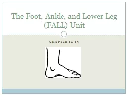
Calcaneus Talus Tarsals Navicular Cuboid Cuneiform medial middle lateral Metatarsals Phalanges Arches of the Foot Metatarsal arch and transverse Medial longitudinal Lateral longitudinal ID: 730187
Download Presentation The PPT/PDF document "Unit 24: The Foot Bony Anatomy" is the property of its rightful owner. Permission is granted to download and print the materials on this web site for personal, non-commercial use only, and to display it on your personal computer provided you do not modify the materials and that you retain all copyright notices contained in the materials. By downloading content from our website, you accept the terms of this agreement.
Slide1
Unit 24: The FootSlide2
Bony Anatomy
Calcaneus
Talus
Tarsals
Navicular
Cuboid
Cuneiform (medial, middle, lateral)
Metatarsals
PhalangesSlide3Slide4
Arches of the Foot
Metatarsal arch and transverse
Medial longitudinal
Lateral longitudinalSlide5
Plantar FasciaSlide6
Soft Tissue Anatomy
Ligaments
Subtalar ligaments
Plantar calcaneonavicular
Shock absorption
Spring ligament
Midtarsal ligaments
Anterior tarsal ligamentsSlide7Slide8Slide9
Muscle of the Foot and Lower LegSlide10Slide11
Muscles and Movements
Eversion –ankle
Abduction – forefootPronation – combination movement
Where are these muscles located?Slide12
Inversion – forefoot
Adduction – forefoot
Supination – combination movement
Where are these muscles located?Slide13
Phalange motion-
Flexion
ExtensionAbduction
AdductionSlide14
Prevention of Foot Injuries
Highly vulnerable area to variety of injuries
Injuries best prevented by selecting appropriate footwear, correcting biomechanical structural deficiencies through orthotics
Foot will adapt to training surfaces over time
Must be aware of potential difficulties associated with non-yielding and absorbent training surfacesSlide15
Foot Assessment
Athletes should be referred to qualified personnel for injury evaluation
History
Generic history questions
Questions specific to the foot
Location of pain - heel, foot, toes, arches?
Training surfaces or changes in footwear?
Changes in training, volume or type?
Does footwear increase discomfort?Slide16
Observations
Does athlete favor a foot, limp, or is unable to bear weight?
Does foot color change w/weight bearing?
Is there
pes
planus
/
cavus
?
How is foot alignment?
Structural deformities?
What does wear pattern look like on the sole of the shoe?
Is the wear symmetrical?Slide17
Posterior viewWhat do you see????Slide18Slide19
Corrected left foot positioning Slide20
Corrected rightSlide21
Palpation
Should assess the bony anatomy first
Checking for deformities and areas of tenderness
Assessment of soft tissue (muscles and tendons) will allow for detection of point tenderness, swelling, muscle spasm or muscle guarding
Circulation must also be monitored using the
dorsal pedal pulse
Located on anterior surface of ankle and footSlide22
Recognition and Management of Specific Injuries
Foot problems are associated with improper footwear, poor hygiene, anatomical structural deviations or abnormal stresses
Sports place exceptional stress on feet
ATC’s must be aware of potential problems and be capable of identifying, ameliorating or preventing themSlide23
Shoe Problems
What examples of improper footware can you think of?
What about flip flops?Long term problems?
What should you wear after an injury?Slide24
Retrocalcaneal Bursitis (Pump Bump)
Cause of Injury
Caused by inflammation of bursa beneath Achilles tendon
Result of pressure and rubbing of shoe heel counter of a shoe
Chronic condition that develops over time and may take extensive time to resolve, exostosis (pump bump) may develop
Must differentiate from Sever’s diseaseSlide25
Sign and Symptoms
Signs of inflammation
Tender, palpable bump on calcaneous
Pain w/palpation superior and anterior to Achilles insertion, swelling on both sides of the heel cord
Care
Routine stretching of Achilles, heel lifts to reduce stress, donut pad to reduce pressure
Select different footwear that results in increasing or decreasing height of heel counter.Slide26
Heel Bruise
Cause of Injury
Caused by sudden starts, stops, or changes of direction, irritation of fat pad
Signs of Injury
Severe pain in heel and unable to withstand stress of weight bearing
May progress to chronic inflammation of bone covering
Care
Reduce weight bearing for 24 hours, RICE and NSAID’s
Resume activity with heel cup or doughnut pad after pain has subsided (be sure to wear shock absorbent shoes)Slide27Slide28
Plantar Fasciitis
Cause of Condition
Increased stress on fascia
Change from rigid supportive footwear to flexible footwear
Poor running technique
Leg length discrepancy, excessive
pronation
, inflexible longitudinal arch, tight
gastroc-soleus
complex
Running on soft surfaces, shoes with poor support
Sign and Symptoms
Pain in anterior medial heel, along medial longitudinal arch
Increased pain in morning, loosens after first few steps
Increased pain with forefoot
dorsiflexionSlide29
CareExtended treatment (8-12 weeks) is required
Orthotic therapy is very useful (soft orthotic with deep heel cup)
Simple arch taping, use of a night splint to stretch
Vigorous heel cord stretching and exercises that increase great toe dorsiflexion
NSAID’s and occasionally steroidal injectionSlide30
Metatarsal Fractures
Cause of Injury
Direct force or by placing
torsional
/twisting stresses on bone
Signs of Injury
Difficult to distinguish fracture from sprain in this case
Generally present with swelling, pain, point tenderness and possible deformity
X-ray will be necessary to distinguish
fx
from sprain
Care
Symptomatic
RICE for swelling
Short leg walking cast once swelling subsides (3-6 weeks)Slide31Slide32
Jones Fracture
Cause of Injury
Fracture of metatarsal caused by inversion or high velocity rotational forces
Most common = base of 5th metatarsal
Sign of Injury
Immediate swelling, pain over 5th metatarsal
May feel a “pop”
High nonunion rate and course of healing is unpredictable
Care
Generally requires 6-8 weeks non-weight bearing with short leg cast if non-displaced
If nonunion occurs, internal fixation may be requiredSlide33Slide34Slide35
Metatarsal Stress Fractures
Cause of Injury
2nd metatarsal fracture (March fracture)Change in running pattern, mileage, hills, or hard surfaces
Often the result of structural deformities of the foot or training errors (terrain, footwear, surfaces)
Often associated with Morton’s toe
Signs of Injury
Pain and tenderness along second metatarsal
Pain with running and walking
Continued pain/aching when non-weight bearingSlide36
CareDetermine cause of injury
Generally good success with modified rest and training modifications (pool running, stationary bike) for 2-4 weeks
Return to running should be gradual over a 2-3 week period with appropriate shoesSlide37Slide38
Metatarsal Arch Strain
Cause of Injury
Hypermobility of metatarsals caused by laxity in ligaments – results in excessive splay of foot
Will appear to have fallen arch
Signs of Injury
Pain or cramping in metatarsal region
Point tenderness (
metatarsalgia
), weakness
Heavy callus may form in area of pain
Care
Pad to elevate metatarsals just behind ball of foot
Strengthening of foot muscles and heel cord stretchingSlide39
Longitudinal Arch Strain
Cause of Injury
Result of increased stress on arch of foot
Flattening of foot during
midsupport
phase causing strain on arch (appear suddenly or develop slowly)
Sign of Injury
Pain with running and jumping, usually below posterior
tibialis
tendon, accompanied by pain and swelling
May also be associated with sprained
calcaneonavicular
ligament and flexor
hallucis
longus
strain
Care
Immediate care, RICE, reduction of weight bearing
Weight bearing must be pain free
Arch taping may be used to allow pain free walkingSlide40Slide41
Fractures and Dislocations of the Phalanges
Cause of Injury
Kicking un-yielding object, stubbing toe, being stepped on
Signs of Injury
Immediate and intense pain
Swelling and discoloration
Obvious deformity with dislocation
Care
Dislocations should be reduced by a physician
Casting may occur with great toe or stiff-soled shoe
Buddy taping is generally sufficient
Shoe with larger toe box may be necessarySlide42Slide43Slide44
Bunion (
Hallux
Valgus Deformity)
Cause of Injury
Exostosis
of 1st metatarsal head; associated with forefoot
varus
; shoes that are too narrow, pointed, or short
Bursa becomes inflamed and thickens, enlarging joint, and causing lateral
malalignment
of great toe
Sign of Injury
Tenderness, swelling, and enlargement of joint initially
As inflammation continues,
angulation
increases causing painful ambulationSlide45
CareWear correctly fitting shoes, appropriate orthotics, pad over 1st metatarsal head, tape splint between 1st and 2nd toe
Surgery may be required during later stages of conditionSlide46
Morton’s Neuroma Cause of Condition
Thickening of nerve sheath (common plantar nerve) at point where nerve divides into digital branches
Commonly occurs between 3rd and 4th met heads where medial and lateral plantar nerves come together
Signs of Condition
Burning paresthesia and severe intermittent pain in forefoot
Pain relieved with non-weight bearing
Toe hyperextension increases symptomsSlide47Slide48
CareTeardrop pad can be placed between met heads to increase space, decreasing pressure on neuroma
Shoes with wider toe box would be appropriateSlide49
Turf Toe
Cause of Injury
Hyperextension injury resulting in sprain of 1st
metatarsophalangeal
joint
May be the result of single or repetitive trauma
Signs and Symptoms
Pain and swelling which increases during push-off in walking, running, and jumping
Care
Increase rigidity of forefoot region in shoe
Taping the toe to prevent
dorsiflexion
Rest and discourage activity until pain free
3-4 weeks may be required for pain to subsideSlide50
Calluses
Cause of Condition
Develop from friction – may be painful as fatty layer loses elasticity and cushioning effect
May be vulnerable to tears and cracks and possible blister development underneath
Care
Emery callus file may be necessary
Massaging with small amounts of lotion may be helpful
Sanding or pumicing – care must be exercised
Can be prevented
Shoes that fit appropriately are recommended
Wear at least one layer of socks
Apply petroleum jelly to reduce frictionSlide51Slide52
Blisters
Cause of Injury
Shearing forces on skin – results in development of fluid accumulation between layers of skin
Wearing appropriate footwear (socks and shoes) and applying lubricants may help to reduce friction
Care
Take action to reduce friction (apply lubricants, cover with tape/band aid/donut pad)
Avoid puncturing in order to prevent infection
Puncturing may be necessary if pressure build-up is too great and is causing excessive painSlide53Slide54
Corns
Cause of Condition
Result of pressure from improperly fitting shoes
Hard corns are often associated with hammer toes
Soft corns result from wearing narrow shoes and excessive foot perspiration
Signs of Condition
Form between 4
th
and 5
th
toes
Circular area of thickened, white macerated skin
Care
For soft corns – good fitting shoes are necessary in conjunction with good foot hygiene
Use of padding or cotton to separate toes is helpful
Soaking in warm soapy water will also aid in softening of cornsSlide55
Ingrown Toenails
Cause of Condition
Leading edge of nail grows into nearby soft tissue
Care
Shoes should be appropriate width and length
Prevent with correct trimming of nails
Nail should be left sufficiently long and not cut to avoid penetrating into soft tissue
Should be cut short enough that it is not irritated by shoes or socks
Treatment may require soaking and packing toenail with cotton in order to lift nail away from soft tissueSlide56Slide57
Subungual Hematoma
Cause of Injury
Direct pressure, dropping an object on toe, kicking another object
Repetitive shearing forces on toenail
Signs of Injury
Accumulation of blood underneath toenail
Likely to produce extreme pain and ultimately loss of nail
Care
RICE immediately to reduce pain and swelling
Relieve pressure within 12-24 hours (lance or drill nail) – must be sterile to prevent infectionSlide58Slide59
1 month post race 2 months post race