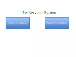
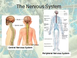
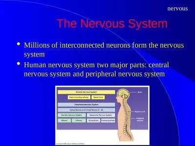
P A R T D Spinal Cord CNS tissue is enclosed within the vertebral column from the foramen magnum to L 1 Provides twoway communication to and from the brain Protected by bone meninges and CSF ID: 774996
Download Presentation The PPT/PDF document " 12 The Central Nervous System" is the property of its rightful owner. Permission is granted to download and print the materials on this web site for personal, non-commercial use only, and to display it on your personal computer provided you do not modify the materials and that you retain all copyright notices contained in the materials. By downloading content from our website, you accept the terms of this agreement.
Slide1
12
The Central Nervous System
P A R T D
Slide2Spinal Cord
CNS tissue is enclosed within the vertebral column from the foramen magnum to L
1
Provides two-way communication to and from the brain
Protected by bone, meninges, and CSF
Epidural space – space between the vertebrae and the dural sheath (dura mater) filled with fat and a network of veins
Slide3Lumbar Tap
Figure 12.30
Slide4Spinal Cord
Figure 12.29a
Slide5Spinal Cord
Conus medullaris – terminal portion of the spinal cord
Filum terminale – fibrous extension of the pia mater; anchors the spinal cord to the coccyx
Denticulate ligaments – delicate shelves of pia mater; attach the spinal cord to the vertebrae
Slide6Spinal Cord
Spinal nerves – 31 pairs attach to the cord by paired roots
Cervical and lumbar enlargements – sites where nerves serving the upper and lower limbs emerge
Cauda equina – collection of nerve roots at the inferior end of the vertebral canal
Slide7Cross-Sectional Anatomy of the Spinal Cord
Anterior median fissure – separates anterior funiculiPosterior median sulcus – divides posterior funiculi
Figure 12.31a
Slide8Gray Matter and Spinal Roots
Gray matter consists of soma, unmyelinated processes, and neuroglia
Gray commissure – connects masses of gray matter; encloses central canal
Posterior (dorsal) horns – interneurons
Anterior (ventral) horns – interneurons and somatic motor neurons
Lateral horns – contain sympathetic nerve fibers
Slide9Gray Matter and Spinal Roots
Figure 12.31b
Slide10Gray Matter: Organization
Dorsal half – sensory roots and ganglia
Ventral half – motor roots
Dorsal and ventral roots fuse laterally to form spinal nerves
Four zones are evident within the gray matter – somatic sensory (SS), visceral sensory (VS), visceral motor (VM), and somatic motor (SM)
Slide11Gray Matter: Organization
Figure 12.32
Slide12White Matter in the Spinal Cord
Fibers run in three directions – ascending, descending, and transversely
Divided into three funiculi (columns) – posterior, lateral, and anterior
Each funiculus contains several fiber tracks
Fiber tract names reveal their origin and destination
Fiber tracts are composed of axons with similar functions
Slide13White Matter: Pathway Generalizations
Pathways decussate
Most consist of two or three neurons
Most exhibit somatotopy (precise spatial relationships)
Pathways are paired (one on each side of the spinal cord or brain)
Slide14White Matter: Pathway Generalizations
Figure 12.33
Slide15Main Ascending Pathways
The central processes of fist-order neurons branch diffusely as they enter the spinal cord and medulla
Some branches take part in spinal cord reflexes
Others synapse with second-order neurons in the cord and medullary nuclei
Fibers from touch and pressure receptors form collateral synapses with interneurons in the dorsal horns
Slide16Three Ascending Pathways
The nonspecific and specific ascending pathways send impulses to the sensory cortex
These pathways are responsible for discriminative touch and conscious proprioception
The spinocerebellar tracts send impulses to the cerebellum and do not contribute to sensory perception
Slide17Nonspecific Ascending Pathway
Nonspecific pathway for pain, temperature, and crude touch within the lateral spinothalamic tract
Figure 12.34b
Slide18Specific and Posterior Spinocerebellar Tracts
Specific ascending pathways within the fasciculus gracilis and fasciculus cuneatus tracts, and their continuation in the medial lemniscal tracts
The posterior spinocerebellar tract
Slide19Figure 12.34a
Slide20Descending (Motor) Pathways
Descending tracts deliver efferent impulses from the brain to the spinal cord, and are divided into two groups
Direct pathways equivalent to the pyramidal tracts
Indirect pathways, essentially all others
Motor pathways involve two neurons (upper and lower)
Slide21The Direct (Pyramidal) System
Direct pathways originate with the pyramidal neurons in the precentral gyri
Impulses are sent through the corticospinal tracts and synapse in the anterior horn
Stimulation of anterior horn neurons activates skeletal muscles
Parts of the direct pathway, called corticobulbar tracts, innervate cranial nerve nuclei
The direct pathway regulates fast and fine (skilled) movements
Slide22Figure 12.35a
Slide23Indirect (Extrapyramidal) System
Includes the brain stem, motor nuclei, and all motor pathways not part of the pyramidal system
This system includes the rubrospinal, vestibulospinal, reticulospinal, and tectospinal tracts
Slide24Indirect (Extrapyramidal) System
These motor pathways are complex and multisynaptic, and regulate:
Axial muscles that maintain balance and posture
Muscles controlling coarse movements of the proximal portions of limbs
Head, neck, and eye movement
Slide25Figure 12.35b
Slide26Extrapyramidal (Multineuronal) Pathways
Reticulospinal tracts – maintain balance
Rubrospinal tracts – control flexor muscles
Superior colliculi and tectospinal tracts mediate head movements
Slide27Spinal Cord Trauma: Paralysis
Paralysis – loss of motor function
Flaccid paralysis – severe damage to the ventral root or anterior horn cells
Lower motor neurons are damaged and impulses do not reach muscles
There is no voluntary or involuntary control of muscles
Slide28Spinal Cord Trauma: Paralysis
Spastic paralysis – only upper motor neurons of the primary motor cortex are damaged
Spinal neurons remain intact and muscles are stimulated irregularly
There is no voluntary control of muscles
Slide29Spinal Cord Trauma: Transection
Cross sectioning of the spinal cord at any level results in total motor and sensory loss in regions inferior to the cut
Paraplegia – transection between T
1
and L
1
Quadriplegia – transection in the cervical region
Slide30Poliomyelitis
Destruction of the anterior horn motor neurons by the poliovirus
Early symptoms – fever, headache, muscle pain and weakness, and loss of somatic reflexes
Vaccines are available and can prevent infection
Slide31Amyotrophic Lateral Sclerosis (ALS)
Lou Gehrig’s disease – neuromuscular condition involving destruction of anterior horn motor neurons and fibers of the pyramidal tract
Symptoms – loss of the ability to speak, swallow, and breathe
Death occurs within five years
Linked to malfunctioning genes for glutamate transporter and/or superoxide dismutase
Slide32Developmental Aspects of the CNS
CNS is established during the first month of development
Gender-specific areas appear in response to testosterone (or lack thereof)
Maternal exposure to radiation, drugs (e.g., alcohol and opiates), or infection can harm the fetus’ developing CNS
Smoking decreases oxygen in the blood, which can lead to neuron death and fetal brain damage
Slide33Developmental Aspects of the CNS
The hypothalamus is one of the last areas of the CNS to develop
Visual cortex develops slowly over the first 11 weeks
Growth and maturation of the nervous system occurs throughout childhood and reflects progressive myelination
Slide34Developmental Aspects of the CNS
Age brings some cognitive declines, but these are not significant in healthy individuals until they reach their 80s
Excessive use of alcohol causes signs of senility unrelated to the aging process