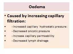
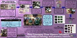
axial Spondyloarthritis when to repeat an MRI R Sengupta 1 H Marzo Ortega 23 D McGonagle 23 A Bennett 4 1 Royal National Hospital for Rheumatic Diseases Bath ID: 759251
Download Presentation The PPT/PDF document "FRI 0403 Exploring the Natural History ..." is the property of its rightful owner. Permission is granted to download and print the materials on this web site for personal, non-commercial use only, and to display it on your personal computer provided you do not modify the materials and that you retain all copyright notices contained in the materials. By downloading content from our website, you accept the terms of this agreement.
Slide1
FRI 0403
Exploring
the Natural History of Bone Marrow Oedema Lesions in axial Spondyloarthritis: when to repeat an MRI?R. Sengupta* 1, H. Marzo Ortega2,3, D. McGonagle2,3, A. Bennett41Royal National Hospital for Rheumatic Diseases, Bath; 2NIHR Leeds Musculoskeletal Biomedical Research Unit, Leeds Teaching Hospitals Trust, and 3Leeds Institute of Rheumatic and Musculoskeletal Medicine, University of Leeds, Leeds 4Defence Medical Rehabilitation Centre, Epsom, United Kingdom
IntroductionMRI determined bone marrow oedema (BMO) lesions in the sacroiliac joints (SIJ) and spine have both diagnostic and prognostic value in axial spondyloarthritis (axSpA). It is known that MRI inflammation fluctuates, however it is unclear over what time period this occurs.MRI scans are often normal in early axSpA and there is no clear evidence to guide the role of repeat MRI to aid diagnosis of axSpA.ObjectivesTo identify the natural change in BMO determined MRI lesions over 12-weeks in patients with with clinically suspected axSpA.To identify if negative MRI scans in patients with clinically suspected axSpA may become positive within a 12-week period.
Methods29 subjects, <40yrs, fulfilling the ASAS Inflammatory Back Pain (IBP) criteria1, with normal SIJ x-rays were recruited from 2 sites in the UK. MRI scans were performed at baseline, 4, 8, and 12 weeks (total= 109 scans). The MRI protocol included sagittal T1 and STIR cervico-thoracic and thoraco-lumbar spine and coronal oblique T1 and STIR sequences of the SIJs. NSAIDs were allowed if either continued at stable doses or discontinued throughout the study. Scans were read by consensus by 2 experienced readers (DMG, HMO) unaware of clinical details or date sequence using the Leeds MRI Scoring system2. In addition, each scan was scored as either “positive” or “negative” according to the ASAS definitions for the SIJ and spine3.
Results
ReferencesSieper et al. New criteria for inflammatory back pain in patients with chronic back pain: a real patient exercise by experts from the Assessment of SpondyloArthritis international Society (ASAS). Ann Rheum Dis. 2009 Jun;68(6):784-8. doi: 10.1136/ard.2008.101501. Epub 2009 Jan 15.Marzo-Ortega H, McGonagle D, O'Connor P, Hensor EM, Bennett AN, Green MJ, Emery. Baseline and 1 year MRI of the sacroiliac joint and lumbar spine in very early inflammatory back pain. Relationship between symptoms, HLA-B27 and disease extent and persistence. Ann RheumDis. 2009 Nov;68(11):1721-7.Rudwaleit M et al. Defining active sacroiliitis on magnetic resonance imaging (MRI) for classification of axial spondyloarthritis: a consensual approach by the ASAS/OMERACT MRI group. Ann Rheum Dis 2009 Oct;68(10):1520-7.
28 patients completed the study10 (36%) met the ASAS clinical criteria for axSpA prior to any imagingSpinal lesions:At baseline, there were n=22 BMO (vertebral corner) and n= 18 fatty corner lesions in the spine.26 new spinal inflammatory corner lesions appeared at any time point when the baseline MRI was normal.SIJ lesions:At baseline, BMO was present in 38 (33%) SIJ quadrants. Between any 2 time points, 16 (14%) BMO quadrant lesions resolved and new BMO lesions appeared in 6 (5%) SIJ quadrants. ASAS imaging status:18 (64%) of the patients were ASAS (spine and SIJ) negative at baseline and remained so during the entire study. 7 (25%) were ASAS SIJ positive, 1 (4%) were ASAS spine only positive and 2 (7%) were ASAS (spine and SIJ) positive. A total of 4 (14%) patients changed from ASAS imaging classification criteria negative to positive (spine (n=3) or SIJ (n=1) during the study. All of these met ASAS clinical arm classification criteria at baseline. MRI inter rater reliability for an ASAS defined positive scan demonstrated a Kappa (SE) = .80 (.19) p = 01 for the SIJs and a Kappa (SE) = 1.00 (.00) p < .0001 for the spine.
Figure 1: MRI changes in 1 patient between baseline and 12 weeks demonstrating the development of osteitis
in the sacroiliac joints
Table 2: ASAS status change
for MRI scans between any 2 time points
Table 1: Baseline demographic characteristics of the study population
Results
Conclusions
There
is little change in inflammatory lesions at the spine or SIJs within a 12 week time frame
However, in a patient with IBP and high BASDAI with an initial negative MRI scan, sequential scanning within 12 weeks may be worth considering as 14% of patients change to a positive MRI, confirming an
axSpA
diagnosis and thus making them eligible for potentially effective biologic therapy