
© 2015 Pearson Education, Inc. In which bone is
CS
Published · 53 slides · 0 views
1 / 1








Description
2015 Pearson Education, Inc. In which bone is the obturator foramen located? os coxa femur sacrum coccyx 2015 Pearson Education, Inc. In which bone is the obturator foramen located? os coxa femur sacrum coccyx 2015 Pearson Education,
Related Topics
Share
Embed code
Download this presentation From Below
"© 2015 Pearson Education, Inc. In which bone is" is the property of its rightful owner. Permission is granted to download and print the materials on this website for personal, non-commercial use only, and to display it on your personal computer provided you do not modify the materials and that you retain all copyright notices contained in the materials. By downloading content from our website, you accept the terms of this agreement.
Presentation Transcript
01
© 2015 Pearson Education, Inc.<br>
02
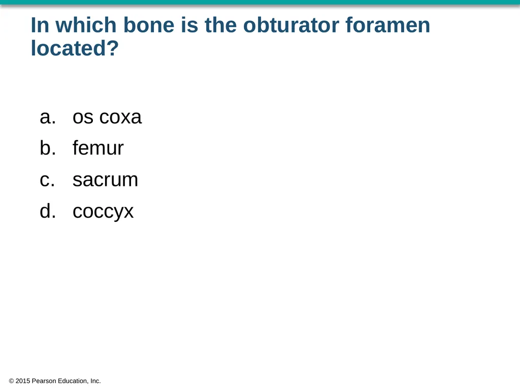
In which bone is the obturator foramen located? os coxa
femur
sacrum
coccyx © 2015 Pearson Education, Inc.<br>
femur
sacrum
coccyx © 2015 Pearson Education, Inc.<br>
03
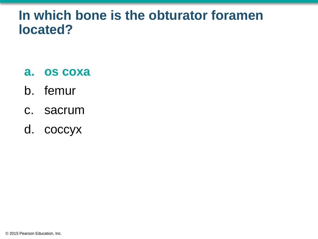
In which bone is the obturator foramen located? os coxa
femur
sacrum
coccyx © 2015 Pearson Education, Inc.<br>
femur
sacrum
coccyx © 2015 Pearson Education, Inc.<br>
04
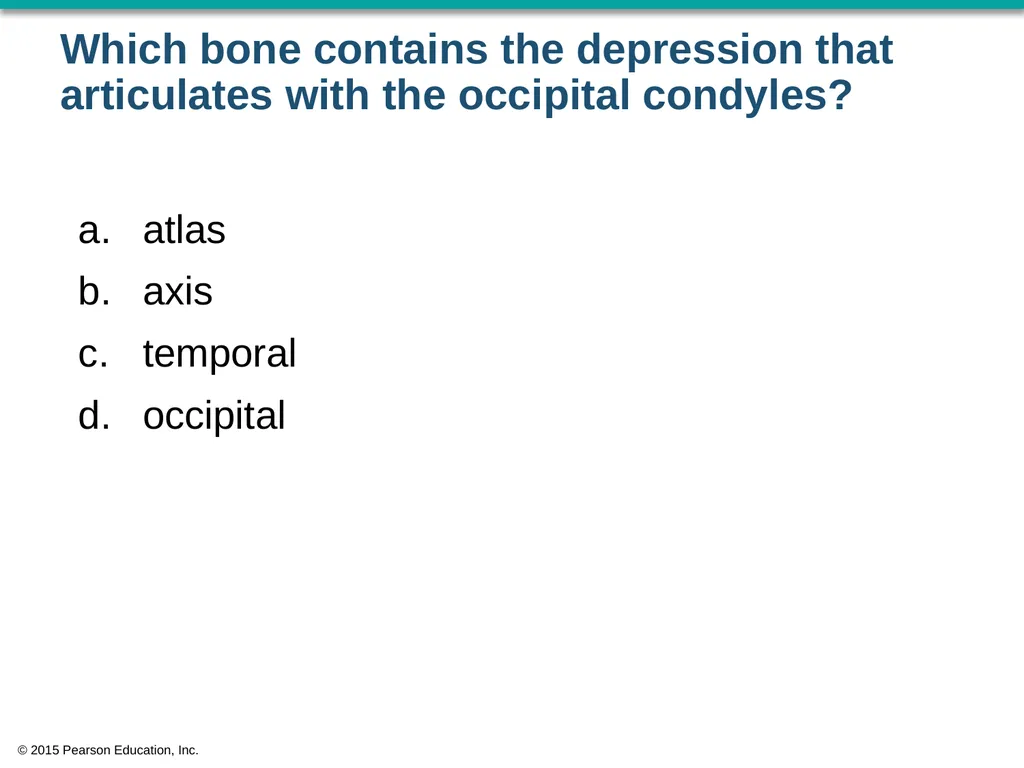
Which bone contains the depression that articulates with the occipital condyles? atlas
axis
temporal
occipital © 2015 Pearson Education, Inc.<br>
axis
temporal
occipital © 2015 Pearson Education, Inc.<br>
05
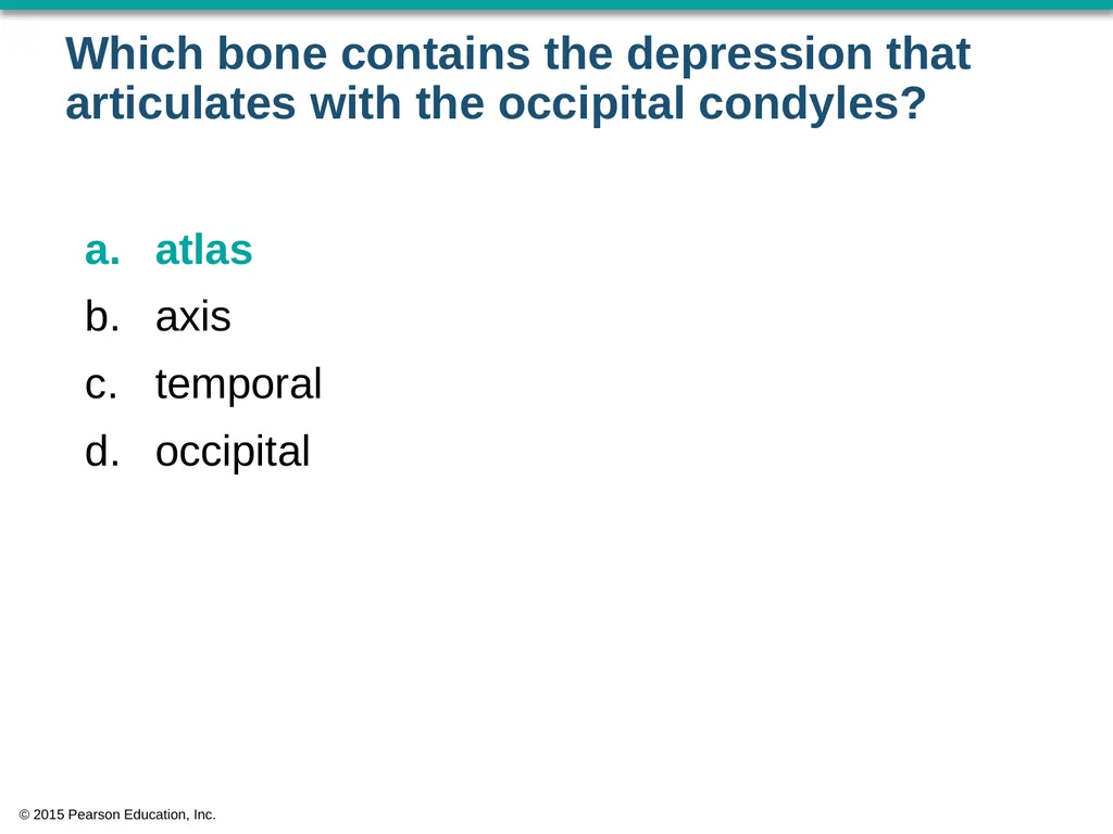
Which bone contains the depression that articulates with the occipital condyles? atlas
axis
temporal
occipital © 2015 Pearson Education, Inc.<br>
axis
temporal
occipital © 2015 Pearson Education, Inc.<br>
06
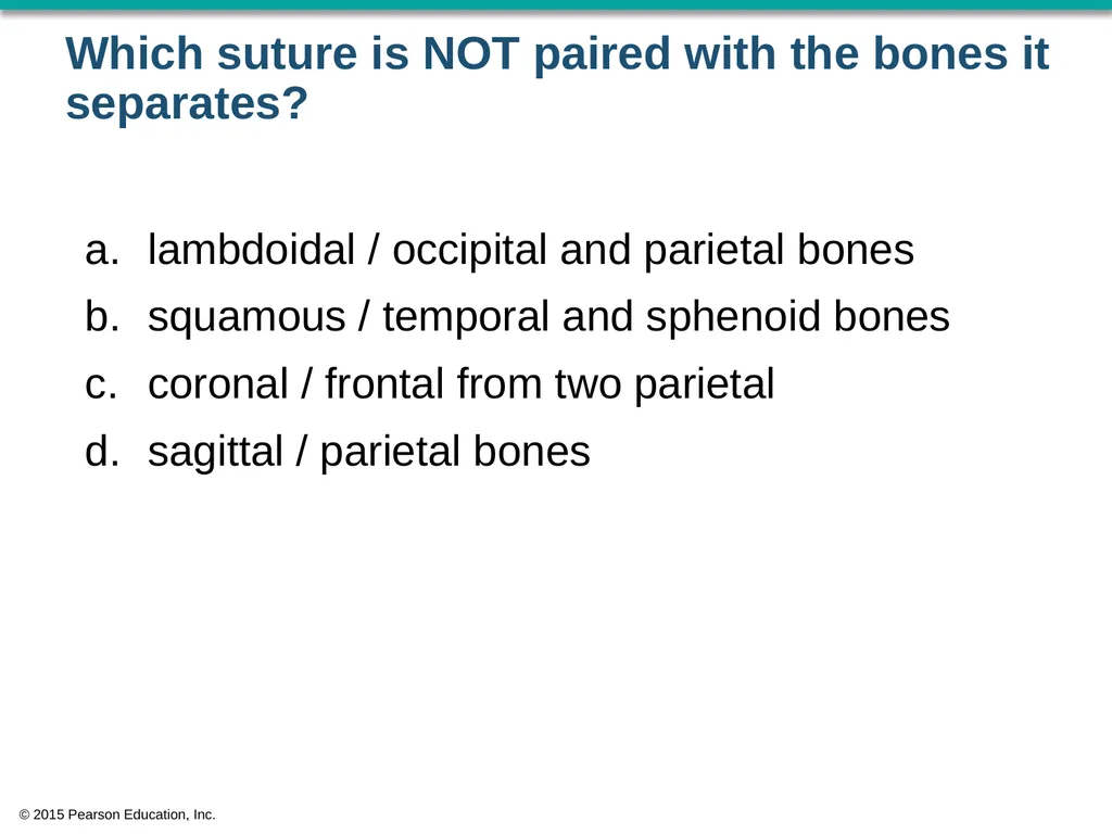
Which suture is NOT paired with the bones it separates? lambdoidal / occipital and parietal bones
squamous / temporal and sphenoid bones
coronal / frontal from two parietal
sagittal / parietal bones © 2015 Pearson Education, Inc.<br>
squamous / temporal and sphenoid bones
coronal / frontal from two parietal
sagittal / parietal bones © 2015 Pearson Education, Inc.<br>
07
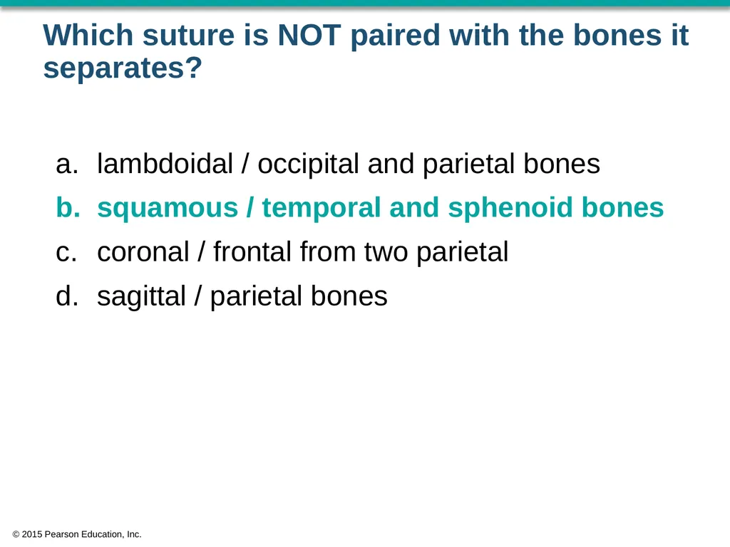
Which suture is NOT paired with the bones it separates? lambdoidal / occipital and parietal bones
squamous / temporal and sphenoid bones
coronal / frontal from two parietal
sagittal / parietal bones © 2015 Pearson Education, Inc.<br>
squamous / temporal and sphenoid bones
coronal / frontal from two parietal
sagittal / parietal bones © 2015 Pearson Education, Inc.<br>
08
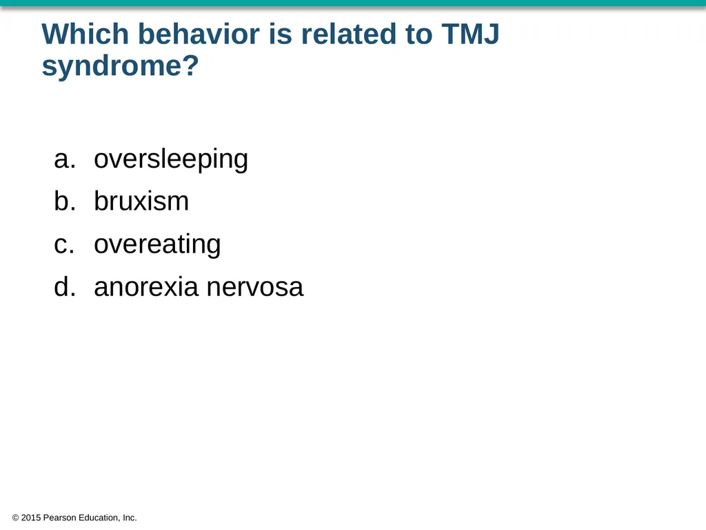
Which behavior is related to TMJ syndrome? oversleeping
bruxism
overeating
anorexia nervosa © 2015 Pearson Education, Inc.<br>
bruxism
overeating
anorexia nervosa © 2015 Pearson Education, Inc.<br>
09
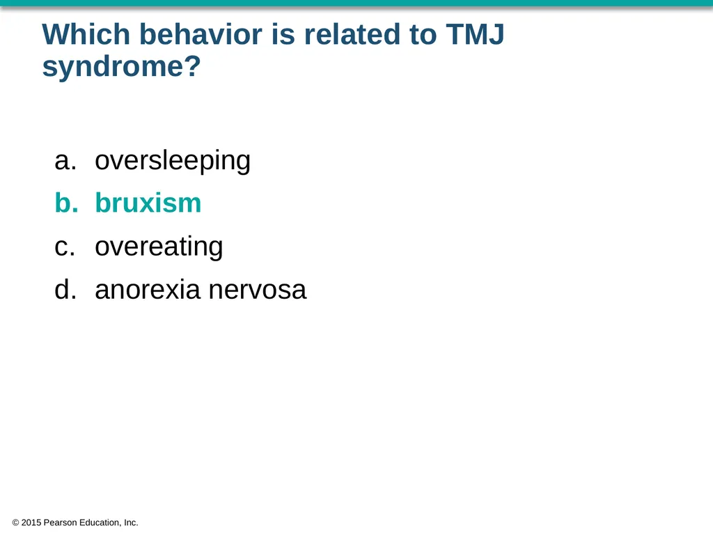
Which behavior is related to TMJ syndrome? oversleeping
bruxism
overeating
anorexia nervosa © 2015 Pearson Education, Inc.<br>
bruxism
overeating
anorexia nervosa © 2015 Pearson Education, Inc.<br>
10
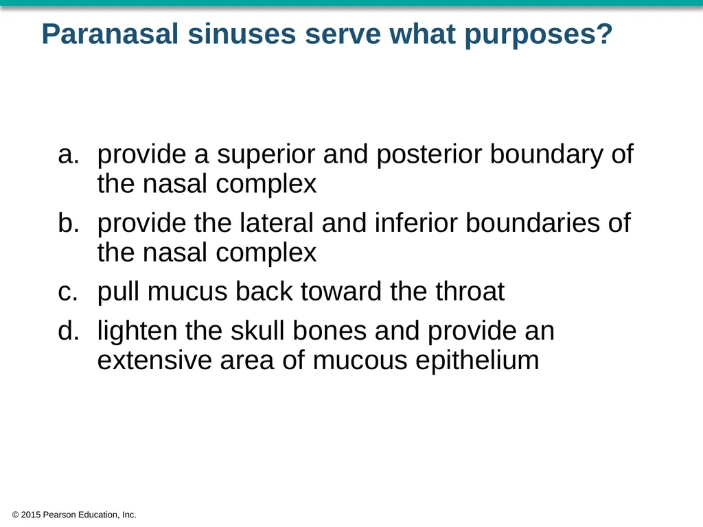
Paranasal sinuses serve what purposes? provide a superior and posterior boundary of the nasal complex
provide the lateral and inferior boundaries of the nasal complex
pull mucus back toward the throat
lighten the skull bones and provide an extensive area of mucous epithelium © 2015 Pearson Education, Inc.<br>
provide the lateral and inferior boundaries of the nasal complex
pull mucus back toward the throat
lighten the skull bones and provide an extensive area of mucous epithelium © 2015 Pearson Education, Inc.<br>
11
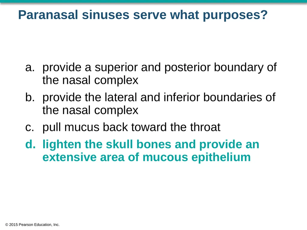
Paranasal sinuses serve what purposes? provide a superior and posterior boundary of the nasal complex
provide the lateral and inferior boundaries of the nasal complex
pull mucus back toward the throat
lighten the skull bones and provide an extensive area of mucous epithelium © 2015 Pearson Education, Inc.<br>
provide the lateral and inferior boundaries of the nasal complex
pull mucus back toward the throat
lighten the skull bones and provide an extensive area of mucous epithelium © 2015 Pearson Education, Inc.<br>
12
Jose suffers a blow to the skull that fractures his nose and breaks the nasal septum superior to the vomer. What bone is broken? frontal bone
right temporal bone
right parietal bone
ethmoid bone © 2015 Pearson Education, Inc.<br>
right temporal bone
right parietal bone
ethmoid bone © 2015 Pearson Education, Inc.<br>
13
Jose suffers a blow to the skull that fractures his nose and breaks the nasal septum superior to the vomer. What bone is broken? frontal bone
right temporal bone
right parietal bone
ethmoid bone © 2015 Pearson Education, Inc.<br>
right temporal bone
right parietal bone
ethmoid bone © 2015 Pearson Education, Inc.<br>
14
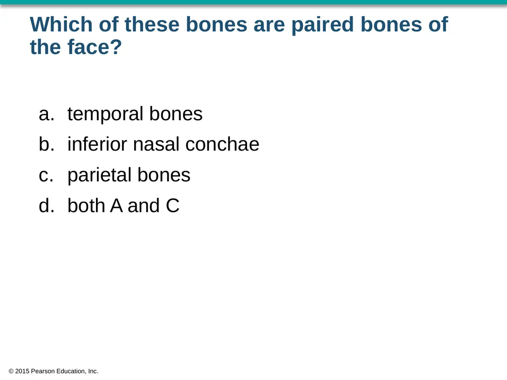
Which of these bones are paired bones of the face? temporal bones
inferior nasal conchae
parietal bones
both A and C © 2015 Pearson Education, Inc.<br>
inferior nasal conchae
parietal bones
both A and C © 2015 Pearson Education, Inc.<br>
15
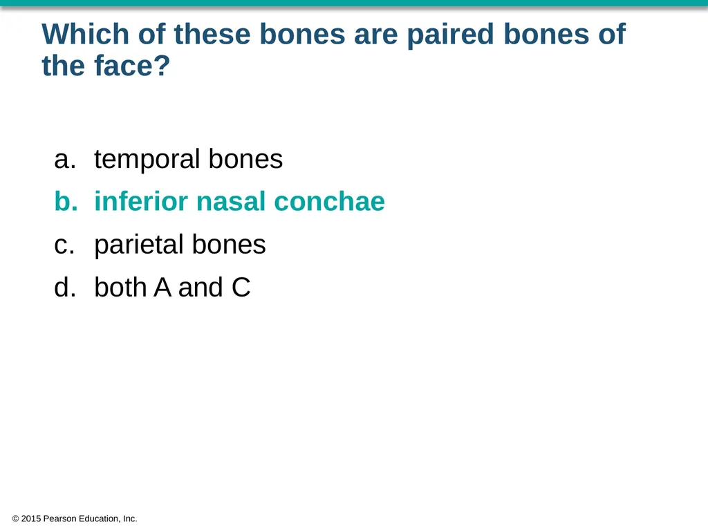
Which of these bones are paired bones of the face? temporal bones
inferior nasal conchae
parietal bones
both A and C © 2015 Pearson Education, Inc.<br>
inferior nasal conchae
parietal bones
both A and C © 2015 Pearson Education, Inc.<br>
16
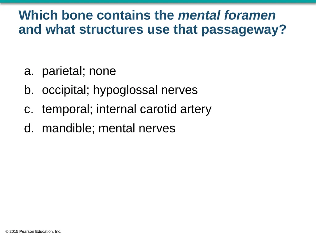
Which bone contains the mental foramen and what structures use that passageway? parietal; none
occipital; hypoglossal nerves
temporal; internal carotid artery
mandible; mental nerves © 2015 Pearson Education, Inc.<br>
occipital; hypoglossal nerves
temporal; internal carotid artery
mandible; mental nerves © 2015 Pearson Education, Inc.<br>
17
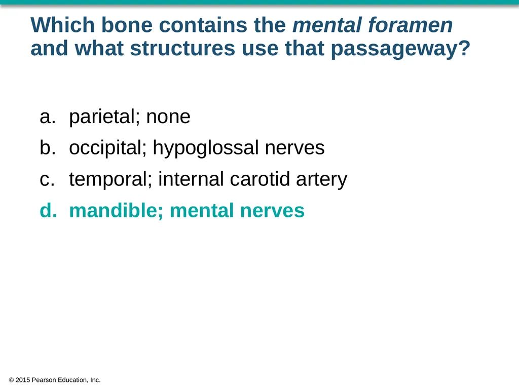
Which bone contains the mental foramen and what structures use that passageway? parietal; none
occipital; hypoglossal nerves
temporal; internal carotid artery
mandible; mental nerves © 2015 Pearson Education, Inc.<br>
occipital; hypoglossal nerves
temporal; internal carotid artery
mandible; mental nerves © 2015 Pearson Education, Inc.<br>
18
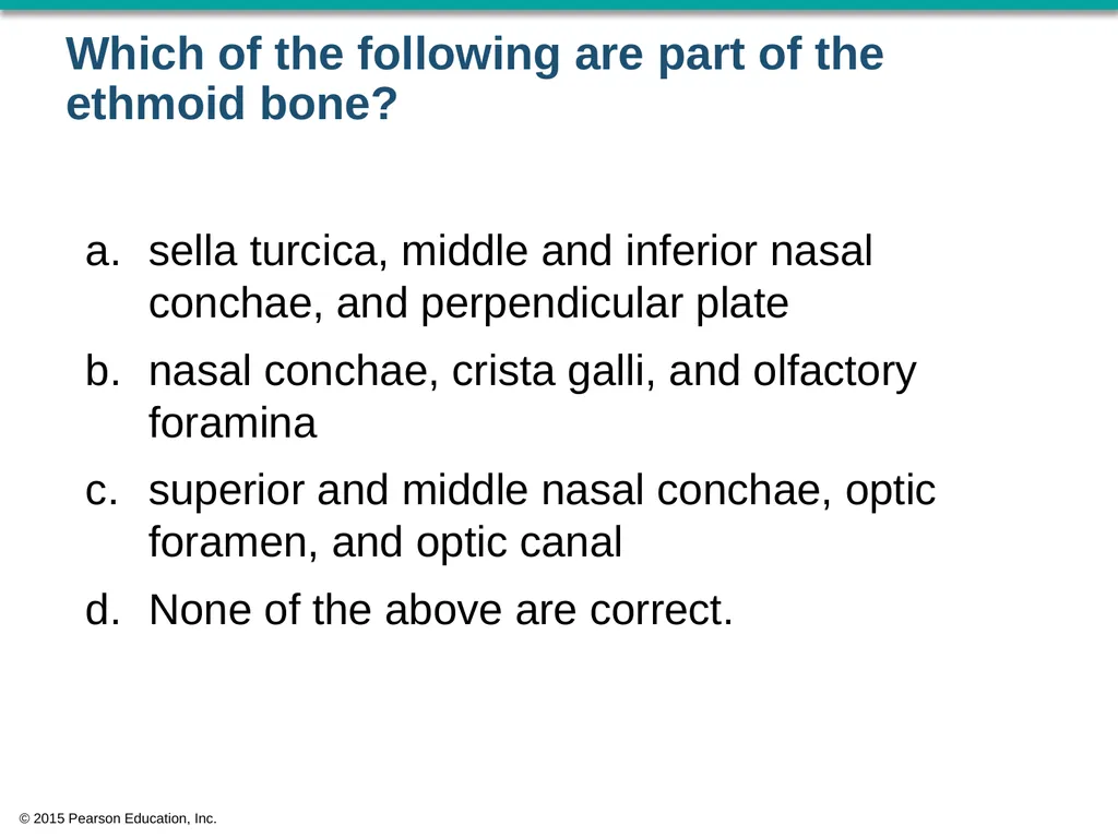
Which of the following are part of the ethmoid bone? sella turcica, middle and inferior nasal conchae, and perpendicular plate
nasal conchae, crista galli, and olfactory foramina
superior and middle nasal conchae, optic foramen, and optic canal
None of the above are correct. © 2015 Pearson Education, Inc.<br>
nasal conchae, crista galli, and olfactory foramina
superior and middle nasal conchae, optic foramen, and optic canal
None of the above are correct. © 2015 Pearson Education, Inc.<br>
19
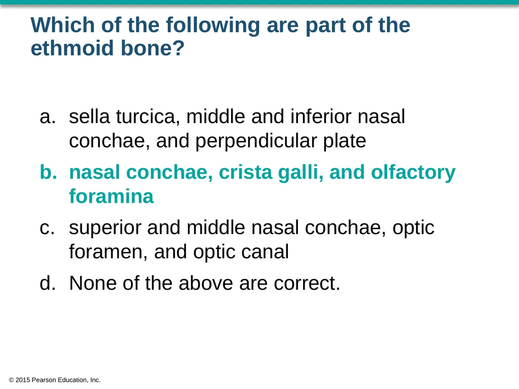
Which of the following are part of the ethmoid bone? sella turcica, middle and inferior nasal conchae, and perpendicular plate
nasal conchae, crista galli, and olfactory foramina
superior and middle nasal conchae, optic foramen, and optic canal
None of the above are correct. © 2015 Pearson Education, Inc.<br>
nasal conchae, crista galli, and olfactory foramina
superior and middle nasal conchae, optic foramen, and optic canal
None of the above are correct. © 2015 Pearson Education, Inc.<br>
20
The significance of the petrous part of the temporal bone is that it serves as ____. the housing for the auditory ossicles
a passage for the jugular vein
sites of attachment for muscles that move the mandible
the housing for the temporal sinuses © 2015 Pearson Education, Inc.<br>
a passage for the jugular vein
sites of attachment for muscles that move the mandible
the housing for the temporal sinuses © 2015 Pearson Education, Inc.<br>
21
The significance of the petrous part of the temporal bone is that it serves as ____. the housing for the auditory ossicles
a passage for the jugular vein
sites of attachment for muscles that move the mandible
the housing for the temporal sinuses © 2015 Pearson Education, Inc.<br>
a passage for the jugular vein
sites of attachment for muscles that move the mandible
the housing for the temporal sinuses © 2015 Pearson Education, Inc.<br>
22
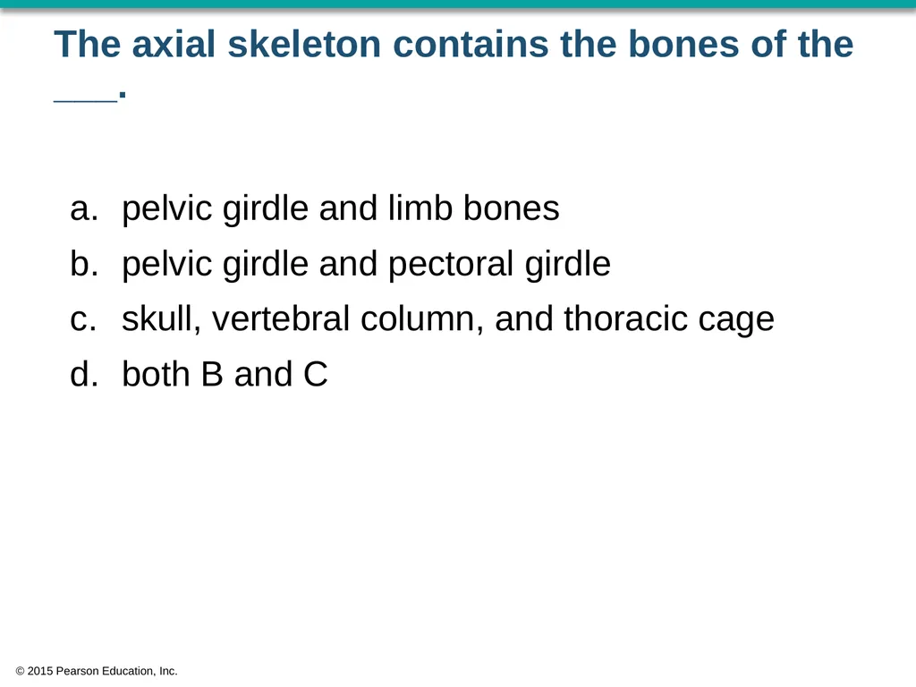
The axial skeleton contains the bones of the ___. pelvic girdle and limb bones
pelvic girdle and pectoral girdle
skull, vertebral column, and thoracic cage
both B and C © 2015 Pearson Education, Inc.<br>
pelvic girdle and pectoral girdle
skull, vertebral column, and thoracic cage
both B and C © 2015 Pearson Education, Inc.<br>
23
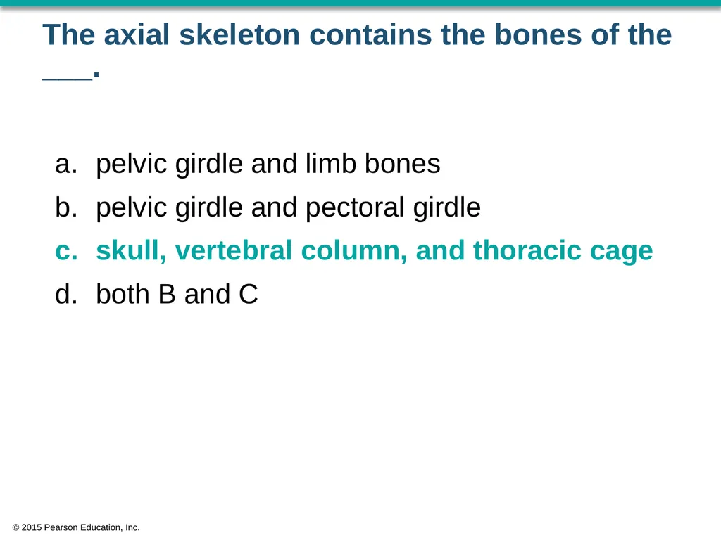
The axial skeleton contains the bones of the ___. pelvic girdle and limb bones
pelvic girdle and pectoral girdle
skull, vertebral column, and thoracic cage
both B and C © 2015 Pearson Education, Inc.<br>
pelvic girdle and pectoral girdle
skull, vertebral column, and thoracic cage
both B and C © 2015 Pearson Education, Inc.<br>
24
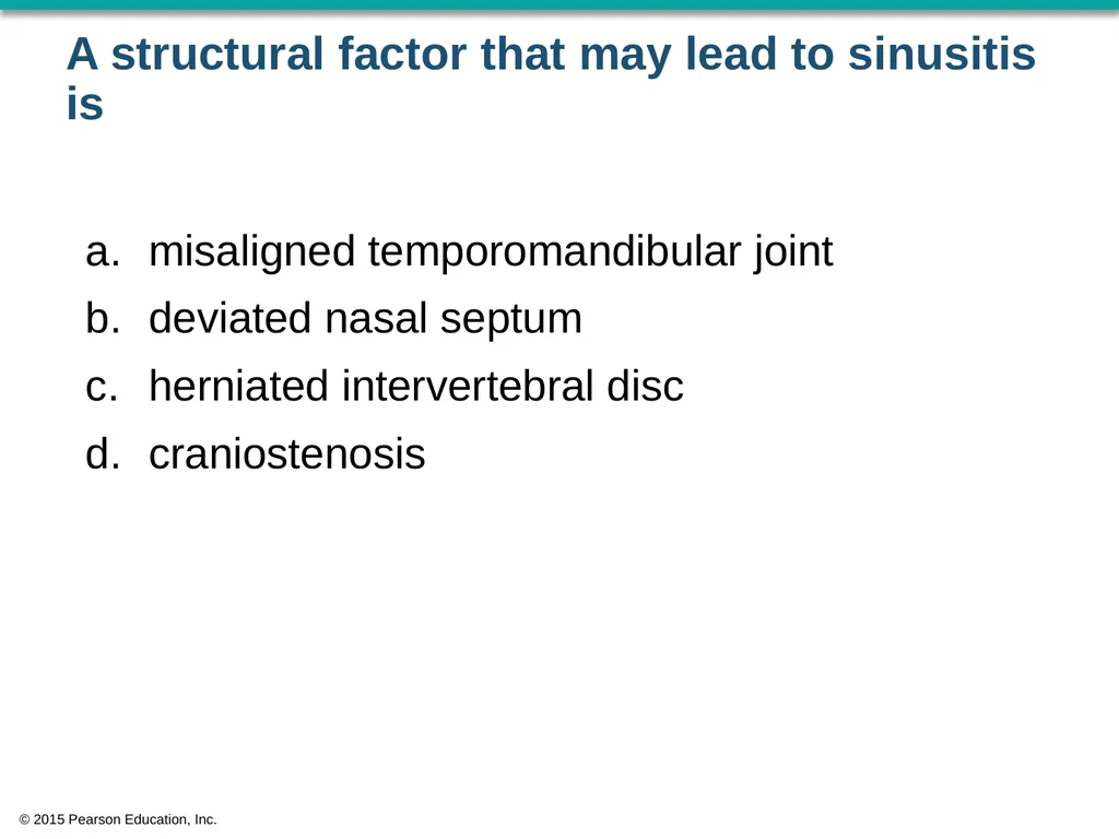
A structural factor that may lead to sinusitis is misaligned temporomandibular joint
deviated nasal septum
herniated intervertebral disc
craniostenosis © 2015 Pearson Education, Inc.<br>
deviated nasal septum
herniated intervertebral disc
craniostenosis © 2015 Pearson Education, Inc.<br>
25
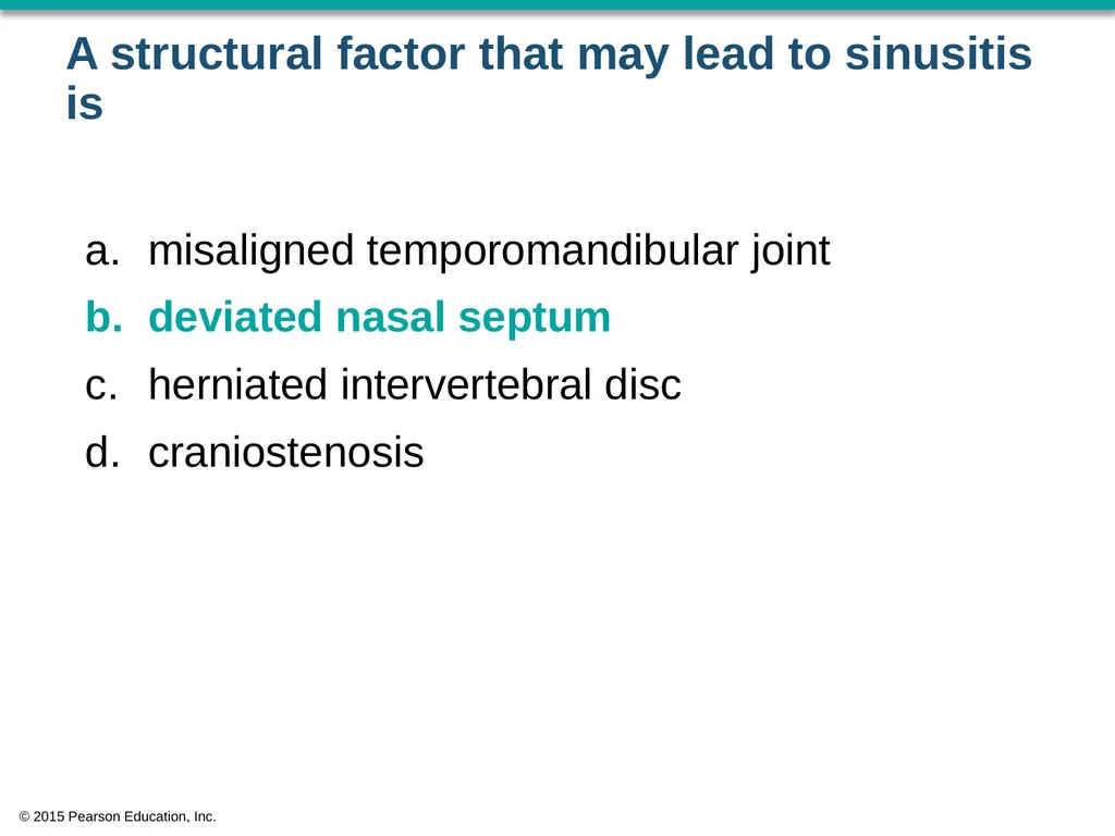
A structural factor that may lead to sinusitis is misaligned temporomandibular joint
deviated nasal septum
herniated intervertebral disc
craniostenosis © 2015 Pearson Education, Inc.<br>
deviated nasal septum
herniated intervertebral disc
craniostenosis © 2015 Pearson Education, Inc.<br>
26
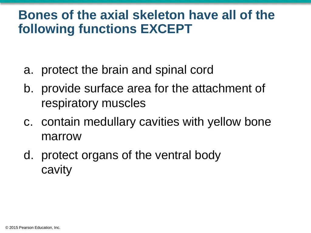
Bones of the axial skeleton have all of the following functions EXCEPT protect the brain and spinal cord
provide surface area for the attachment of respiratory muscles
contain medullary cavities with yellow bone marrow
protect organs of the ventral body cavity © 2015 Pearson Education, Inc.<br>
provide surface area for the attachment of respiratory muscles
contain medullary cavities with yellow bone marrow
protect organs of the ventral body cavity © 2015 Pearson Education, Inc.<br>
27
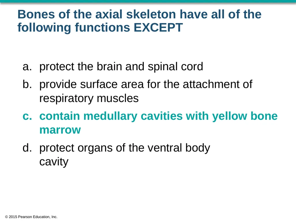
Bones of the axial skeleton have all of the following functions EXCEPT protect the brain and spinal cord
provide surface area for the attachment of respiratory muscles
contain medullary cavities with yellow bone marrow
protect organs of the ventral body cavity © 2015 Pearson Education, Inc.<br>
provide surface area for the attachment of respiratory muscles
contain medullary cavities with yellow bone marrow
protect organs of the ventral body cavity © 2015 Pearson Education, Inc.<br>
28
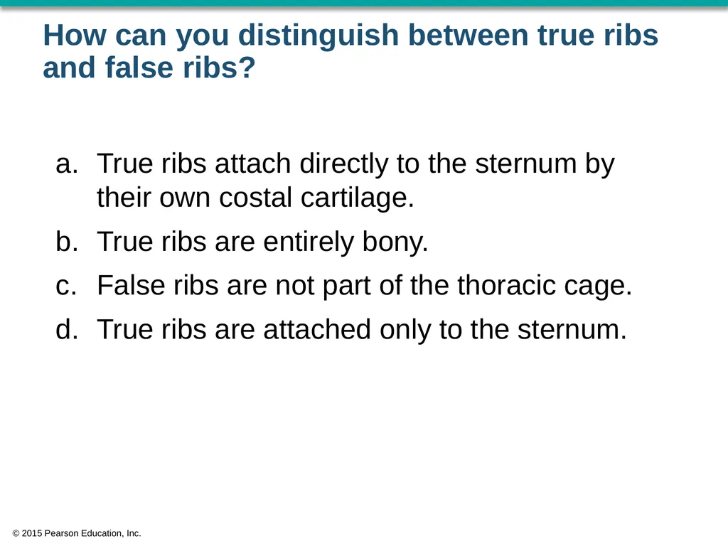
How can you distinguish between true ribs and false ribs? True ribs attach directly to the sternum by their own costal cartilage.
True ribs are entirely bony.
False ribs are not part of the thoracic cage.
True ribs are attached only to the sternum. © 2015 Pearson Education, Inc.<br>
True ribs are entirely bony.
False ribs are not part of the thoracic cage.
True ribs are attached only to the sternum. © 2015 Pearson Education, Inc.<br>
29
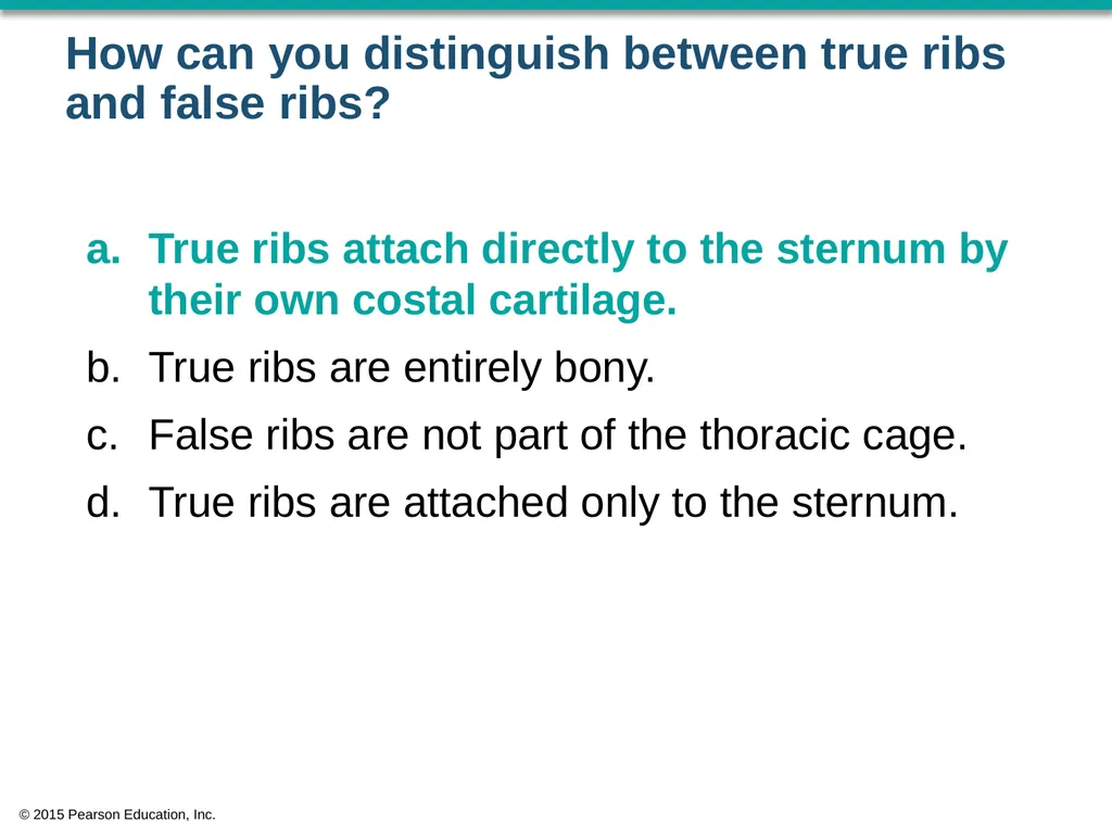
How can you distinguish between true ribs and false ribs? True ribs attach directly to the sternum by their own costal cartilage.
True ribs are entirely bony.
False ribs are not part of the thoracic cage.
True ribs are attached only to the sternum. © 2015 Pearson Education, Inc.<br>
True ribs are entirely bony.
False ribs are not part of the thoracic cage.
True ribs are attached only to the sternum. © 2015 Pearson Education, Inc.<br>
30
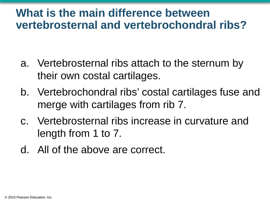
What is the main difference between vertebrosternal and vertebrochondral ribs? Vertebrosternal ribs attach to the sternum by their own costal cartilages.
Vertebrochondral ribs’ costal cartilages fuse and merge with cartilages from rib 7.
Vertebrosternal ribs increase in curvature and length from 1 to 7.
All of the above are correct. © 2015 Pearson Education, Inc.<br>
Vertebrochondral ribs’ costal cartilages fuse and merge with cartilages from rib 7.
Vertebrosternal ribs increase in curvature and length from 1 to 7.
All of the above are correct. © 2015 Pearson Education, Inc.<br>
31
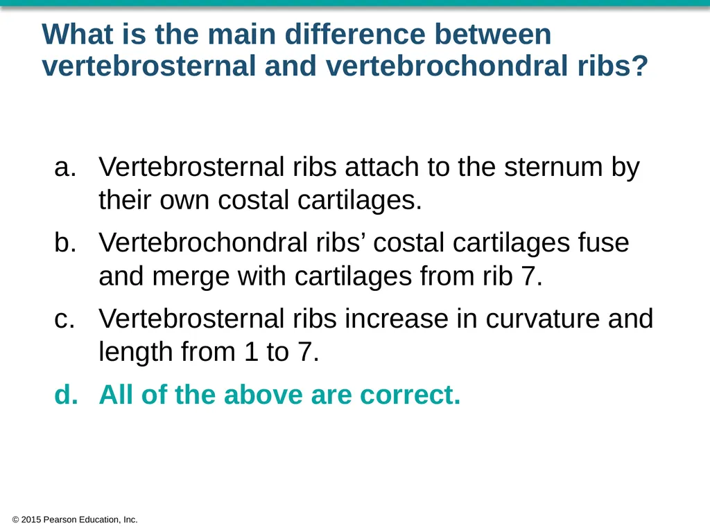
What is the main difference between vertebrosternal and vertebrochondral ribs? Vertebrosternal ribs attach to the sternum by their own costal cartilages.
Vertebrochondral ribs’ costal cartilages fuse and merge with cartilages from rib 7.
Vertebrosternal ribs increase in curvature and length from 1 to 7.
All of the above are correct. © 2015 Pearson Education, Inc.<br>
Vertebrochondral ribs’ costal cartilages fuse and merge with cartilages from rib 7.
Vertebrosternal ribs increase in curvature and length from 1 to 7.
All of the above are correct. © 2015 Pearson Education, Inc.<br>
32
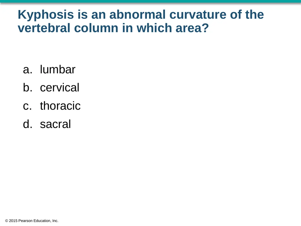
Kyphosis is an abnormal curvature of the vertebral column in which area? lumbar
cervical
thoracic
sacral © 2015 Pearson Education, Inc.<br>
cervical
thoracic
sacral © 2015 Pearson Education, Inc.<br>
33
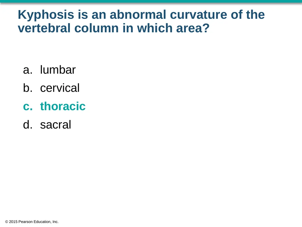
Kyphosis is an abnormal curvature of the vertebral column in which area? lumbar
cervical
thoracic
sacral © 2015 Pearson Education, Inc.<br>
cervical
thoracic
sacral © 2015 Pearson Education, Inc.<br>
34
Why are the bodies of lumbar vertebrae so large? They develop first and therefore have longer to grow.
to provide more flexibility
They support more body mass than any other vertebrae.
to provide greater protection to the lumbar spinal nerves © 2015 Pearson Education, Inc.<br>
to provide more flexibility
They support more body mass than any other vertebrae.
to provide greater protection to the lumbar spinal nerves © 2015 Pearson Education, Inc.<br>
35
Why are the bodies of lumbar vertebrae so large? They develop first and therefore have longer to grow.
to provide more flexibility
They support more body mass than any other vertebrae.
to provide greater protection to the lumbar spinal nerves © 2015 Pearson Education, Inc.<br>
to provide more flexibility
They support more body mass than any other vertebrae.
to provide greater protection to the lumbar spinal nerves © 2015 Pearson Education, Inc.<br>
36
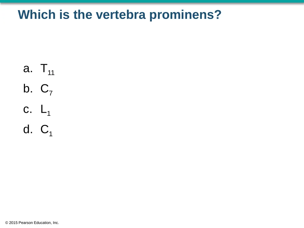
Which is the vertebra prominens? T11
C7
L1
C1 © 2015 Pearson Education, Inc.<br>
C7
L1
C1 © 2015 Pearson Education, Inc.<br>
37
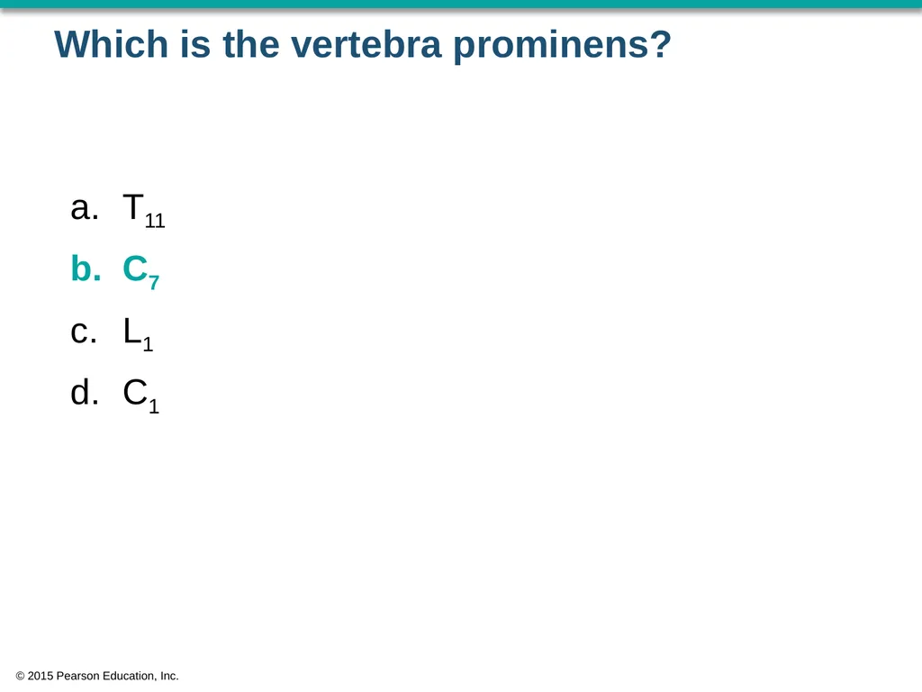
Which is the vertebra prominens? T11
C7
L1
C1 © 2015 Pearson Education, Inc.<br>
C7
L1
C1 © 2015 Pearson Education, Inc.<br>
38
Joe suffered a hairline fracture at the base of the dens. Which bone is fractured, and where is it located? second cervical vertebra; posterior neck
first cervical vertebra; posterior neck
occipital bone; posterior base of skull
sacrum; posterior pelvis © 2015 Pearson Education, Inc.<br>
first cervical vertebra; posterior neck
occipital bone; posterior base of skull
sacrum; posterior pelvis © 2015 Pearson Education, Inc.<br>
39
Joe suffered a hairline fracture at the base of the dens. Which bone is fractured, and where is it located? second cervical vertebra; posterior neck
first cervical vertebra; posterior neck
occipital bone; posterior base of skull
sacrum; posterior pelvis © 2015 Pearson Education, Inc.<br>
first cervical vertebra; posterior neck
occipital bone; posterior base of skull
sacrum; posterior pelvis © 2015 Pearson Education, Inc.<br>
40
The secondary curves of the spine are the _______. lumbar and thoracic
cervical and sacral
lumbar and sacral
cervical and lumbar © 2015 Pearson Education, Inc.<br>
cervical and sacral
lumbar and sacral
cervical and lumbar © 2015 Pearson Education, Inc.<br>
41
The secondary curves of the spine are the _______. lumbar and thoracic
cervical and sacral
lumbar and sacral
cervical and lumbar © 2015 Pearson Education, Inc.<br>
cervical and sacral
lumbar and sacral
cervical and lumbar © 2015 Pearson Education, Inc.<br>
42
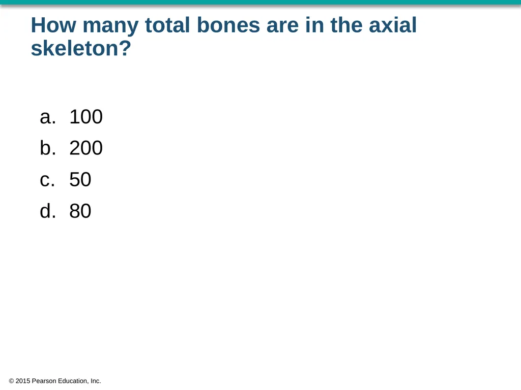
How many total bones are in the axial skeleton? 100
200
50
80 © 2015 Pearson Education, Inc.<br>
200
50
80 © 2015 Pearson Education, Inc.<br>
43
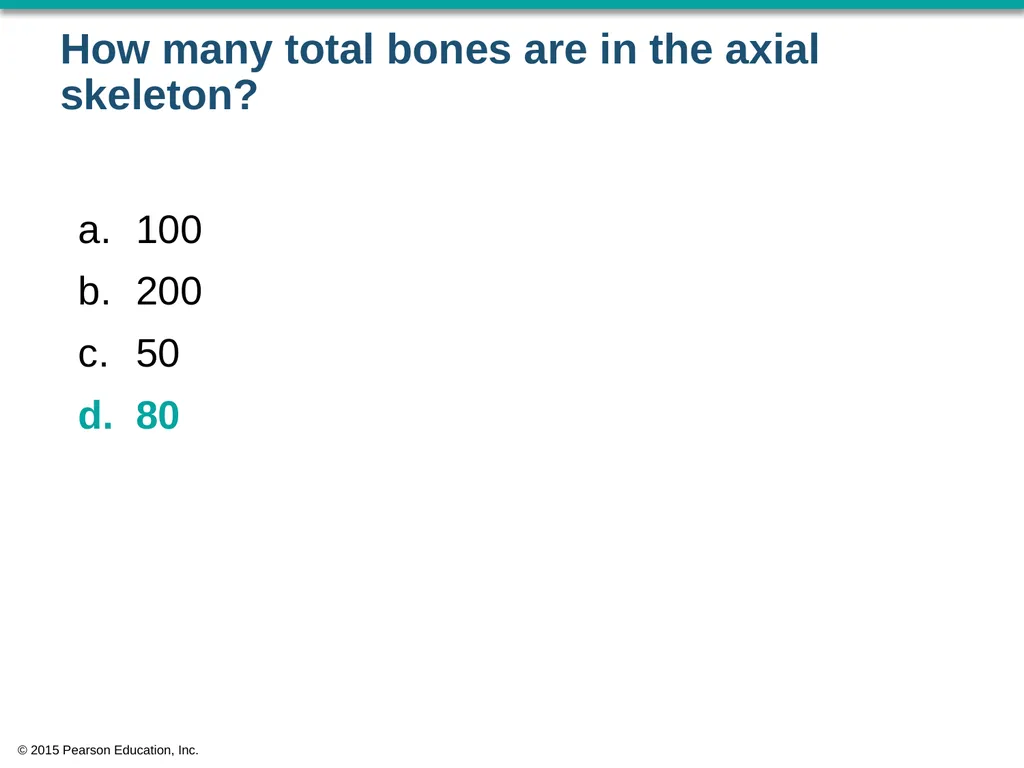
How many total bones are in the axial skeleton? 100
200
50
80 © 2015 Pearson Education, Inc.<br>
200
50
80 © 2015 Pearson Education, Inc.<br>
44
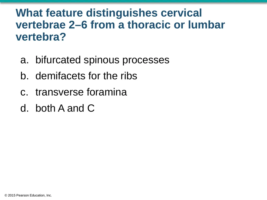
What feature distinguishes cervical vertebrae 2–6 from a thoracic or lumbar vertebra? bifurcated spinous processes
demifacets for the ribs
transverse foramina
both A and C © 2015 Pearson Education, Inc.<br>
demifacets for the ribs
transverse foramina
both A and C © 2015 Pearson Education, Inc.<br>
45
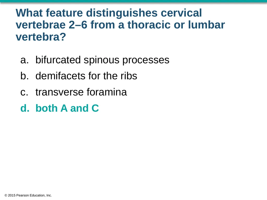
What feature distinguishes cervical vertebrae 2–6 from a thoracic or lumbar vertebra? bifurcated spinous processes
demifacets for the ribs
transverse foramina
both A and C © 2015 Pearson Education, Inc.<br>
demifacets for the ribs
transverse foramina
both A and C © 2015 Pearson Education, Inc.<br>
46
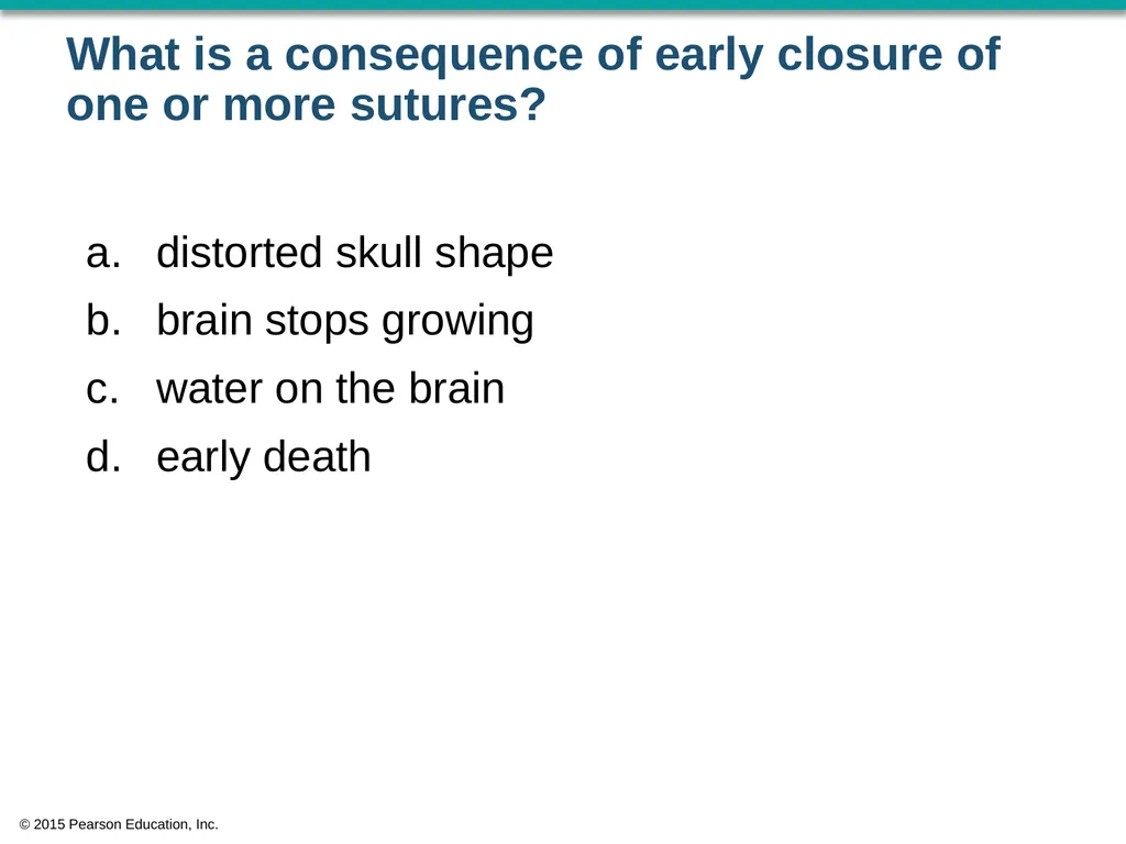
What is a consequence of early closure of one or more sutures? distorted skull shape
brain stops growing
water on the brain
early death © 2015 Pearson Education, Inc.<br>
brain stops growing
water on the brain
early death © 2015 Pearson Education, Inc.<br>
47
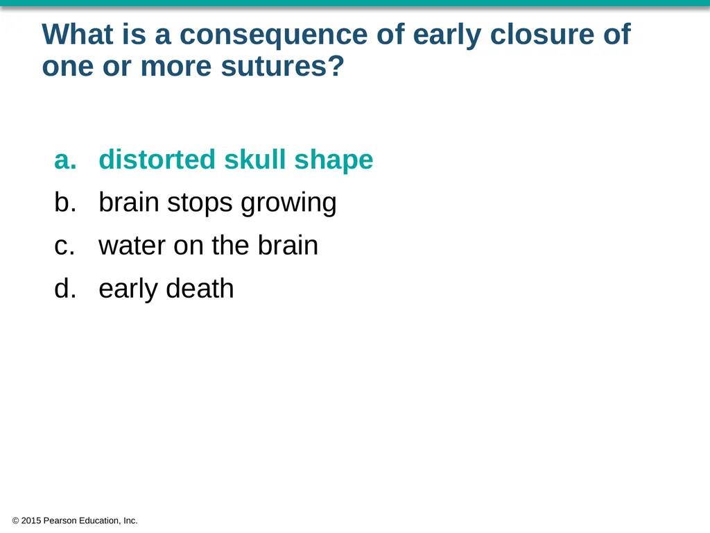
What is a consequence of early closure of one or more sutures? distorted skull shape
brain stops growing
water on the brain
early death © 2015 Pearson Education, Inc.<br>
brain stops growing
water on the brain
early death © 2015 Pearson Education, Inc.<br>
48
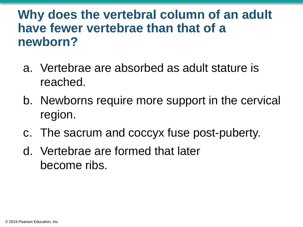
Why does the vertebral column of an adult have fewer vertebrae than that of a newborn? Vertebrae are absorbed as adult stature is reached.
Newborns require more support in the cervical region.
The sacrum and coccyx fuse post-puberty.
Vertebrae are formed that later become ribs. © 2015 Pearson Education, Inc.<br>
Newborns require more support in the cervical region.
The sacrum and coccyx fuse post-puberty.
Vertebrae are formed that later become ribs. © 2015 Pearson Education, Inc.<br>
49
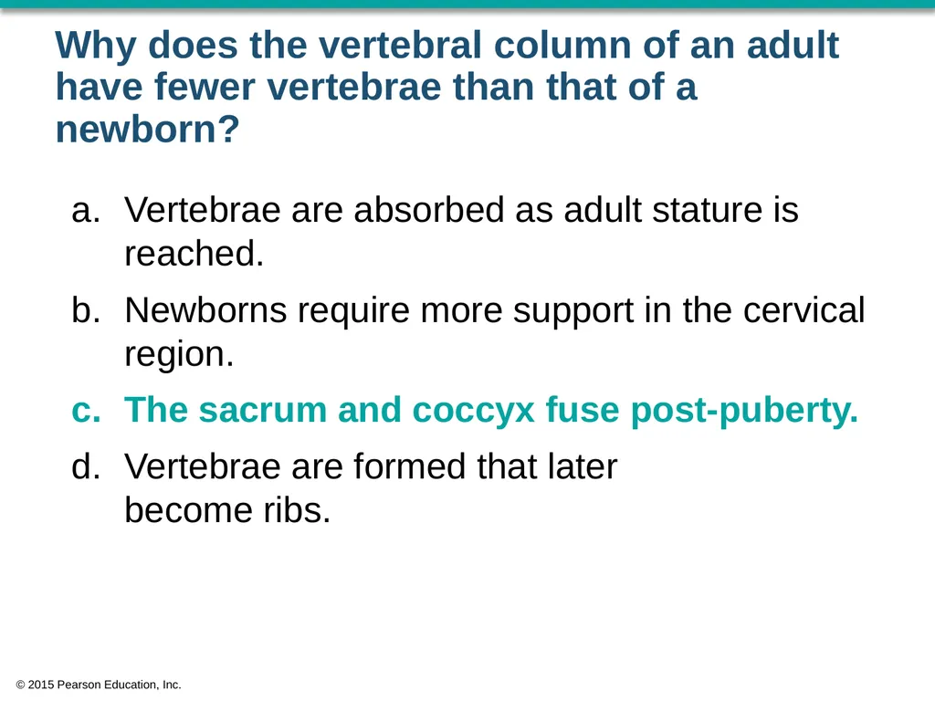
Why does the vertebral column of an adult have fewer vertebrae than that of a newborn? Vertebrae are absorbed as adult stature is reached.
Newborns require more support in the cervical region.
The sacrum and coccyx fuse post-puberty.
Vertebrae are formed that later become ribs. © 2015 Pearson Education, Inc.<br>
Newborns require more support in the cervical region.
The sacrum and coccyx fuse post-puberty.
Vertebrae are formed that later become ribs. © 2015 Pearson Education, Inc.<br>
50
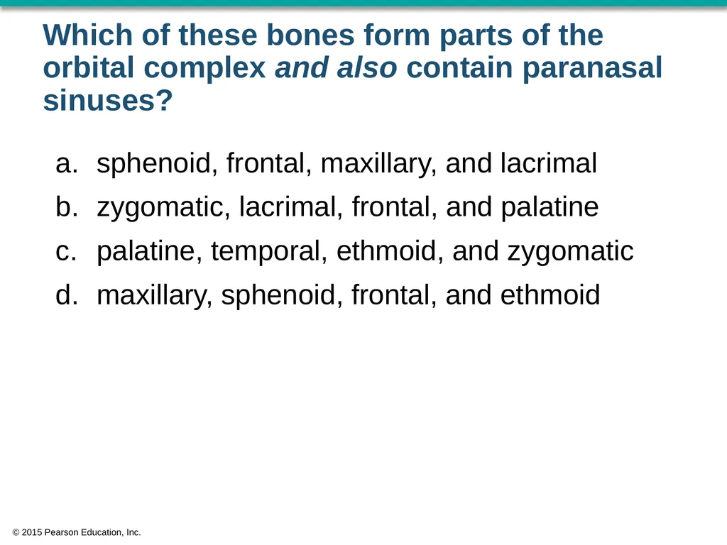
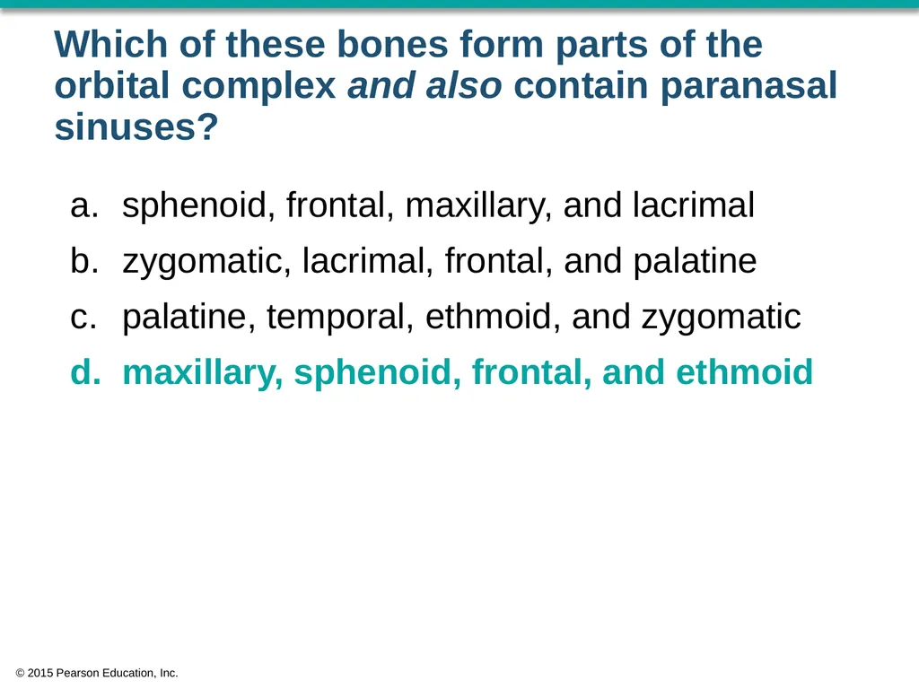
Which of these bones form parts of the orbital complex and also contain paranasal sinuses? sphenoid, frontal, maxillary, and lacrimal
zygomatic, lacrimal, frontal, and palatine
palatine, temporal, ethmoid, and zygomatic
maxillary, sphenoid, frontal, and ethmoid © 2015 Pearson Education, Inc.<br>
zygomatic, lacrimal, frontal, and palatine
palatine, temporal, ethmoid, and zygomatic
maxillary, sphenoid, frontal, and ethmoid © 2015 Pearson Education, Inc.<br>
51
Which of these bones form parts of the orbital complex and also contain paranasal sinuses? sphenoid, frontal, maxillary, and lacrimal
zygomatic, lacrimal, frontal, and palatine
palatine, temporal, ethmoid, and zygomatic
maxillary, sphenoid, frontal, and ethmoid © 2015 Pearson Education, Inc.<br>
zygomatic, lacrimal, frontal, and palatine
palatine, temporal, ethmoid, and zygomatic
maxillary, sphenoid, frontal, and ethmoid © 2015 Pearson Education, Inc.<br>
52
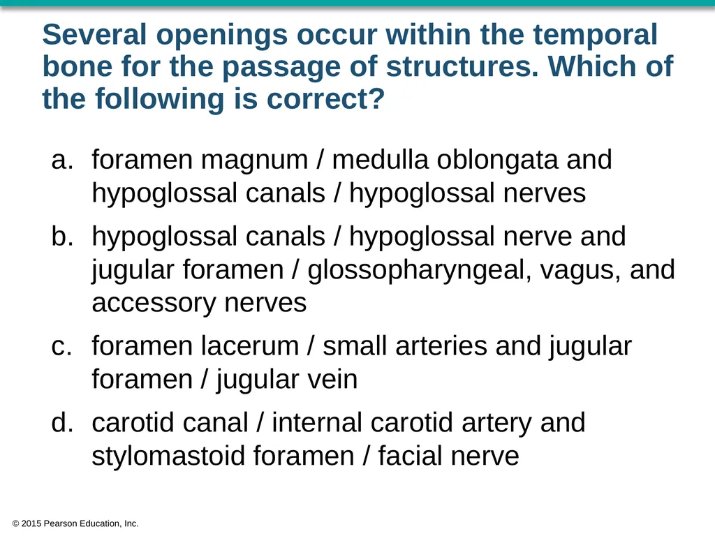
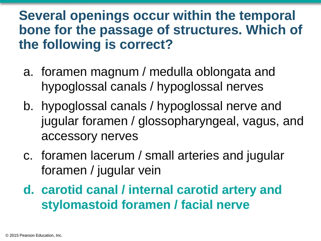
Several openings occur within the temporal bone for the passage of structures. Which of the following is correct? foramen magnum / medulla oblongata and hypoglossal canals / hypoglossal nerves
hypoglossal canals / hypoglossal nerve and jugular foramen / glossopharyngeal, vagus, and accessory nerves
foramen lacerum / small arteries and jugular foramen / jugular vein
carotid canal / internal carotid artery and stylomastoid foramen / facial nerve © 2015 Pearson Education, Inc.<br>
hypoglossal canals / hypoglossal nerve and jugular foramen / glossopharyngeal, vagus, and accessory nerves
foramen lacerum / small arteries and jugular foramen / jugular vein
carotid canal / internal carotid artery and stylomastoid foramen / facial nerve © 2015 Pearson Education, Inc.<br>
53
Several openings occur within the temporal bone for the passage of structures. Which of the following is correct? foramen magnum / medulla oblongata and hypoglossal canals / hypoglossal nerves
hypoglossal canals / hypoglossal nerve and jugular foramen / glossopharyngeal, vagus, and accessory nerves
foramen lacerum / small arteries and jugular foramen / jugular vein
carotid canal / internal carotid artery and stylomastoid foramen / facial nerve © 2015 Pearson Education, Inc.<br>
hypoglossal canals / hypoglossal nerve and jugular foramen / glossopharyngeal, vagus, and accessory nerves
foramen lacerum / small arteries and jugular foramen / jugular vein
carotid canal / internal carotid artery and stylomastoid foramen / facial nerve © 2015 Pearson Education, Inc.<br>