03
Bacillus anthracis Naturally occurring in the environment.
Zoonotic disease that occurs most frequently in herbivorous animals (e.g., cattle, sheep, and goats), which acquire endospores from contaminated soil.
Human disease is less common and results from contact with infected animals or with commercial products derived from them, such as wool and hides.
Five naturally occurring cases of anthrax have been reported since 2006: gastrointestinal, cutaneous, and inhalational.<br>
04
Infection can occur in one of four forms: Cutanous - via broken skin
Gastrointestinal - via ingestion
Inhalation anthrax – via breathing in anthrax spores. Commonly know as “woolsorter disease” due to its prevalence in the textile mills by workers handling wool and other contaminated animal products.<br>
05
Infection can occur in one of four forms: Injection anthrax - Recently, another type of anthrax infection has been identified in heroin-injecting drug users in northern Europe. This type of infection has never been reported in the United States. Symptoms may be similar to those of cutaneous anthrax, but there may be infection deep under the skin or in the muscle where the drug was injected. Injection anthrax can spread throughout the body faster and be harder to recognize and treat.<br>
06
Sentinel Laboratory Procedures for B. anthracis In a Sentinel Laboratory, you could encounter
this organism in:
Cutaneous - Vesicles (early), Eschars (late), (unroof vesicle and aspirate fluid)
Inhalational - Sputum, Blood culture or CSF culture (inhalational, septicemic)
Gastrointestinal - Stool and Blood culture
Postmortem Tissue - Tissue pieces
Environmental or non-clinical specimens are not to be processed by Sentinel Labs<br>
07
Sentinel Laboratory Procedures for B. anthracis Gram stain
Culture characteristics
Hemolysis
Catalase
Motility<br>
08
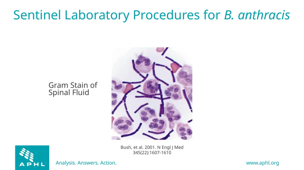
Sentinel Laboratory Procedures for B. anthracis Gram stain morphology – in clinical specimen smears
Large Gram positive bacilli in short chains, 2 - 4 cells
Spores are NOT usually present<br>
09
Gram Stain of Spinal Fluid Sentinel Laboratory Procedures for B. anthracis<br>
10
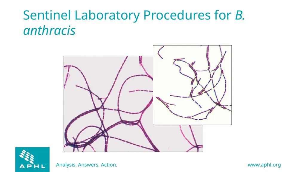
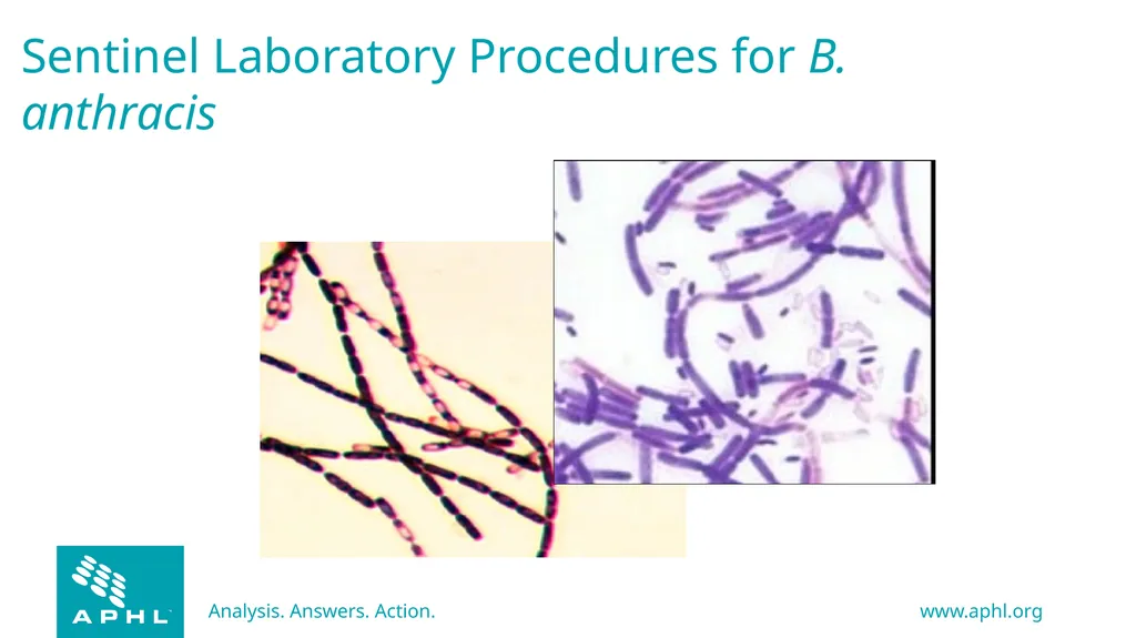
Sentinel Laboratory Procedures for B. anthracis Gram stain morphology – from culture
Large Gram positive bacilli
Nonencapsulated, often in long chains
Cells are more readily decolorized with age
Central to subterminal oval spores, with no swelling of cell
Presence of spores increases with age of culture<br>
11
Sentinel Laboratory Procedures for B. anthracis<br>
12
Sentinel Laboratory Procedures for B. anthracis<br>
13
Sentinel Laboratory Procedures for B. anthracis Culture characteristics
Organism will grow on most routine culture media – BAP, CHOC, but NOT MAC or EMB
Will grow in routine blood culture systems
Growth on plate media may be evident as early as 8 hours<br>
14
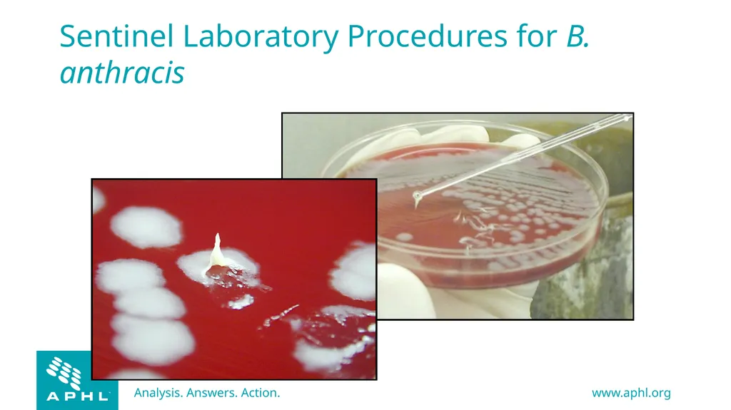
Sentinel Laboratory Procedures for B. anthracis Colony on BAP @ 35°C, 18-24 hr
Flat or slightly raised, white to grey
Undulate edge may show curling resembling a Medusa head
Surface has “ground glass” appearance
Tenacious or sticky consistency
B. anthracis is non-hemolytic<br>
15
B. anthracis on sheep blood agar @35C for 18 hrs Sentinel Laboratory Procedures for B. anthracis<br>
16
Sentinel Laboratory Procedures for B. anthracis<br>
17
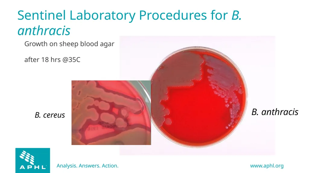
B. cereus B. anthracis Growth on sheep blood agar after 18 hrs @35C Sentinel Laboratory Procedures for B. anthracis<br>
18
Screening Tests: Motility Motility can be detected by microscopic wet preparation (using TSB)—not recommended due to safety concerns and difficulty interpreting.
Motility test medium is more reliable but results take longer<br>
19
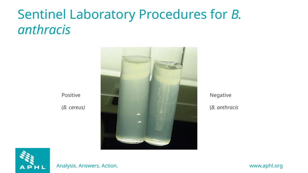
Positive
(B. cereus)) Negative
(B. anthracis) Sentinel Laboratory Procedures for B. anthracis<br>
20
Screening Tests: Catalase Catalase - Positive
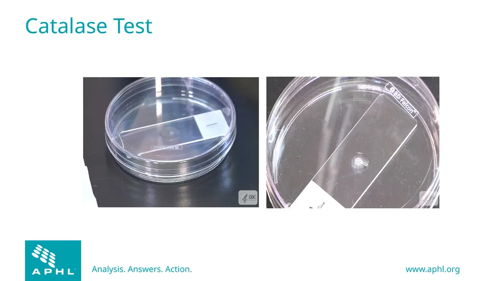
Catalase test is very dangerous—do not perform as a slide test on the open bench
Recommend doing tube test or doing it in a BSC or doing the slide test in a covered petri dish<br>
21
Catalase Test Negative Positive<br>
22
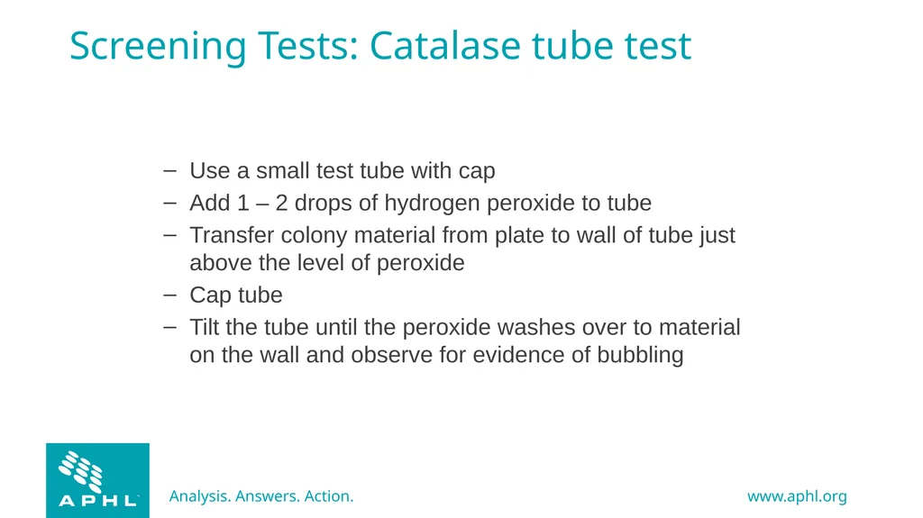
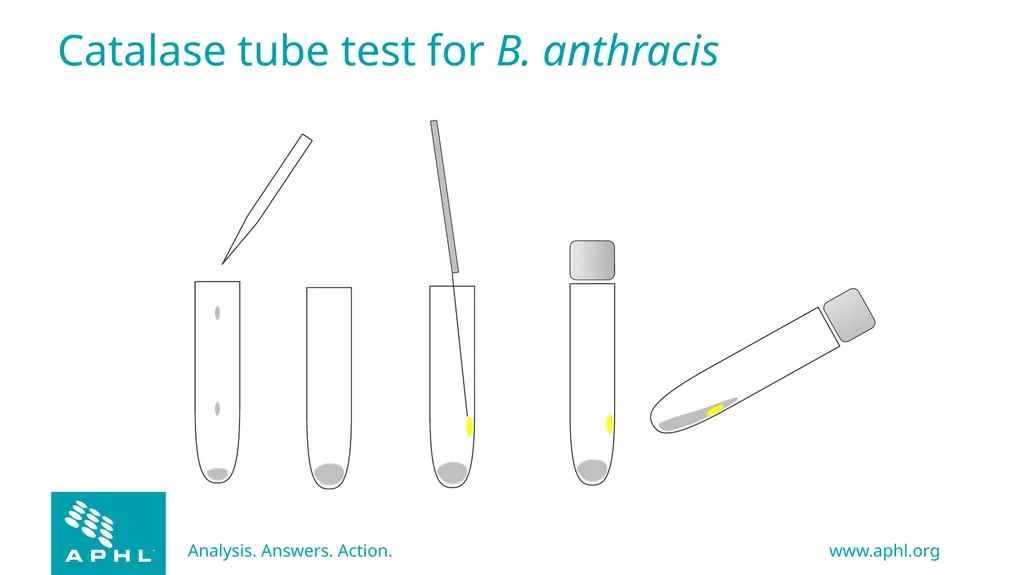
Screening Tests: Catalase tube test Use a small test tube with cap
Add 1 – 2 drops of hydrogen peroxide to tube
Transfer colony material from plate to wall of tube just above the level of peroxide
Cap tube
Tilt the tube until the peroxide washes over to material on the wall and observe for evidence of bubbling<br>
23
Catalase tube test for B. anthracis<br>
24
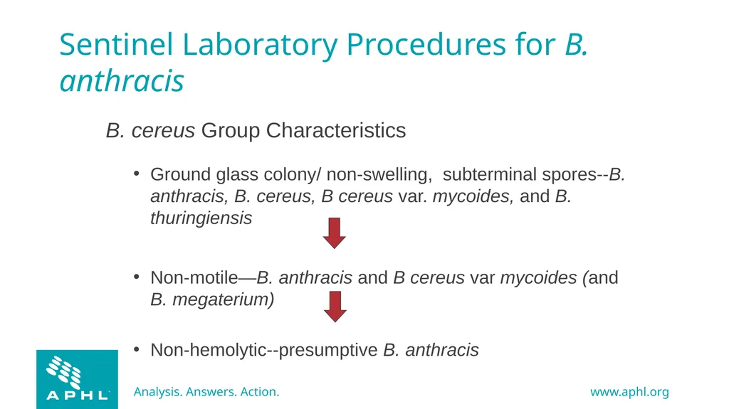
Sentinel Laboratory Procedures for B. anthracis B. cereus Group Characteristics
Ground glass colony/ non-swelling, subterminal spores--B. anthracis, B. cereus, B cereus var. mycoides, and B. thuringiensis
Non-motile—B. anthracis and B cereus var mycoides (and B. megaterium)
Non-hemolytic--presumptive B. anthracis<br>
25
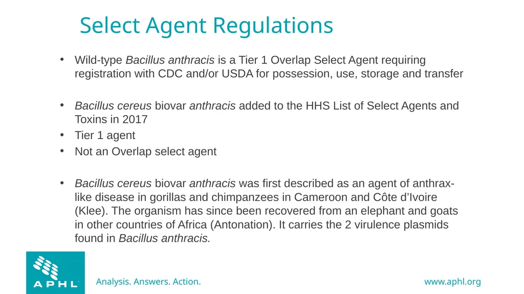
Select Agent Regulations Wild-type Bacillus anthracis is a Tier 1 Overlap Select Agent requiring registration with CDC and/or USDA for possession, use, storage and transfer
Bacillus cereus biovar anthracis added to the HHS List of Select Agents and Toxins in 2017
Tier 1 agent
Not an Overlap select agent
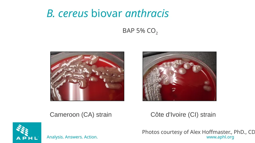
Bacillus cereus biovar anthracis was first described as an agent of anthrax-like disease in gorillas and chimpanzees in Cameroon and Côte d’Ivoire (Klee). The organism has since been recovered from an elephant and goats in other countries of Africa (Antonation). It carries the 2 virulence plasmids found in Bacillus anthracis.<br>
26
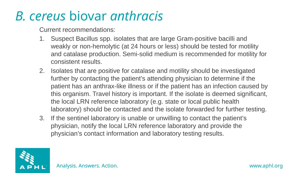
B. cereus biovar anthracis Current recommendations:
Suspect Bacillus spp. isolates that are large Gram-positive bacilli and weakly or non-hemolytic (at 24 hours or less) should be tested for motility and catalase production. Semi-solid medium is recommended for motility for consistent results.
Isolates that are positive for catalase and motility should be investigated further by contacting the patient’s attending physician to determine if the patient has an anthrax-like illness or if the patient has an infection caused by this organism. Travel history is important. If the isolate is deemed significant, the local LRN reference laboratory (e.g. state or local public health laboratory) should be contacted and the isolate forwarded for further testing.
If the sentinel laboratory is unable or unwilling to contact the patient’s physician, notify the local LRN reference laboratory and provide the physician’s contact information and laboratory testing results.<br>
27
B. cereus biovar anthracis Cameroon (CA) strain Côte d'Ivoire (CI) strain BAP 5% CO2 Photos courtesy of Alex Hoffmaster., PhD., CDC<br>
28
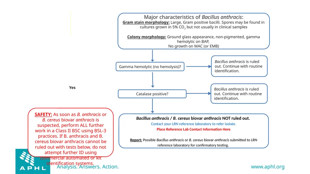
Bacillus anthracis�Identification Flowchart Yes SAFETY: As soon as B. anthracis or B. cereus biovar anthracis is suspected, perform ALL further work in a Class II BSC using BSL-3 practices. If B. anthracis and B. cereus biovar anthracis cannot be ruled out with tests below, do not attempt further ID using commercial automated or kit identification systems.<br>
29
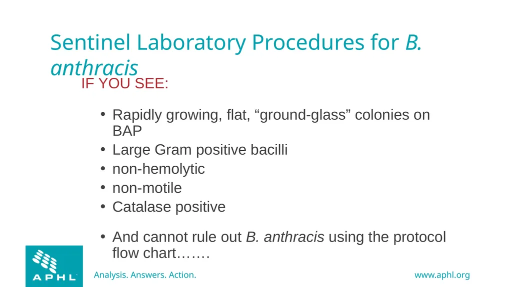
Sentinel Laboratory Procedures for B. anthracis IF YOU SEE:
Rapidly growing, flat, “ground-glass” colonies on BAP
Large Gram positive bacilli
non-hemolytic
non-motile
Catalase positive
And cannot rule out B. anthracis using the protocol flow chart…….<br>
30
CONTACT YOUR LRN
reference laboratory<br>