Background Spatial Frequency Domain Imaging as a
Description: Background Spatial Frequency Domain Imaging as a Novel Method to Quantify Skin Changes in Scleroderma Hung Vo MD1; Aarohi M. Mehendale2; Anahita Pilvar2, Michael York MD1, Marcin Trojanowski MD1, Eugene Kissin MD1, Darren Roblyer PhD2, and
Related Topics
Download Presentation
"Background Spatial Frequency Domain Imaging as a" is the property of its rightful owner. Permission is granted to download and print the materials on this website for personal, non-commercial use only, and to display it on your personal computer provided you do not modify the materials and that you retain all copyright notices contained in the materials. By downloading content from our website, you accept the terms of this agreement.
Presentation Transcript
slide1. Background Spatial Frequency Domain Imaging as a Novel Method to Quantify Skin Changes in Scleroderma Hung Vo MD1; Aarohi M. Mehendale2; Anahita Pilvar2, Michael York MD1, Marcin Trojanowski MD1, Eugene Kissin MD1, Darren Roblyer PhD2, and Andreea M. Bujor MD, PhD1
1. Division of Rheumatology, Boston University Chobanian & Avedisian School of Medicine, Boston, MA 02118, USA
2. Department of Biomedical Engineering, Boston University, Boston, MA 02215, USA Future Works Validate SFDI in a larger cohort with more enrolled patients
Refine our protocol to enhance sensitivity for assessing skin changes in arms, hands, and fingers
Create a portable handheld device for easier imaging acquisition Systemic sclerosis (SSc) is an autoimmune disease characterized by excessive collagen deposition
Gold standard for evaluating skin fibrosis in SSc is the modified Rodnan skin score (mRSS) which has notable limitations
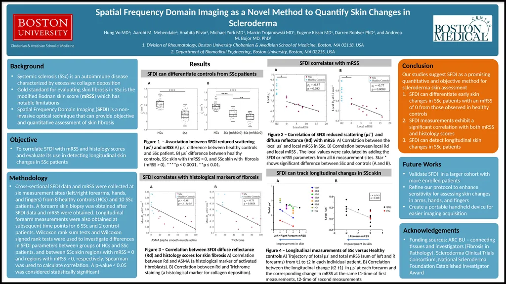
Spatial Frequency Domain Imaging (SFDI) is a non-invasive optical technique that can provide objective and quantitative assessment of skin fibrosis SFDI can track longitudinal changes in SSc skin Objective To correlate SFDI with mRSS and histology scores and evaluate its use in detecting longitudinal skin changes in SSc patients Methodology Cross-sectional SFDI data and mRSS were collected at six measurement sites (left/right forearms, hands, and fingers) from 8 healthy controls (HCs) and 10 SSc patients. A forearm skin biopsy was obtained after SFDI data and mRSS were obtained. Longitudinal forearm measurements were also obtained at subsequent time points for 6 SSc and 2 control patients. Wilcoxon rank sum tests and Wilcoxon signed rank tests were used to investigate differences in SFDI parameters between groups of HCs and SSc patients, and between SSc skin regions with mRSS = 0 and regions with mRSS > 0, respectively. Spearman was used to calculate correlation. A p-value < 0.05 was considered statistically significant SFDI can differentiate controls from SSc patients SFDI correlates with histological markers of fibrosis Conclusion Our studies suggest SFDI as a promising quantitative and objective method for scleroderma skin assessment
SFDI can differentiate early skin changes in SSc patients with an mRSS of 0 from those observed in healthy controls
SFDI measurements exhibit a significant correlation with both mRSS and histology scores
SFDI can detect longitudinal skin changes in SSc patients Figure 4 – Longitudinal measurements of SSc versus Healthy controls A) Trajectory of total μs' and total mRSS (sum of left and R forearms) from t1 to t2 in each individual patient. B) Correlation between the longitudinal change (t2-t1) in μs' at each forearm and the corresponding change in mRSS at the same t1-time of first measurements, t2-time of second measurements SFDI correlates with mRSS Figure 1 – Association between SFDI reduced scattering (μs’) and mRSS A) µs´ difference between healthy controls and SSc patient. B) µs´ difference between healthy controls, SSc skin with (mRSS = 0, and SSc skin with fibrosis (mRSS > 0). ****p < 0.0001, **p ≤ 0.01. Figure 2 – Correlation of SFDI reduced scattering (μs’) and diffuse reflectance (Rd) with mRSS A) Correlation between the local µs´ and local mRSS in SSc. B) Correlation between local Rd and local mRSS . The local values were calculated by adding the SFDI or mRSS parameters from all 6 measurement sites. Star * shows significant difference between SSc and controls (A and B). Figure 3 – Correlation between SFDI diffuse reflectance (Rd) and histology scores for skin fibrosis A) Correlation between Rd and ASMA (a histological marker of activated fibroblasts). B) Correlation between Rd and Trichrome staining (a histological marker for collagen deposition). Acknowledgements Funding sources: ARC BU – connecting tissues and investigators (Fibrosis in Pathology), Scleroderma Clinical Trials Consortium, National Scleroderma Foundation Established Investigator Award Results<br>

1. Division of Rheumatology, Boston University Chobanian & Avedisian School of Medicine, Boston, MA 02118, USA
2. Department of Biomedical Engineering, Boston University, Boston, MA 02215, USA Future Works Validate SFDI in a larger cohort with more enrolled patients
Refine our protocol to enhance sensitivity for assessing skin changes in arms, hands, and fingers
Create a portable handheld device for easier imaging acquisition Systemic sclerosis (SSc) is an autoimmune disease characterized by excessive collagen deposition
Gold standard for evaluating skin fibrosis in SSc is the modified Rodnan skin score (mRSS) which has notable limitations
Spatial Frequency Domain Imaging (SFDI) is a non-invasive optical technique that can provide objective and quantitative assessment of skin fibrosis SFDI can track longitudinal changes in SSc skin Objective To correlate SFDI with mRSS and histology scores and evaluate its use in detecting longitudinal skin changes in SSc patients Methodology Cross-sectional SFDI data and mRSS were collected at six measurement sites (left/right forearms, hands, and fingers) from 8 healthy controls (HCs) and 10 SSc patients. A forearm skin biopsy was obtained after SFDI data and mRSS were obtained. Longitudinal forearm measurements were also obtained at subsequent time points for 6 SSc and 2 control patients. Wilcoxon rank sum tests and Wilcoxon signed rank tests were used to investigate differences in SFDI parameters between groups of HCs and SSc patients, and between SSc skin regions with mRSS = 0 and regions with mRSS > 0, respectively. Spearman was used to calculate correlation. A p-value < 0.05 was considered statistically significant SFDI can differentiate controls from SSc patients SFDI correlates with histological markers of fibrosis Conclusion Our studies suggest SFDI as a promising quantitative and objective method for scleroderma skin assessment
SFDI can differentiate early skin changes in SSc patients with an mRSS of 0 from those observed in healthy controls
SFDI measurements exhibit a significant correlation with both mRSS and histology scores
SFDI can detect longitudinal skin changes in SSc patients Figure 4 – Longitudinal measurements of SSc versus Healthy controls A) Trajectory of total μs' and total mRSS (sum of left and R forearms) from t1 to t2 in each individual patient. B) Correlation between the longitudinal change (t2-t1) in μs' at each forearm and the corresponding change in mRSS at the same t1-time of first measurements, t2-time of second measurements SFDI correlates with mRSS Figure 1 – Association between SFDI reduced scattering (μs’) and mRSS A) µs´ difference between healthy controls and SSc patient. B) µs´ difference between healthy controls, SSc skin with (mRSS = 0, and SSc skin with fibrosis (mRSS > 0). ****p < 0.0001, **p ≤ 0.01. Figure 2 – Correlation of SFDI reduced scattering (μs’) and diffuse reflectance (Rd) with mRSS A) Correlation between the local µs´ and local mRSS in SSc. B) Correlation between local Rd and local mRSS . The local values were calculated by adding the SFDI or mRSS parameters from all 6 measurement sites. Star * shows significant difference between SSc and controls (A and B). Figure 3 – Correlation between SFDI diffuse reflectance (Rd) and histology scores for skin fibrosis A) Correlation between Rd and ASMA (a histological marker of activated fibroblasts). B) Correlation between Rd and Trichrome staining (a histological marker for collagen deposition). Acknowledgements Funding sources: ARC BU – connecting tissues and investigators (Fibrosis in Pathology), Scleroderma Clinical Trials Consortium, National Scleroderma Foundation Established Investigator Award Results<br>