
Blair Upper Cervical Research Hour 3 Blair
TF
Published · 40 slides · 0 views

1 / 1






Description
Blair Upper Cervical Research Hour 3 Blair Research The slides are properties of Dr. Thomas Forest, Dr. Dennis Campbell, Dr. Todd Hubbard. Certified Advanced Blair Instructors They are not to be copied nor distributed Absence of Symmetry
Related Topics
Share
Embed code
Download this presentation From Below
"Blair Upper Cervical Research Hour 3 Blair" is the property of its rightful owner. Permission is granted to download and print the materials on this website for personal, non-commercial use only, and to display it on your personal computer provided you do not modify the materials and that you retain all copyright notices contained in the materials. By downloading content from our website, you accept the terms of this agreement.
Presentation Transcript
01
Blair Upper Cervical Research Hour 3 Blair Research
The slides are properties of
Dr. Thomas Forest, Dr. Dennis Campbell, &
Dr. Todd Hubbard.
Certified Advanced Blair Instructors
They are not to be copied nor distributed<br>
The slides are properties of
Dr. Thomas Forest, Dr. Dennis Campbell, &
Dr. Todd Hubbard.
Certified Advanced Blair Instructors
They are not to be copied nor distributed<br>
02
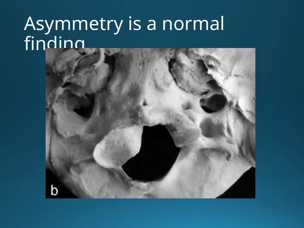
Absence of Symmetry in Superior Articular Facets on the First Cervical Vertebra
JMPT 1994 17(5):314-320 Gottlieb MS The shape, size, angle, texture, border, and/or number of superior articular facets were measured to determine symmetry.<br>
03
Absence of Symmetry in Superior Articular Facets on the First Cervical Vertebra
JMPT 1994 17(5):314-320 Gottlieb MS The shape, size, angle, texture, border, and/or number of superior articular facets were measured to determine symmetry.
Results: On comparison of right and left sides, 0% of the facets were mirror images while 63% of the atlases had grossly asymmetrical facets<br>
Results: On comparison of right and left sides, 0% of the facets were mirror images while 63% of the atlases had grossly asymmetrical facets<br>
04
Absence of Symmetry in Superior Articular Facets on the First Cervical Vertebra
JMPT 1994 17(5):314-320 Gottlieb MS The shape, size, angle, texture, border, and/or number of superior articular facets were measured to determine symmetry.
Results: On comparison of right and left sides, 0% of the facets were mirror images while 63% of the atlases had grossly asymmetrical facets
Three atlases had two facets on the left and one on the right, while two atlases had two facets on the right with a single facet on the left. Two atlases had four superior facets, two on each side.<br>
Results: On comparison of right and left sides, 0% of the facets were mirror images while 63% of the atlases had grossly asymmetrical facets
Three atlases had two facets on the left and one on the right, while two atlases had two facets on the right with a single facet on the left. Two atlases had four superior facets, two on each side.<br>
05
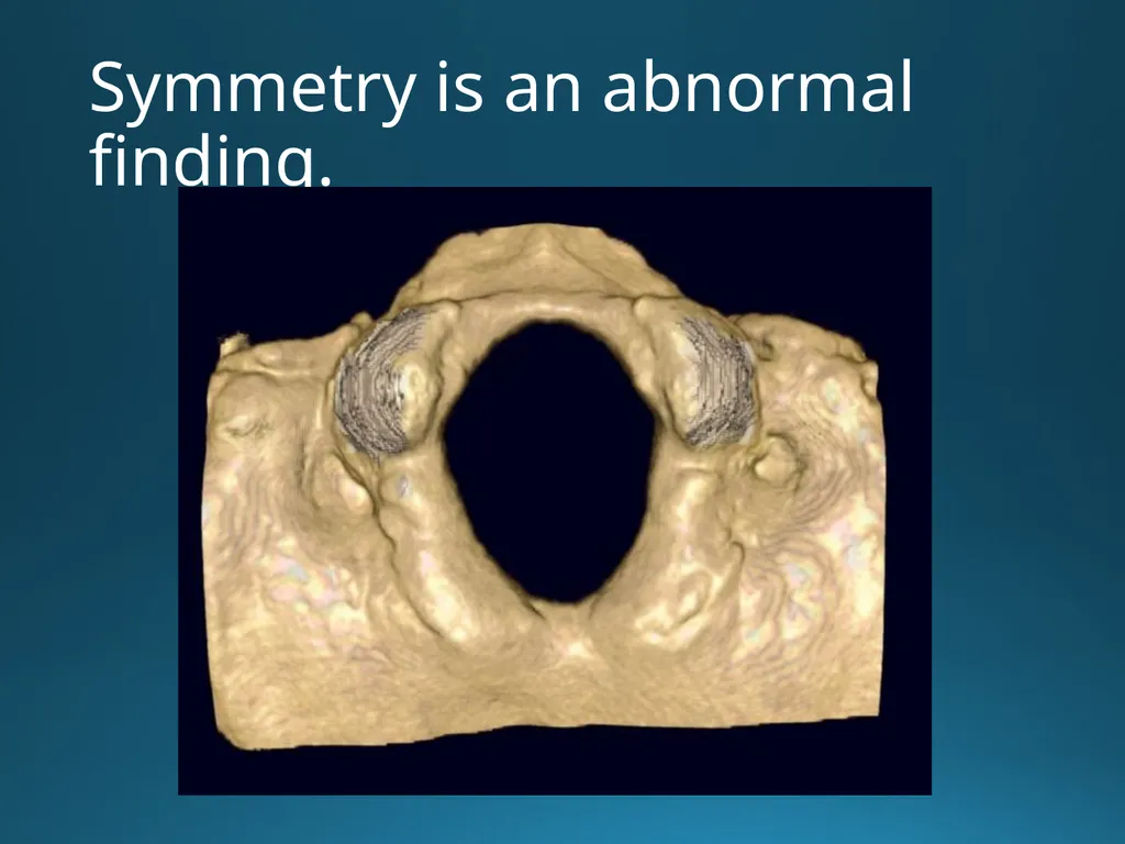
Symmetry is an abnormal finding.<br>
06
Asymmetry is a normal finding.<br>
07
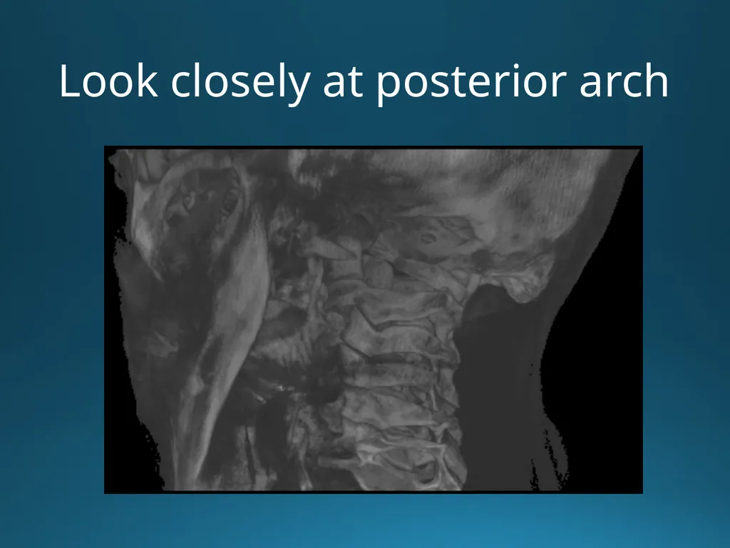
Look closely at posterior arch<br>
08
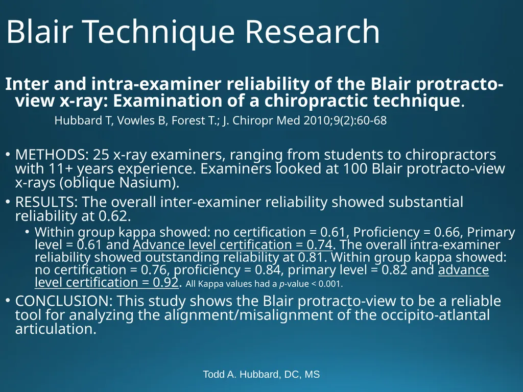
Todd A. Hubbard, DC, MS Blair Technique Research Inter and intra-examiner reliability of the Blair protracto-view x-ray: Examination of a chiropractic technique.
Hubbard T, Vowles B, Forest T.; J. Chiropr Med 2010;9(2):60-68
METHODS: 25 x-ray examiners, ranging from students to chiropractors with 11+ years experience. Examiners looked at 100 Blair protracto-view x-rays (oblique Nasium).
RESULTS: The overall inter-examiner reliability showed substantial reliability at 0.62.
Within group kappa showed: no certification = 0.61, Proficiency = 0.66, Primary level = 0.61 and Advance level certification = 0.74. The overall intra-examiner reliability showed outstanding reliability at 0.81. Within group kappa showed: no certification = 0.76, proficiency = 0.84, primary level = 0.82 and advance level certification = 0.92. All Kappa values had a p-value < 0.001.
CONCLUSION: This study shows the Blair protracto-view to be a reliable tool for analyzing the alignment/misalignment of the occipito-atlantal articulation.<br>
Hubbard T, Vowles B, Forest T.; J. Chiropr Med 2010;9(2):60-68
METHODS: 25 x-ray examiners, ranging from students to chiropractors with 11+ years experience. Examiners looked at 100 Blair protracto-view x-rays (oblique Nasium).
RESULTS: The overall inter-examiner reliability showed substantial reliability at 0.62.
Within group kappa showed: no certification = 0.61, Proficiency = 0.66, Primary level = 0.61 and Advance level certification = 0.74. The overall intra-examiner reliability showed outstanding reliability at 0.81. Within group kappa showed: no certification = 0.76, proficiency = 0.84, primary level = 0.82 and advance level certification = 0.92. All Kappa values had a p-value < 0.001.
CONCLUSION: This study shows the Blair protracto-view to be a reliable tool for analyzing the alignment/misalignment of the occipito-atlantal articulation.<br>
09
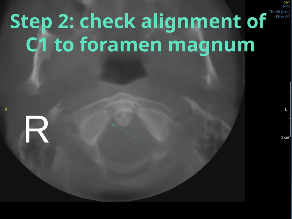
Analysis step 1 (image reve R Step 2: check alignment of
C1 to foramen magnum<br>
C1 to foramen magnum<br>
10
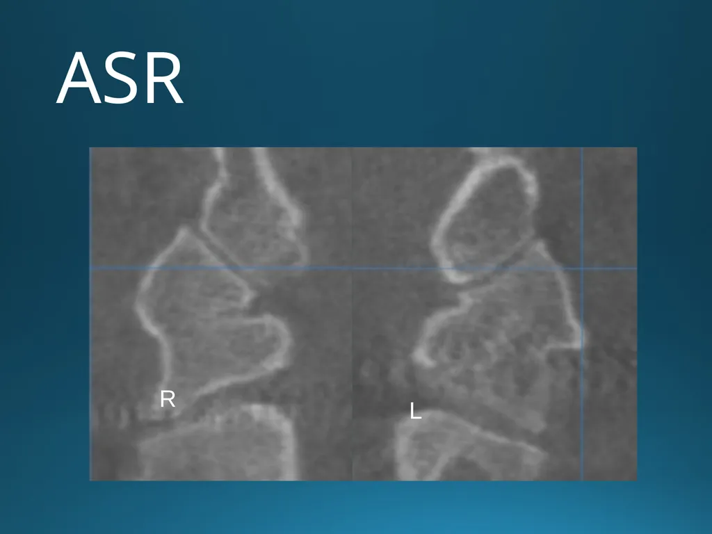
ASR R L<br>
11
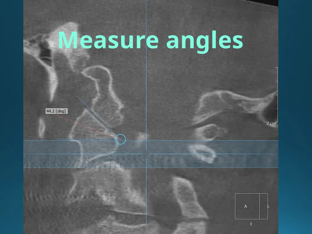
Measure angles<br>
12
Upper Cervical Chiropractic Care for a 10-year-old Patient with Complex Regional Pain Syndrome
Lauren Millman, D.C., Thomas Forest, D.C., Todd Hubbard, D.D., M.S. Journal of Upper Cervical Chiropractic Research – October 10, 2012-pages 85-91 Objective- To review the effectiveness of chiropractic care using a specific upper cervical technique in the case of a 10-year-old male with Complex Regional Pain Syndrome.<br>
13
Upper Cervical Chiropractic Care for a 10-year-old Patient with Complex Regional Pain Syndrome
Lauren Millman, D.C., Thomas Forest, D.C., Todd Hubbard, D.D., M.S. Journal of Upper Cervical Chiropractic Research – October 10, 2012-pages 85-91 Objective- To review the effectiveness of chiropractic care using a specific upper cervical technique in the case of a 10-year-old male with Complex Regional Pain Syndrome.
Intervention: Upon initial examination, indications of upper cervical subluxation were evident, based upon radiographic findings, thermography, and leg length inequality. Chiropractic care utilizing a specific upper cervical technique protocol (Blair upper cervical technique) was administered in order to correct upper cervical subluxation.<br>
Intervention: Upon initial examination, indications of upper cervical subluxation were evident, based upon radiographic findings, thermography, and leg length inequality. Chiropractic care utilizing a specific upper cervical technique protocol (Blair upper cervical technique) was administered in order to correct upper cervical subluxation.<br>
14
Upper Cervical Chiropractic Care for a 10-year-old Patient with Complex Regional Pain Syndrome
Lauren Millman, D.C., Thomas Forest, D.C., Todd Hubbard, D.D., M.S Journal of Upper Cervical Chiropractic Research – October 10, 2012-pages 85-91 Objective- To review the effectiveness of chiropractic care using a specific upper cervical technique in the case of a 10-year-old male with Complex Regional Pain Syndrome.
Intervention: Upon initial examination, indications of upper cervical subluxation were evident, based upon radiographic findings, thermography, and leg length inequality. Chiropractic care utilizing a specific upper cervical technique protocol (Blair upper cervical technique) was administered in order to correct upper cervical subluxation.
Outcome: although still ongoing case, the following report is a detailed 10-month record of the 1-year-old boy’s response to his upper cervical technique, including a decrease in symptoms associated with CRPS Type 1, noting them as less frequent, of less duration and decreased severity.<br>
Intervention: Upon initial examination, indications of upper cervical subluxation were evident, based upon radiographic findings, thermography, and leg length inequality. Chiropractic care utilizing a specific upper cervical technique protocol (Blair upper cervical technique) was administered in order to correct upper cervical subluxation.
Outcome: although still ongoing case, the following report is a detailed 10-month record of the 1-year-old boy’s response to his upper cervical technique, including a decrease in symptoms associated with CRPS Type 1, noting them as less frequent, of less duration and decreased severity.<br>
15
Upper Cervical Chiropractic Care for a 10-year-old Patient with Complex Regional Pain Syndrome
Lauren Millman, D.C., Thomas Forest, D.C., Todd Hubbard, D.D., M.S. Journal of Upper Cervical Chiropractic Research – October 10, 2012-pages 85-91 Objective- To review the effectiveness of chiropractic care using a specific upper cervical technique in the case of a 10-year-old male with Complex Regional Pain Syndrome.
Intervention: Upon initial examination, indications of upper cervical subluxation were evident, based upon radiographic findings, thermography, and leg length inequality. Chiropractic care utilizing a specific upper cervical technique protocol (Blair upper cervical technique) was administered in order to correct upper cervical subluxation.
Outcome: although still ongoing case, the following report is a detailed 10-month record of the 1-year-old boy’s response to his upper cervical technique, including a decrease in symptoms associated with CRPS Type 1, noting them as less frequent, of less duration and decreased severity.
Conclusion: Due to the marked and timely improvement of this patient, this case report exhibits the need for further investigation in the use of the non-invasive, light –force, cost-effective, and highly specific approach to correcting upper cervical subluxations which have an effect on this condition.<br>
Intervention: Upon initial examination, indications of upper cervical subluxation were evident, based upon radiographic findings, thermography, and leg length inequality. Chiropractic care utilizing a specific upper cervical technique protocol (Blair upper cervical technique) was administered in order to correct upper cervical subluxation.
Outcome: although still ongoing case, the following report is a detailed 10-month record of the 1-year-old boy’s response to his upper cervical technique, including a decrease in symptoms associated with CRPS Type 1, noting them as less frequent, of less duration and decreased severity.
Conclusion: Due to the marked and timely improvement of this patient, this case report exhibits the need for further investigation in the use of the non-invasive, light –force, cost-effective, and highly specific approach to correcting upper cervical subluxations which have an effect on this condition.<br>
16
Todd A. Hubbard, DC, MS Cyclic Vomiting in a 7 year old female.
Hubbard T, Crisp C. Journal of Chiropractic Medicine
Received 15 July 2010; accepted 19 July 2010 Clinical Features: A 7-yr-old girl had a history of cyclic vomiting episodes for the past 4 1/2 years. She also complained of a 2-month history of constant headaches and stomach ache.
Intervention and Outcome: The patient received low-force chiropractic spinal manipulation to her upper cervical spine. There was improvement in her symptoms within an hour after the chiropractic adjustment. Her symptoms only returned after direct trauma to her neck. The recurring symptoms again disappeared immediately after chiropractic adjustment.
Conclusion: This case study suggests there may be a role for the use of chiropractic spinal manipulative therapy for treating cyclic vomiting syndrome.<br>
Intervention and Outcome: The patient received low-force chiropractic spinal manipulation to her upper cervical spine. There was improvement in her symptoms within an hour after the chiropractic adjustment. Her symptoms only returned after direct trauma to her neck. The recurring symptoms again disappeared immediately after chiropractic adjustment.
Conclusion: This case study suggests there may be a role for the use of chiropractic spinal manipulative therapy for treating cyclic vomiting syndrome.<br>
17
Upper Cervical Protocol For Ten Meniere’s Patients By Michael Burcon, D.C.
Copyrighted Journal of Vertebral Subluxation Res.
Meniere’s is characterized by vertigo, nausea, inner ear pressure, low frequency hearing loss, and tinnitus. HA’s,depression, neck stiffness, and “brain fog” are associated symptoms. A Blair series of cervical x-rays were taken along with standard upper cervical testing procedures. It is noteworthy that each of the Meniere’s patients had a posterior inferior displacement (subluxation) of the C-1 segment. Conclusion: All patients responded favorably to side posture spinal adjustments. The odds that ten consecutive Meniere’s patients would have a posterior and inferior atlas listing with laterality on the opposite side of ear involvement is over one million to one!<br>
Copyrighted Journal of Vertebral Subluxation Res.
Meniere’s is characterized by vertigo, nausea, inner ear pressure, low frequency hearing loss, and tinnitus. HA’s,depression, neck stiffness, and “brain fog” are associated symptoms. A Blair series of cervical x-rays were taken along with standard upper cervical testing procedures. It is noteworthy that each of the Meniere’s patients had a posterior inferior displacement (subluxation) of the C-1 segment. Conclusion: All patients responded favorably to side posture spinal adjustments. The odds that ten consecutive Meniere’s patients would have a posterior and inferior atlas listing with laterality on the opposite side of ear involvement is over one million to one!<br>
18
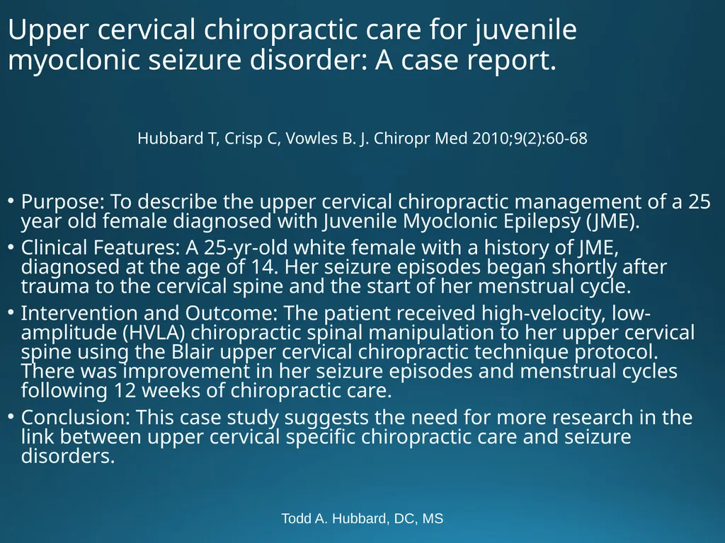
Todd A. Hubbard, DC, MS Upper cervical chiropractic care for juvenile myoclonic seizure disorder: A case report. Hubbard T, Crisp C, Vowles B. J. Chiropr Med 2010;9(2):60-68
Purpose: To describe the upper cervical chiropractic management of a 25 year old female diagnosed with Juvenile Myoclonic Epilepsy (JME).
Clinical Features: A 25-yr-old white female with a history of JME, diagnosed at the age of 14. Her seizure episodes began shortly after trauma to the cervical spine and the start of her menstrual cycle.
Intervention and Outcome: The patient received high-velocity, low-amplitude (HVLA) chiropractic spinal manipulation to her upper cervical spine using the Blair upper cervical chiropractic technique protocol. There was improvement in her seizure episodes and menstrual cycles following 12 weeks of chiropractic care.
Conclusion: This case study suggests the need for more research in the link between upper cervical specific chiropractic care and seizure disorders.<br>
Purpose: To describe the upper cervical chiropractic management of a 25 year old female diagnosed with Juvenile Myoclonic Epilepsy (JME).
Clinical Features: A 25-yr-old white female with a history of JME, diagnosed at the age of 14. Her seizure episodes began shortly after trauma to the cervical spine and the start of her menstrual cycle.
Intervention and Outcome: The patient received high-velocity, low-amplitude (HVLA) chiropractic spinal manipulation to her upper cervical spine using the Blair upper cervical chiropractic technique protocol. There was improvement in her seizure episodes and menstrual cycles following 12 weeks of chiropractic care.
Conclusion: This case study suggests the need for more research in the link between upper cervical specific chiropractic care and seizure disorders.<br>
19
Upper Cervical Chiropractic Management of an Adult with
Ankylosing Spondylitis: A Case Report
Cailen E Wells DC1 & John Williams DC2 Clinical Features: A 52 year old male presented with postural stiffness in multiple joints, mid-back pain, neck pain, bilateral shoulder pain, bilateral leg and hip pain. Past history included three motor vehicle accidents prior to the diagnosis of Ankylosing Spondylitis.<br>
20
Upper Cervical Chiropractic Management of an Adult with
Ankylosing Spondylitis: A Case Report
Cailen E Wells DC1 & John Williams DC2 Clinical Features: A 52 year old male presented with postural stiffness in multiple joints, mid-back pain, neck pain, bilateral shoulder pain, bilateral leg and hip pain. Past history included three motor vehicle accidents prior to the diagnosis of Ankylosing Spondylitis.
Intervention and Outcomes: Subluxations were confirmed with upper cervical radiographs, paraspinal digital infrared imaging, and a Grostic leg check analysis. Blair and Knee Chest techniques were utilized over a 4 month period. Positive improvements were also seen in blood pressure.<br>
Intervention and Outcomes: Subluxations were confirmed with upper cervical radiographs, paraspinal digital infrared imaging, and a Grostic leg check analysis. Blair and Knee Chest techniques were utilized over a 4 month period. Positive improvements were also seen in blood pressure.<br>
21
Upper Cervical Chiropractic Management of an Adult with
Ankylosing Spondylitis: A Case Report
Cailen E Wells DC1 & John Williams DC2 Clinical Features: A 52 year old male presented with postural stiffness in multiple joints, mid-back pain, neck pain, bilateral shoulder pain, bilateral leg and hip pain. Past history included three motor vehicle accidents prior to the diagnosis of Ankylosing Spondylitis.
Intervention and Outcomes: Subluxations were confirmed with upper cervical radiographs, paraspinal digital infrared imaging, and a Grostic leg check analysis. Blair and Knee Chest techniques were utilized over a 4 month period. Positive improvements were also seen in blood pressure.
Conclusion: This case report exhibits the need for further study of both the Knee Chest and Blair corrections of an upper cervical vertebral subluxation in a patient with AS.
Key Words: Upper cervical chiropractic, Blair technique,<br>
Intervention and Outcomes: Subluxations were confirmed with upper cervical radiographs, paraspinal digital infrared imaging, and a Grostic leg check analysis. Blair and Knee Chest techniques were utilized over a 4 month period. Positive improvements were also seen in blood pressure.
Conclusion: This case report exhibits the need for further study of both the Knee Chest and Blair corrections of an upper cervical vertebral subluxation in a patient with AS.
Key Words: Upper cervical chiropractic, Blair technique,<br>
22
Harvey Lillard (?)<br>
23
Hearing Loss After Blunt Neck Trauma
Otology and Neurotology 2003: 24(5):734-737 Segal, et al Objective-to report for the 1st time hearing impairment from blunt neck trauma involving 83 patients. 51.5% had hearing loss, 55.4% noted tinnitus within three months of the trauma.
A reflexogenic disturbance of the neck via divergent afferent proprioceptive activity to the central auditory nervous system might be an explanation.”
The medical prognosis is poor with only 10% showing spontaneous improvement within a year.<br>
A reflexogenic disturbance of the neck via divergent afferent proprioceptive activity to the central auditory nervous system might be an explanation.”
The medical prognosis is poor with only 10% showing spontaneous improvement within a year.<br>
24
Cervicogenic Hearing Loss
M. Hulse, Professor of ENT, Heidelberg University
Source: Greg Buchanan info@upspine.com Findings in 62 patients suffering from vertebragenic hearing disorders are reported before and after chiropractic management.
Results indicate that hearing disorders are reversible… the therapy of choice is chiropractic manipulation of the upper cervical spine.<br>
Results indicate that hearing disorders are reversible… the therapy of choice is chiropractic manipulation of the upper cervical spine.<br>
25
There is ample research confirming the efficacy of Blair Upper Cervical care. Join this group of dedicated precision oriented doctors who feel that EVERYONE deserves to be neurologically clear. A brain stem free of neurological interference will impact society physically, socially, and mentally. As BJ has told us, “Get the Big Idea, and all else follows.”<br>
26
The End<br>
27
OPTIONAL<br>
28
What does the LEG CHECK research say Knutson GA. J Manipulative Physiol Ther 2002 Jul-Aug;25(6):425. Incidence of foot rotation, pelvic crest unleveling, and supine leg length alignment asymmetry and their relationship to self-reported back pain.
the supine leg length alignment check had clinical validity as a stand-alone test for recurring back pain 28<br>
the supine leg length alignment check had clinical validity as a stand-alone test for recurring back pain 28<br>
29
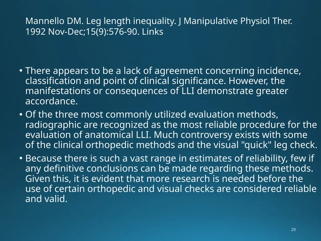
Mannello DM. Leg length inequality. J Manipulative Physiol Ther. 1992 Nov-Dec;15(9):576-90. Links There appears to be a lack of agreement concerning incidence, classification and point of clinical significance. However, the manifestations or consequences of LLI demonstrate greater accordance.
Of the three most commonly utilized evaluation methods, radiographic are recognized as the most reliable procedure for the evaluation of anatomical LLI. Much controversy exists with some of the clinical orthopedic methods and the visual "quick" leg check.
Because there is such a vast range in estimates of reliability, few if any definitive conclusions can be made regarding these methods. Given this, it is evident that more research is needed before the use of certain orthopedic and visual checks are considered reliable and valid. 29<br>
Of the three most commonly utilized evaluation methods, radiographic are recognized as the most reliable procedure for the evaluation of anatomical LLI. Much controversy exists with some of the clinical orthopedic methods and the visual "quick" leg check.
Because there is such a vast range in estimates of reliability, few if any definitive conclusions can be made regarding these methods. Given this, it is evident that more research is needed before the use of certain orthopedic and visual checks are considered reliable and valid. 29<br>
30
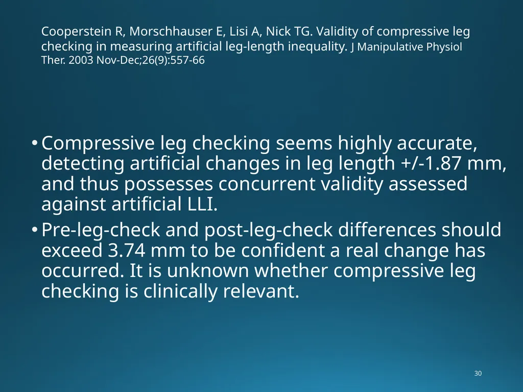
Cooperstein R, Morschhauser E, Lisi A, Nick TG. Validity of compressive leg checking in measuring artificial leg-length inequality. J Manipulative Physiol Ther. 2003 Nov-Dec;26(9):557-66 Compressive leg checking seems highly accurate, detecting artificial changes in leg length +/-1.87 mm, and thus possesses concurrent validity assessed against artificial LLI.
Pre-leg-check and post-leg-check differences should exceed 3.74 mm to be confident a real change has occurred. It is unknown whether compressive leg checking is clinically relevant. 30<br>
Pre-leg-check and post-leg-check differences should exceed 3.74 mm to be confident a real change has occurred. It is unknown whether compressive leg checking is clinically relevant. 30<br>
31
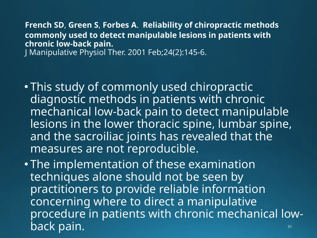
French SD, Green S, Forbes A. Reliability of chiropractic methods commonly used to detect manipulable lesions in patients with chronic low-back pain.
J Manipulative Physiol Ther. 2001 Feb;24(2):145-6. This study of commonly used chiropractic diagnostic methods in patients with chronic mechanical low-back pain to detect manipulable lesions in the lower thoracic spine, lumbar spine, and the sacroiliac joints has revealed that the measures are not reproducible.
The implementation of these examination techniques alone should not be seen by practitioners to provide reliable information concerning where to direct a manipulative procedure in patients with chronic mechanical low-back pain. 31<br>
The implementation of these examination techniques alone should not be seen by practitioners to provide reliable information concerning where to direct a manipulative procedure in patients with chronic mechanical low-back pain. 31<br>
32
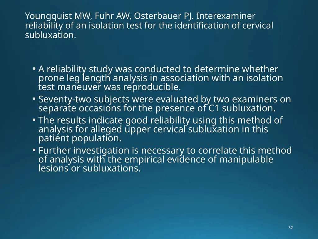
Youngquist MW, Fuhr AW, Osterbauer PJ. Interexaminer reliability of an isolation test for the identification of cervical subluxation. A reliability study was conducted to determine whether prone leg length analysis in association with an isolation test maneuver was reproducible.
Seventy-two subjects were evaluated by two examiners on separate occasions for the presence of C1 subluxation.
The results indicate good reliability using this method of analysis for alleged upper cervical subluxation in this patient population.
Further investigation is necessary to correlate this method of analysis with the empirical evidence of manipulable lesions or subluxations. 32<br>
Seventy-two subjects were evaluated by two examiners on separate occasions for the presence of C1 subluxation.
The results indicate good reliability using this method of analysis for alleged upper cervical subluxation in this patient population.
Further investigation is necessary to correlate this method of analysis with the empirical evidence of manipulable lesions or subluxations. 32<br>
33
Shambaugh P, Sclafani L, Fanselow D. Reliability of the Derifield-Thompson test for leg length inequality, and use of the test to demonstrate cervical adjusting efficacy. J Manipulative Physiol Ther. 1989 Apr;12(2):151. The Derifield-Thompson test for leg length inequality (LLI) is commonly used by chiropractors to assess a need for adjustment and to evaluate the results of adjustment.
The two previous studies testing the reliability of the technique reported conflicting results.
This study demonstrated that clinicians could reliably measure a LLI to less than 3 mm (both inter- and intraobserver), and also detect a change in LLI when the head was rotated.
Neither cervical adjustment nor gluteal massage produced a significant change in observed LLI. 33<br>
The two previous studies testing the reliability of the technique reported conflicting results.
This study demonstrated that clinicians could reliably measure a LLI to less than 3 mm (both inter- and intraobserver), and also detect a change in LLI when the head was rotated.
Neither cervical adjustment nor gluteal massage produced a significant change in observed LLI. 33<br>
34
COOPERSTEIN R, LISI AJ, MORSCHHAUSER E
CROSS-SECTIONAL VALIDY STUDY OF COMPRESSIVE LEG CHECKING IN MEASURING ARTIFICIALLY CREATED LEG LENGTH INEQUALITY J Chiropr Med: SEP 2004(3:3)Page(s)91-95Author(s) Conclusions: Instrumented, compressive leg checking seems highly accurate, detecting artificial changes in leg length of 2-3 mm, and thus possesses concurrent validity assessed against artificial LLI.
Pre- and post leg check differences should exceed about 4-6 mm to be highly confident a real change has occurred.
It is unknown whether compressive leg checking is clinically relevant. 34<br>
Pre- and post leg check differences should exceed about 4-6 mm to be highly confident a real change has occurred.
It is unknown whether compressive leg checking is clinically relevant. 34<br>
35
Prill Leg Checks Dr. Prill Used Supine leg and arm balance checks. The Blair Technique uses a modified Prill leg check with the patient prone. This modification was used to flow with the other prone leg checks used.
When using the muscle testing procedures, it is important that the patient use very little pressure against the doctors resistance. Too much pressure by the patient will cause false positives. We do not want the large skeletal muscles involved, only the smaller proprioception muscles. 35<br>
When using the muscle testing procedures, it is important that the patient use very little pressure against the doctors resistance. Too much pressure by the patient will cause false positives. We do not want the large skeletal muscles involved, only the smaller proprioception muscles. 35<br>
36
Prill Leg Checks Prill Leg Checks are based off of the Kinesiology or muscle testing principle.
S.O.T. uses a Subluxation Complex protocol that incorporate spinal segments that biomechanically work together.
(as a side note) these subluxation complexes follow acupuncture meridians. 36<br>
S.O.T. uses a Subluxation Complex protocol that incorporate spinal segments that biomechanically work together.
(as a side note) these subluxation complexes follow acupuncture meridians. 36<br>
37
Prill Leg Checks Subluxation Complexes:
The theory behind the muscle testing used in the subluxation complex system is that if your patient has a subluxation at any spinal level in the complex, it will be indicated when testing any muscle that is neurologically supplied to a vertebral level in the complex. 37<br>
The theory behind the muscle testing used in the subluxation complex system is that if your patient has a subluxation at any spinal level in the complex, it will be indicated when testing any muscle that is neurologically supplied to a vertebral level in the complex. 37<br>
38
Subluxation Complexes Both DeJarnette and Prill advocate clearing out the upper spinal levels in the complex first before adjusting the lower spinal levels.
When muscle testing, the patient should start the movement and the force should be minimal. Barely enough to even feel the pressure applied. 38<br>
When muscle testing, the patient should start the movement and the force should be minimal. Barely enough to even feel the pressure applied. 38<br>
39
(Scientific Explanation)
Corticospinal tracts and the tracts responsible for tonal changes in leg/limb musculature. The Dorsal Column Medial Lemniscus systems play the part in delivering sensory input to the brain from the body. The Pontomedullary Reticular Formation (PMRF) is the center in the brain (specifically brainstem) that allows us to stay upright and resist the overall effect of gravity. The Ipsilateral Cerebellum refines movement and is thought to be the brain center that "actions" the information gathered by the three areas mentioned above. The VSC impacts these at any number of levels depending upon the practice model of UC or Full Spine work. The effect is believed to be multifaceted; ranging from neurological dysafferentation or dysefferentation, a direct impact of muscle tone due to the misalignment, and even histochemical changes that create localized pockets of inflammation in afferent/efferent pathways.
Ramneek S. Bhogal, DC, DABCI Professor / Chair of Technique Life Chiropractic College West (03-16-19) 39<br>
Corticospinal tracts and the tracts responsible for tonal changes in leg/limb musculature. The Dorsal Column Medial Lemniscus systems play the part in delivering sensory input to the brain from the body. The Pontomedullary Reticular Formation (PMRF) is the center in the brain (specifically brainstem) that allows us to stay upright and resist the overall effect of gravity. The Ipsilateral Cerebellum refines movement and is thought to be the brain center that "actions" the information gathered by the three areas mentioned above. The VSC impacts these at any number of levels depending upon the practice model of UC or Full Spine work. The effect is believed to be multifaceted; ranging from neurological dysafferentation or dysefferentation, a direct impact of muscle tone due to the misalignment, and even histochemical changes that create localized pockets of inflammation in afferent/efferent pathways.
Ramneek S. Bhogal, DC, DABCI Professor / Chair of Technique Life Chiropractic College West (03-16-19) 39<br>
40
Using the Prill Leg Checks Do Not use too much pressure. 1pound of pressure for the patient is enough to elicit a response.
Resistance should be held for 3-4 seconds
These muscle reflexes will only appear for a few seconds. Therefore, while you don’t have to rush, they should be done in a timely manner.
Once a positive test is found, Dr. Prill recommends a physical challenge of the joint for confirmation. (will cover later)
The Prill Leg Length Iniquity tests will be covered later in the class. 40<br>
Resistance should be held for 3-4 seconds
These muscle reflexes will only appear for a few seconds. Therefore, while you don’t have to rush, they should be done in a timely manner.
Once a positive test is found, Dr. Prill recommends a physical challenge of the joint for confirmation. (will cover later)
The Prill Leg Length Iniquity tests will be covered later in the class. 40<br>