BVDMucosal Disease Dr. Bipin Kumar Assistant Professor Department of Veterinary Medicine Bihar Veterinary College, Patna (Bihar Animal Sciences University, Patna) Bovine Virus Diarrhea and mucosal disease are clinically dissimilar disease
"BVD/Mucosal Disease Dr. Bipin Kumar Assistant" is the property of its rightful owner. Permission is granted to
download and print the materials on this website for personal, non-commercial use only, and to display it
on your personal computer provided you do not modify the materials and that you retain all copyright
notices contained in the materials. By downloading content from our website, you accept the terms of this
agreement.
Presentation Transcript
01
BVD/Mucosal Disease Dr. Bipin Kumar
Assistant Professor Department of Veterinary Medicine
Bihar Veterinary College, Patna
(Bihar Animal Sciences University, Patna)<br>
02
Bovine Virus Diarrhea and mucosal disease are clinically dissimilar disease syndromes, and were originally described as separate diseases.
But they are now known to have a common viral etiology.
Mucosal Disease (MD) is usually sporadic, progressive and fatal.
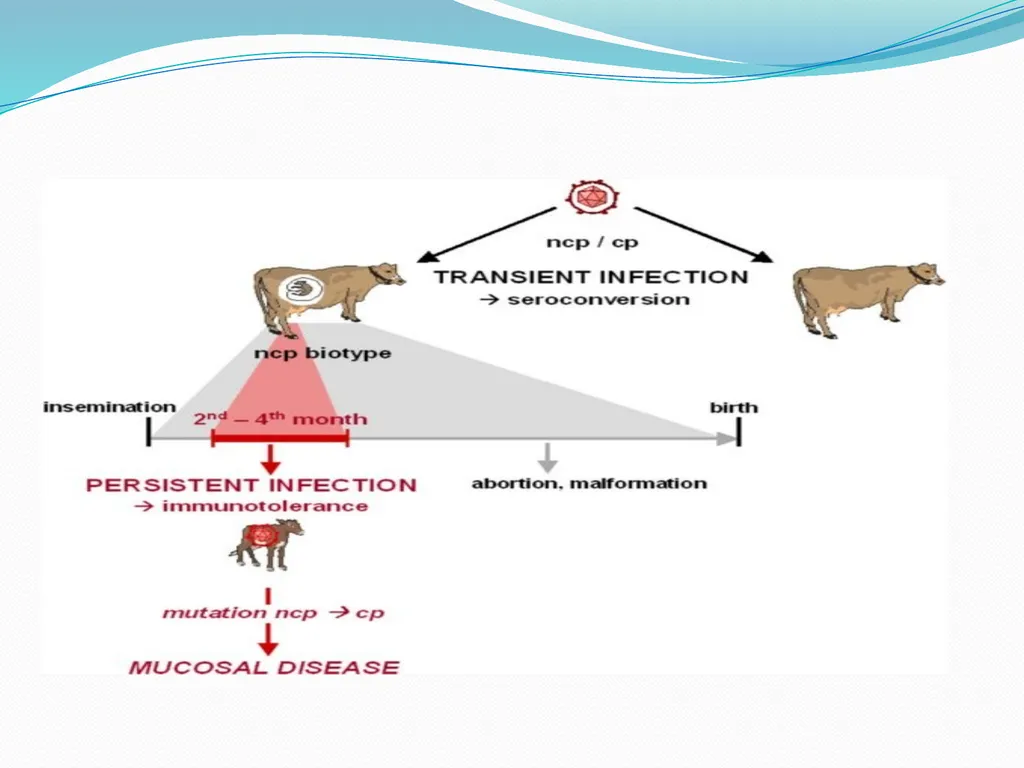
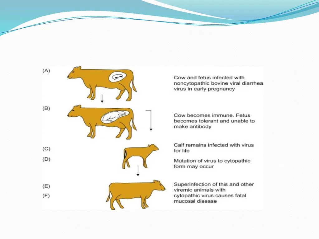
It would seem to occur in a small number of congenitally infected animals which are immunotolerant and harbor virus in all their tissues without showing any clinical symptoms.<br>
03
Etiology Cuased by Pestivirus of Flaviviridae family.
Virus is grouped into two genotypes, Type 1 and Type 2, based on genomic characteristics and the severity of disease they produce in cattle.
Each genotype, 1 and 2, are divided into two biotypes- cytopathic (cp) and non-cytopathic (ncp) based on how they replicate in cell culture.
Cytopathogenicity does not correlate with the severity of disease in vivo (high and low virulence strains) Some cp strains are recovered from animals with mucosal disease (MD), but most of the time ncp isolates are recovered from infected animals.<br>
04
The classic virus is ncp; cp isolates are generated by mutations or genome rearrangements in the original/parental ncp strain.
Most (>95%) of the field isolates are ncp.
Young calf persistently infected with BVD (right) compared to similarly- aged normal herd mate.<br>
05
Epidemiology It is normally an infection of cattle, but it has the ability to cause infections in pigs, sheep, goats,, deer, reindeer, bison and other wild ruminants.
The virus may be present in various secretions and semen.
Spread is by direct and indirect contact.
The mode of infection is by ingestion and inhalation.
Transplacental infections are frequent and result in serious consequences for the embryo/fetus.
Bulls may be persistently infected and the virus in semen is spread by coitus an artificial insemination.
BVD seen predominantly in 6-18 month old cattle as a primary infection.<br>
06
Pathogenesis Infection is via the oropharyngeal route with primary replication in the epithelium of the oropharynx
Rapid uptake into the drainage lymph nodes.
Viremia involves infection of lymphocytes giving rise to a leucopenia,
Spreads to other lymphoid tissues, particularly Peyer's patches.
Concurrent replication in the epithelium of the alimentary tract results in discrete erosions.
which in the oral cavity cause excessive salivation, and in the small and large intestine induce diarrhea.
Lesions in the nasal cavity and conjunctiva may also occur and be associated with discharges.<br>
07
Clinical signs BVDV infection can result in a wide spectrum of clinical disease varying from sub-clinical infection to fatal disease.
Different clinical signs can be seen simultaneously in a herd.
The most common clinical signs are: Infertility or abortion – foetal abnormalities can occur with infections later in pregnancy resulting in brain abnormalities.
Calf with cerebellar hypoplasia unable to stand and “stargazing”
Diarrhoea<br>
08
Mucosal disease – a condition that can vary in severity from mild to severe.
Signs include ill-thrift, diarrhoea, ulceration in mouth and gastro-intestinal tract and lameness (from ulceration of feet).
Death can ensue after a variable period of time. • Failure to conceive • Early embryonic death / abortion / congenital deformity • Foetal loss • Persistently infected calves<br>
09
Diagnosis Diagnosed in laboratories via serology, antigen detection assays, virus isolation, and by viral RNA amplification such as polymerase chain reaction (PCR).
Sample from Buffy coat or nasal swab immunohistochemistry, and antigen-capture ELISA assays.
Serological evaluation of acute BVDV infection is most commonly performed via serum neutralization in paired sera.<br>
10
Treatment Currently, there is no treatment available to cure BVD Virus.
Antibiotics for Secondary Infections Pneumonia etc.
If the animal develops Mucosal Disease, there are treatments to alleviate the symptoms, but the animal will eventually die.
IV fluids and electrolyte for diarrhea and water loss.
PI’s should be euthanized to prevent further contamination and potential infection to the rest of the herd.<br>
11
Prevention and control Vaccination does not provide complete protection against BVDV infection.
It can help to reduce the number of infections
Vaccination is not long lasting.
Regular booster shots should be kept up to date according to label
Vaccinated cattle can thus become infected and show some symptoms but death will not occur.
Pregnant cattle that are vaccinated have less chance if infection and transmission of the virus to the fetus.
There is still a chance of abortions in those cattle that are vaccinated.<br>