04
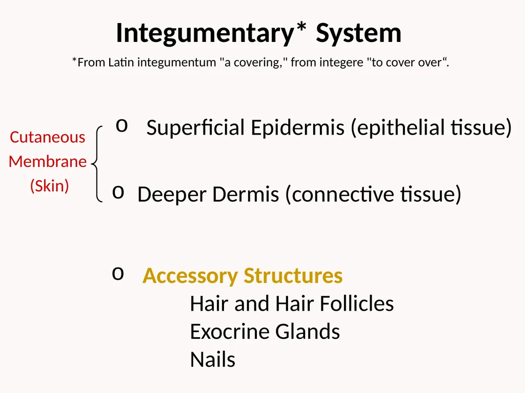
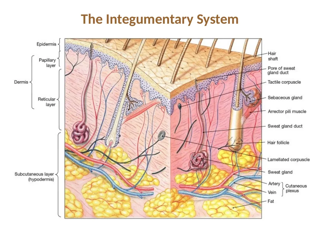
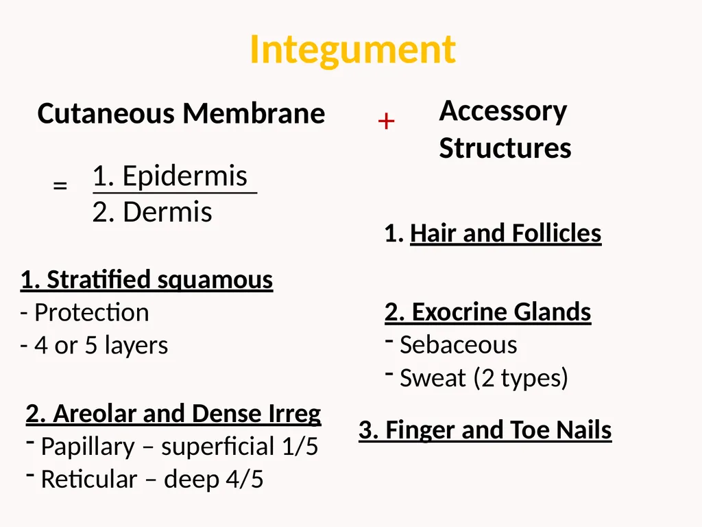
Integument 1. Epidermis Cutaneous Membrane 2. Dermis = 1. Stratified squamous
- Protection
- 4 or 5 layers 2. Areolar and Dense Irreg
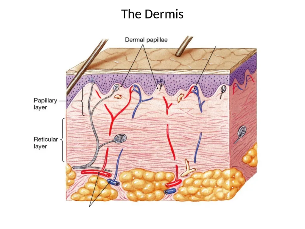
Papillary – superficial 1/5
Reticular – deep 4/5 Accessory
Structures Hair and Follicles 2. Exocrine Glands
Sebaceous
Sweat (2 types) 3. Finger and Toe Nails +<br>
05
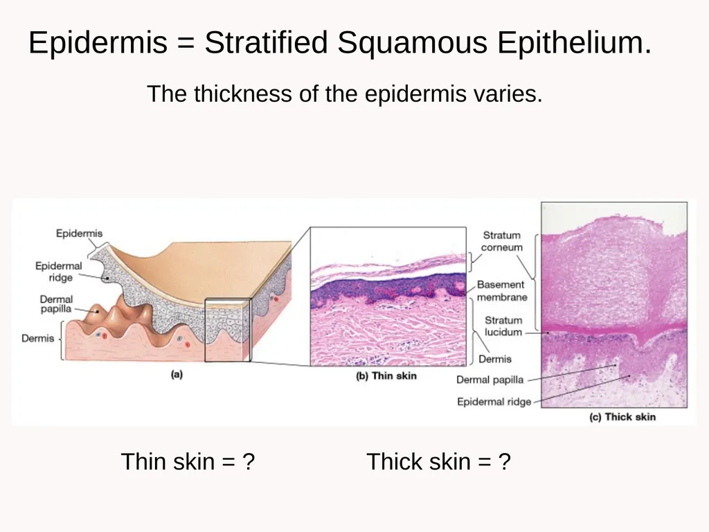
Epidermis = Stratified Squamous Epithelium. The thickness of the epidermis varies. Thin skin = ? Thick skin = ?<br>
06
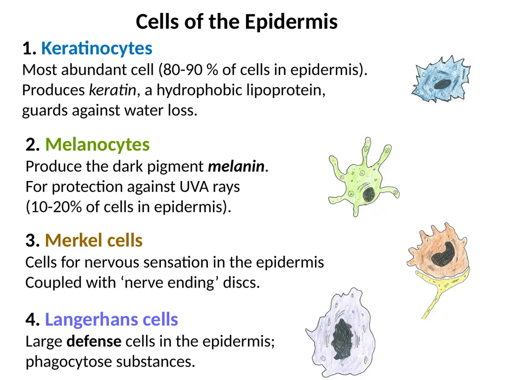
1. Keratinocytes
Most abundant cell (80-90 % of cells in epidermis).
Produces keratin, a hydrophobic lipoprotein,
guards against water loss. 2. Melanocytes
Produce the dark pigment melanin.
For protection against UVA rays
(10-20% of cells in epidermis). 3. Merkel cells
Cells for nervous sensation in the epidermis
Coupled with ‘nerve ending’ discs. 4. Langerhans cells
Large defense cells in the epidermis;
phagocytose substances. Cells of the Epidermis<br>
07
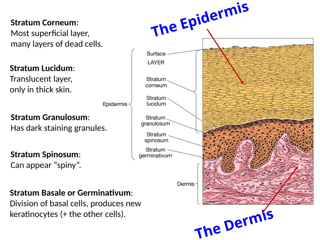
Stratum Corneum:
Most superficial layer,
many layers of dead cells. Stratum Lucidum:
Translucent layer,
only in thick skin. Stratum Granulosum:
Has dark staining granules. Stratum Spinosum:
Can appear “spiny”. Stratum Basale or Germinativum:
Division of basal cells, produces new keratinocytes (+ the other cells). The Epidermis The Dermis<br>
08
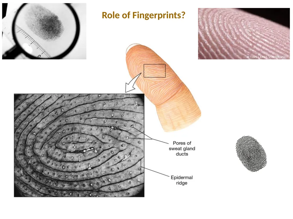
Role of Fingerprints?<br>
09
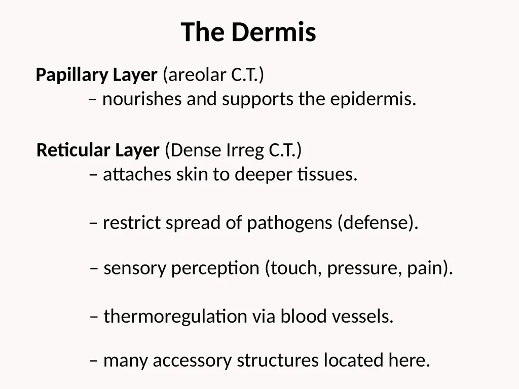
Reticular Layer (Dense Irreg C.T.)
– attaches skin to deeper tissues.
– restrict spread of pathogens (defense). Papillary Layer (areolar C.T.)
– nourishes and supports the epidermis. – sensory perception (touch, pressure, pain).
– thermoregulation via blood vessels. The Dermis – many accessory structures located here.<br>
11
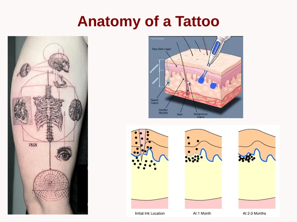
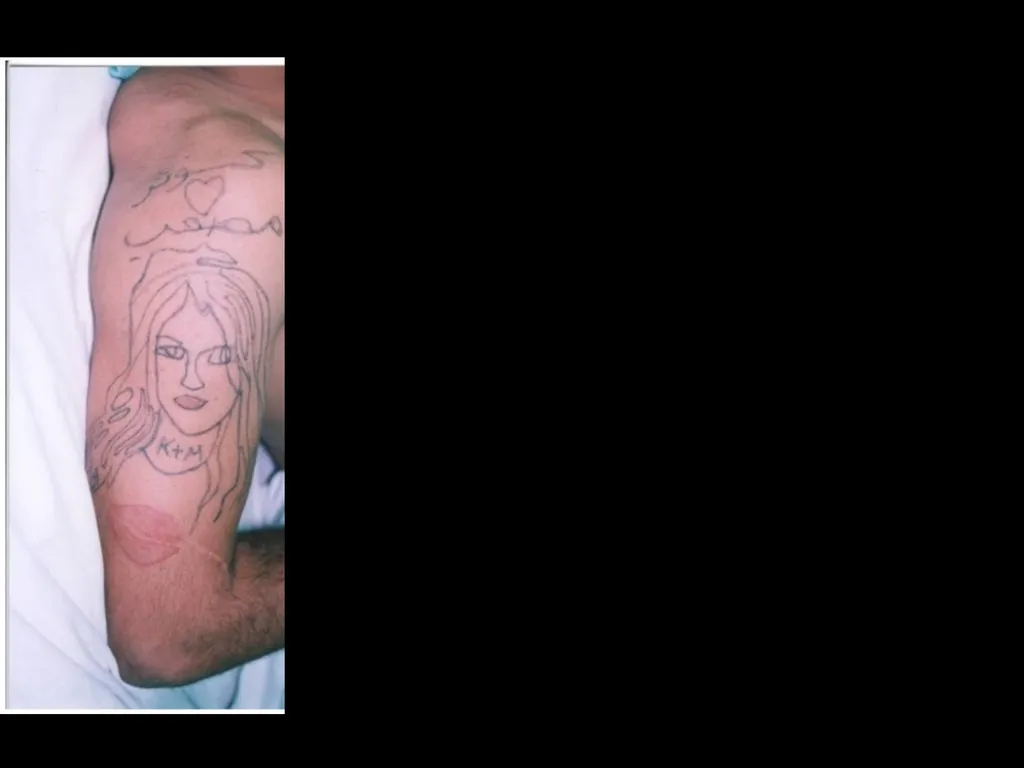
Anatomy of a Tattoo<br>
12
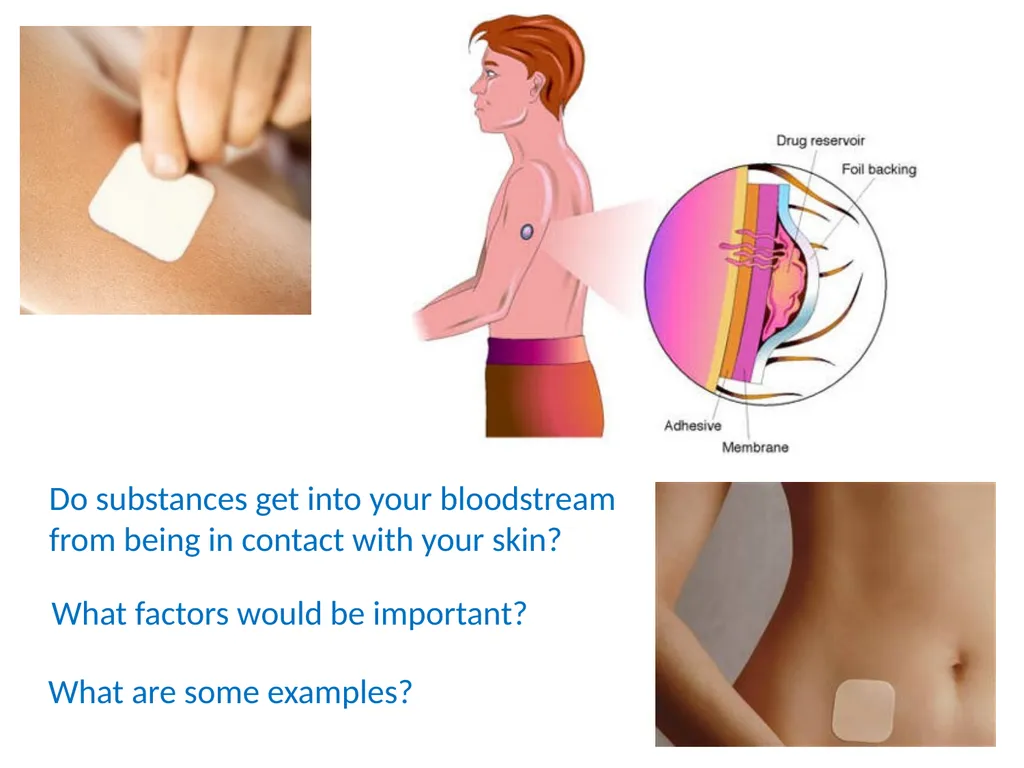
Do substances get into your bloodstream
from being in contact with your skin? What factors would be important? What are some examples?<br>
13
Roles
Stabilizes skin’s position. The Subcutaneous Layer Hypodermis Superficial Fascia - Permits limited independent movement. Provides insulation and protection.<br>
14
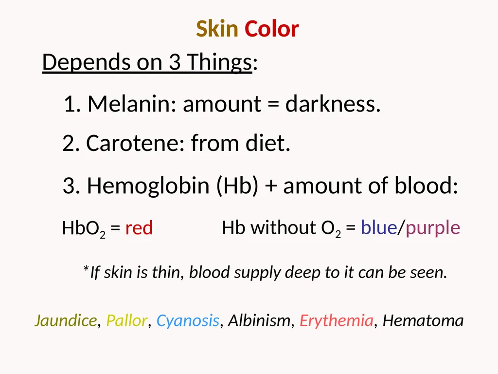
Skin Color 1. Melanin: amount = darkness. 2. Carotene: from diet. 3. Hemoglobin (Hb) + amount of blood: HbO2 = red *If skin is thin, blood supply deep to it can be seen. Depends on 3 Things: Hb without O2 = blue/purple Jaundice, Pallor, Cyanosis, Albinism, Erythemia, Hematoma<br>
15
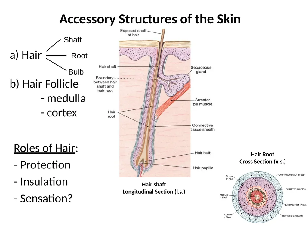
Accessory Structures of the Skin Roles of Hair:
- Protection
- Insulation
- Sensation? a) Hair
b) Hair Follicle
- medulla
- cortex Shaft Root Bulb Hair Root
Cross Section (x.s.) Hair shaft
Longitudinal Section (l.s.)<br>
16
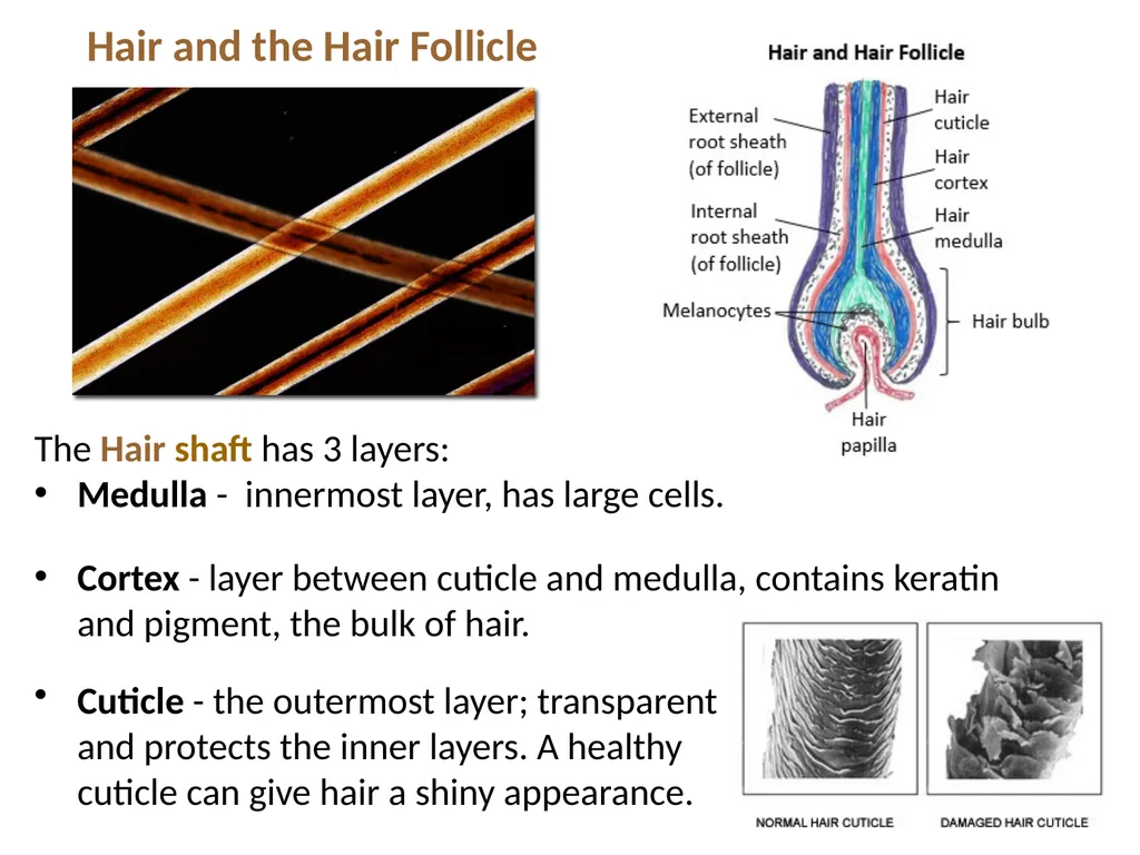
The Hair shaft has 3 layers:
Medulla - innermost layer, has large cells.
Cortex - layer between cuticle and medulla, contains keratin and pigment, the bulk of hair. Hair and the Hair Follicle Cuticle - the outermost layer; transparent and protects the inner layers. A healthy cuticle can give hair a shiny appearance.<br>
17
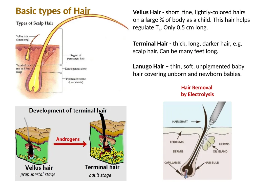
Vellus Hair - short, fine, lightly-colored hairs on a large % of body as a child. This hair helps regulate Tb. Only 0.5 cm long.
Terminal Hair - thick, long, darker hair, e.g. scalp hair. Can be many feet long.
Lanugo Hair – thin, soft, unpigmented baby hair covering unborn and newborn babies. Hair Removal
by Electrolysis Basic types of Hair<br>
18
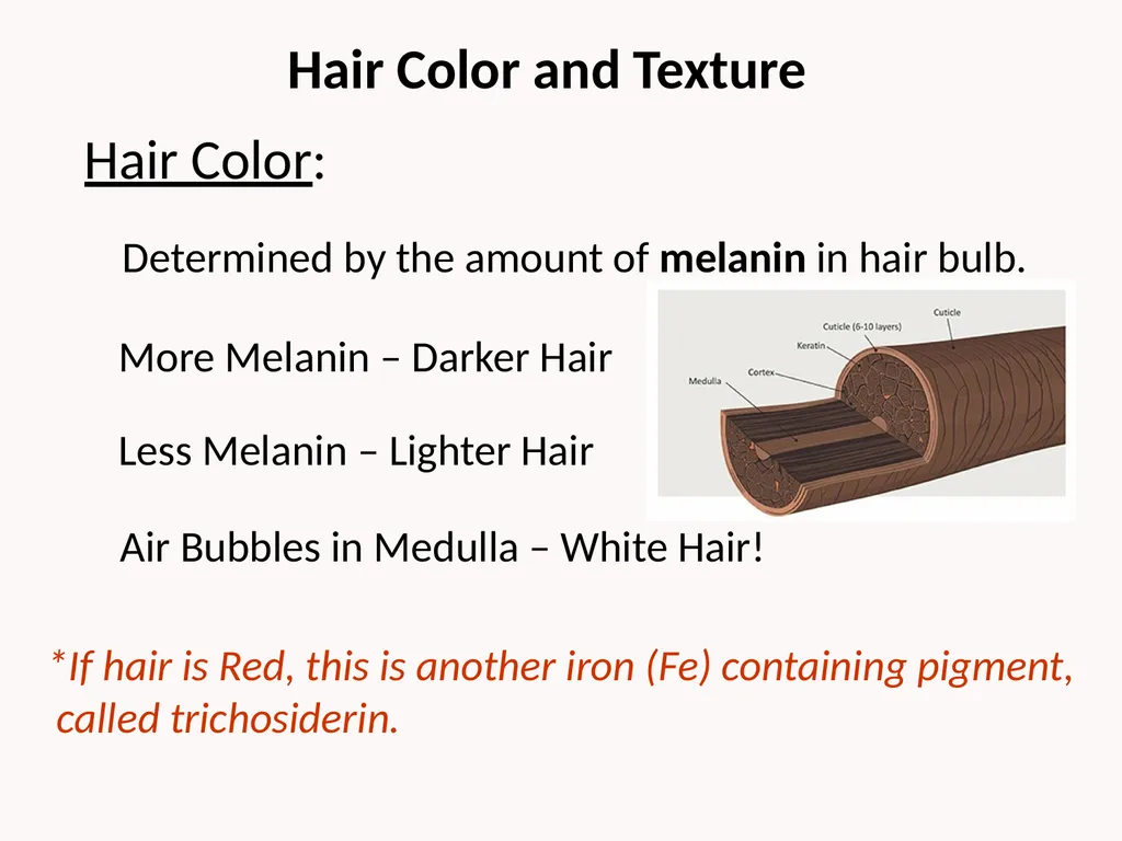
Hair Color and Texture Determined by the amount of melanin in hair bulb. More Melanin – Darker Hair Less Melanin – Lighter Hair Air Bubbles in Medulla – White Hair! *If hair is Red, this is another iron (Fe) containing pigment,
called trichosiderin. Hair Color:<br>
19
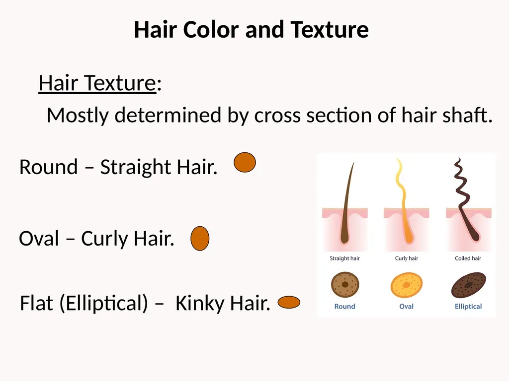
Hair Color and Texture Mostly determined by cross section of hair shaft. Round – Straight Hair. Oval – Curly Hair. Flat (Elliptical) – Kinky Hair. Hair Texture:<br>
20
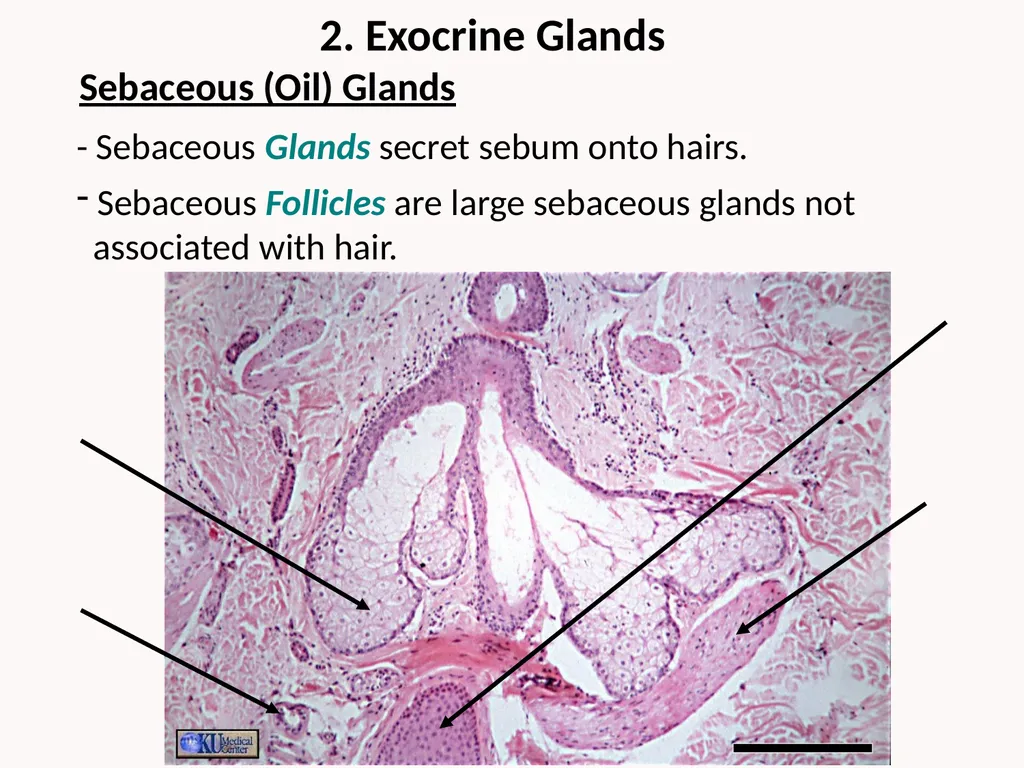
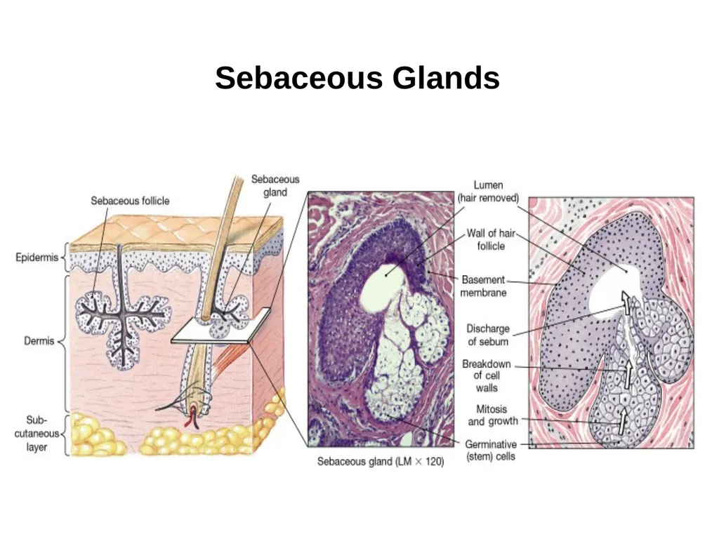
2. Exocrine Glands - Sebaceous Glands secret sebum onto hairs. Sebaceous Follicles are large sebaceous glands not
associated with hair. Sebaceous (Oil) Glands<br>
22
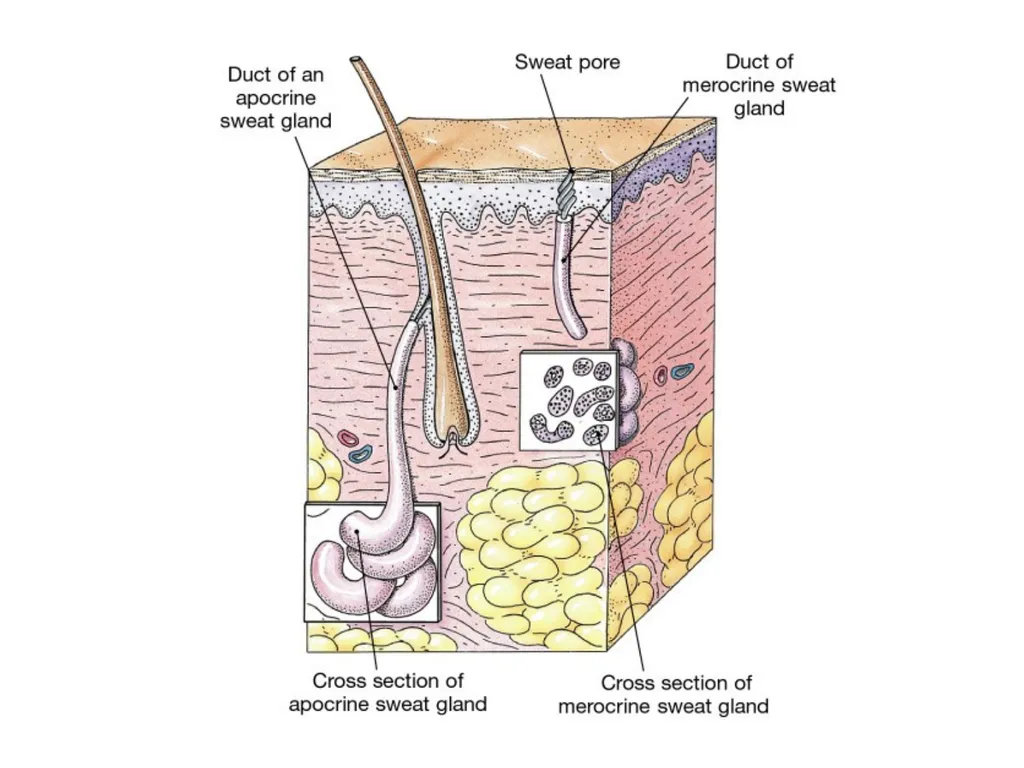
2) Apocrine Sweat Glands
Thicker, lipid rich secretion, only in specific regions of the body, e.g., axillary and inguinal regions. 1) Merocrine Sweat Glands
Thin, watery, sensible perspiration, more numerous than apocrine, highest density on palms and soles. Sudoriferous (Sweat) Glands Mammary Glands
Large, complex apocrine sweat glands, produce milk as nourishment for babies. Ceruminous Glands
Produces waxy cerumen (ear wax), found in ear canal to keep eardrum protection and pliable. Modified Apocrine Sweat Glands<br>
23
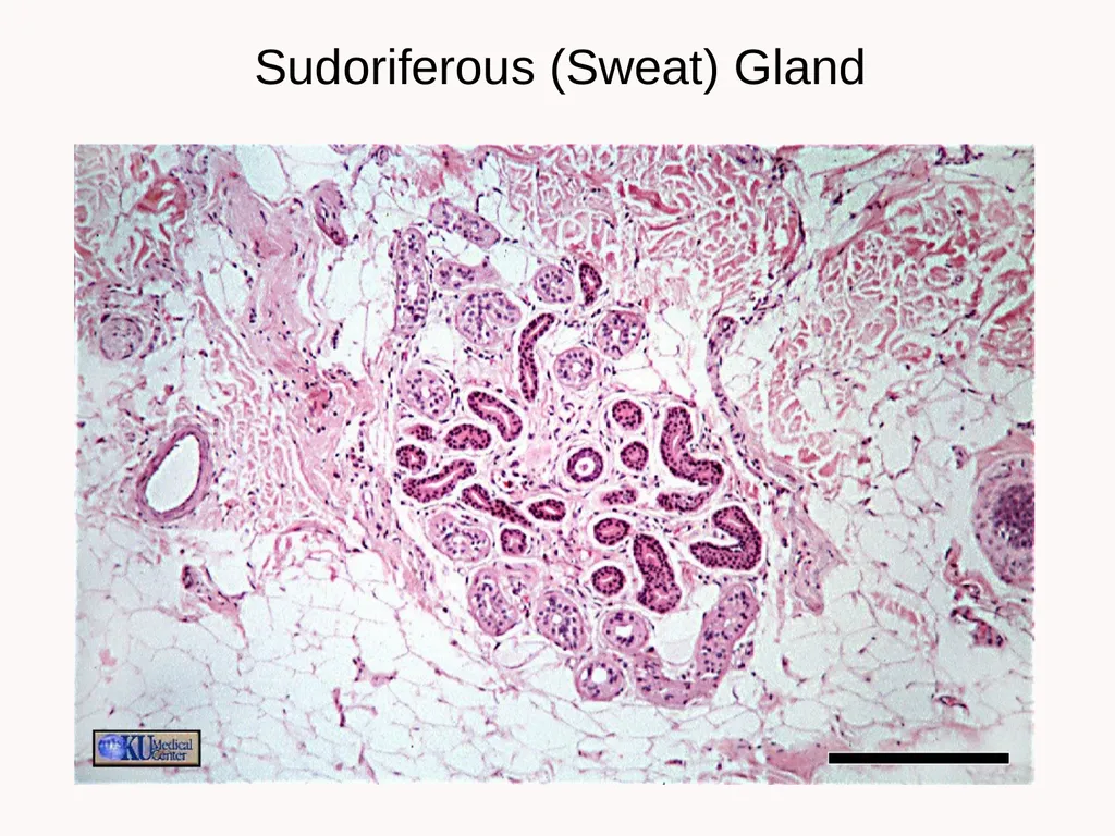
Sudoriferous (Sweat) Gland<br>
24
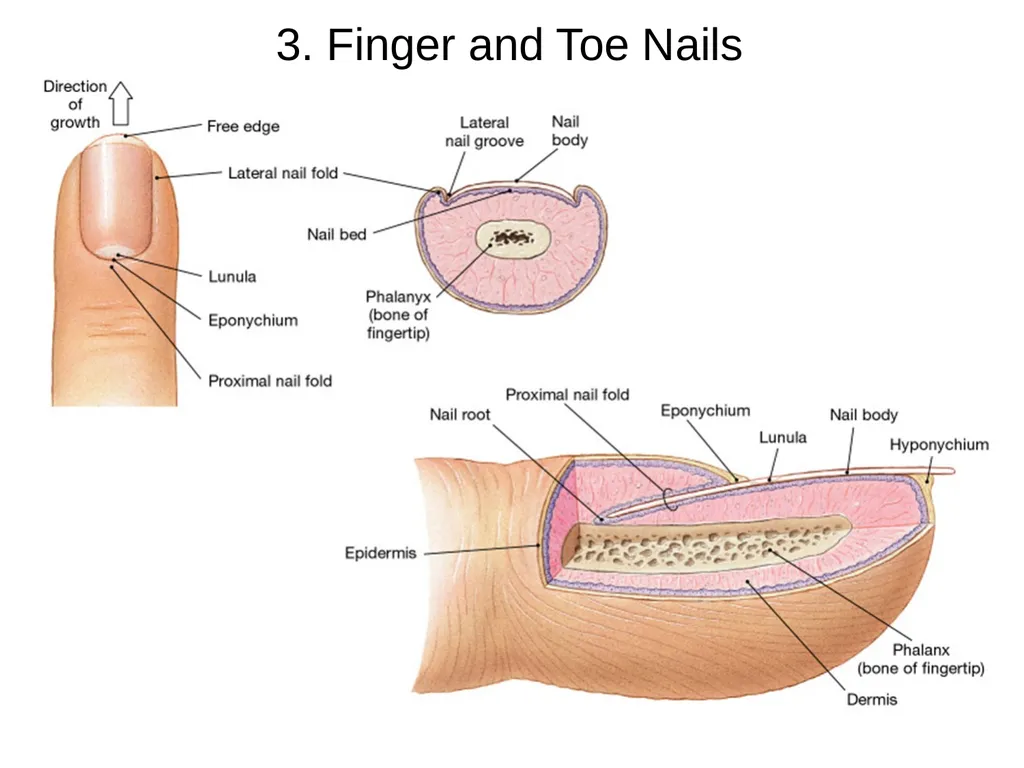
3. Finger and Toe Nails<br>
25
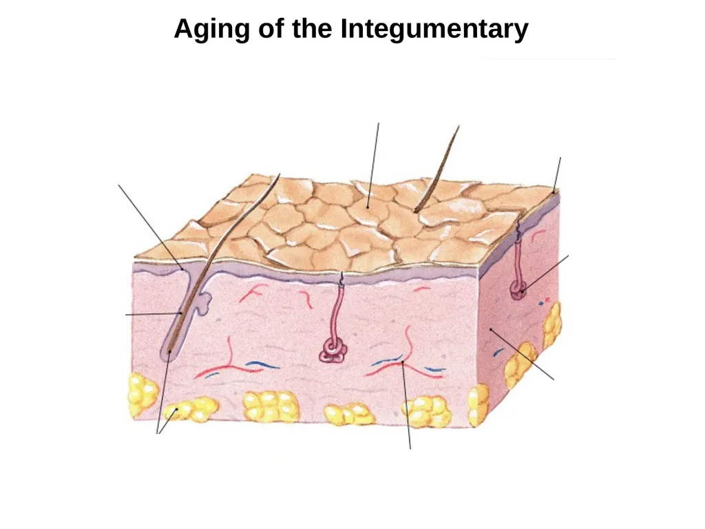
Aging of the Integumentary<br>
26
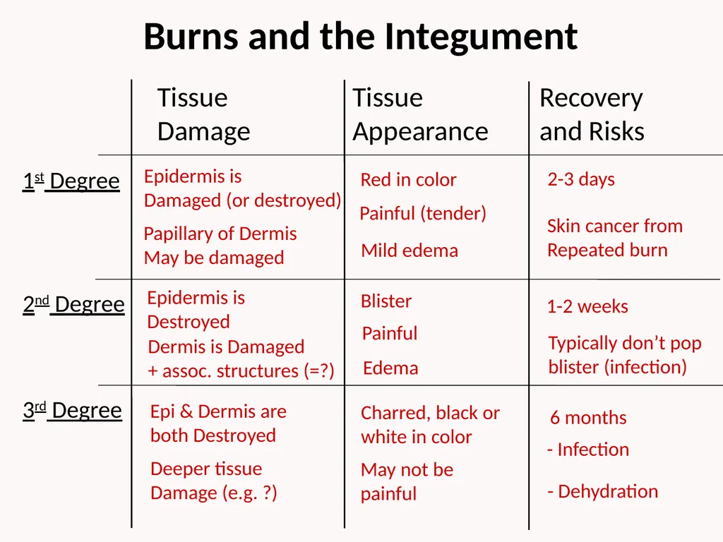
Burns and the Integument Tissue
Damage 1st Degree 2nd Degree 3rd Degree Tissue
Appearance Recovery
and Risks Epidermis is
Damaged (or destroyed) Epidermis is
Destroyed Epi & Dermis are
both Destroyed Papillary of Dermis
May be damaged Dermis is Damaged
+ assoc. structures (=?) Red in color Blister Charred, black or
white in color Painful (tender) Painful 2-3 days 1-2 weeks 6 months Skin cancer from
Repeated burn Typically don’t pop
blister (infection) Deeper tissue
Damage (e.g. ?) May not be
painful - Infection - Dehydration Mild edema Edema<br>
27
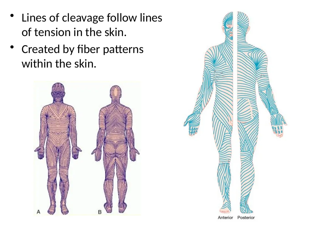
Lines of cleavage follow lines of tension in the skin.
Created by fiber patterns within the skin.<br>
28
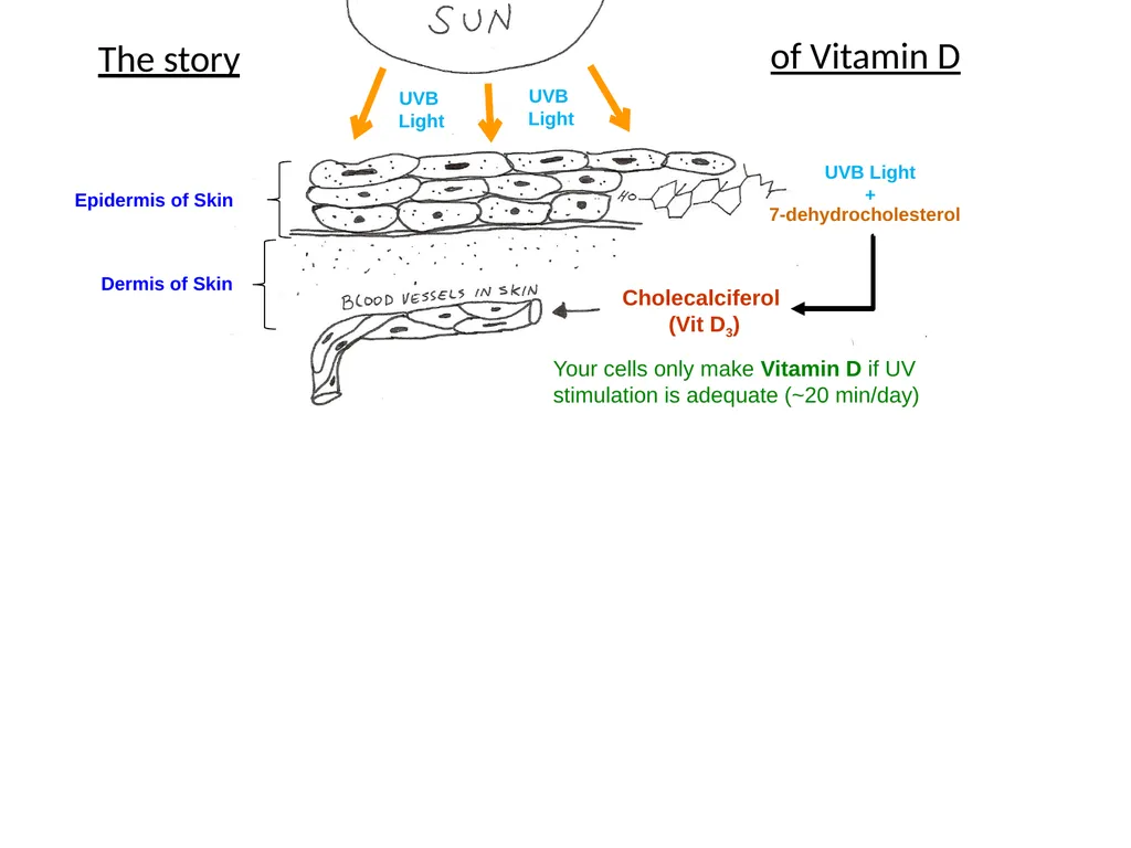
Epidermis of Skin Dermis of Skin 7-dehydrocholesterol UVB Light
+ Cholecalciferol
(Vit D3) Your cells only make Vitamin D if UV stimulation is adequate (~20 min/day) “Calcidiol”
25-Hydroxycholecalciferol
(25-Hydroxy Vit D3) Allow your healthy Liver to process Vit D3 into Calcidiol. “Calcitriol”
1,25-Hydroxycholecalciferol
(1,25-Hydroxy Vit D3) ACTIVE FORM Allow your healthy Kidneys to process Calcidiol into the
most active form, Calcitriol. UVB
Light UVB
Light The story of Vitamin D<br>