
DNA sequencing is the process of determining the


Description: DNA sequencing is the process of determining the nucleic acid sequence the order of nucleotides in DNA. It includes any method or technology that is used to determine the order of the four bases: adenine, guanine, cytosine, and thymine.
Related Topics
Download Presentation
"DNA sequencing is the process of determining the" is the property of its rightful owner. Permission is granted to download and print the materials on this website for personal, non-commercial use only, and to display it on your personal computer provided you do not modify the materials and that you retain all copyright notices contained in the materials. By downloading content from our website, you accept the terms of this agreement.
Presentation Transcript
slide1. DNA sequencing is the process of determining the nucleic acid sequence – the order of nucleotides in DNA. It includes any method or technology that is used to determine the order of the four bases: adenine, guanine, cytosine, and thymine. DNA sequencing The rapid speed of sequencing attained with modern DNA sequencing technology has been instrumental in the sequencing of complete DNA sequences, or genomes, of numerous types and species of life, including the human genome and other complete DNA sequences of many animal, plant, and microbial species.<br>
slide2. Frederick Sanger, a pioneer of sequencing. Sanger is one of the few scientists who was awarded two Nobel prizes, one for the sequencing of proteins, and the other for the sequencing of DNA.<br>
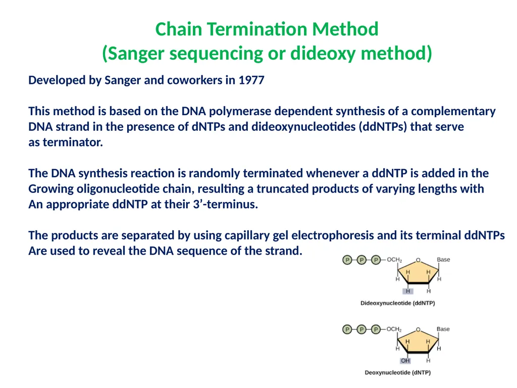
slide3. Chain Termination Method
(Sanger sequencing or dideoxy method) Developed by Sanger and coworkers in 1977
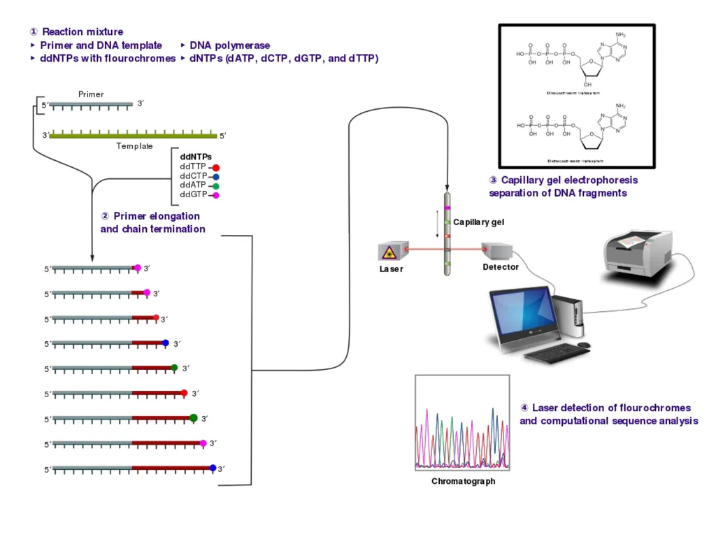
This method is based on the DNA polymerase dependent synthesis of a complementary
DNA strand in the presence of dNTPs and dideoxynucleotides (ddNTPs) that serve
as terminator.
The DNA synthesis reaction is randomly terminated whenever a ddNTP is added in the
Growing oligonucleotide chain, resulting a truncated products of varying lengths with
An appropriate ddNTP at their 3’-terminus.
The products are separated by using capillary gel electrophoresis and its terminal ddNTPs
Are used to reveal the DNA sequence of the strand.<br>
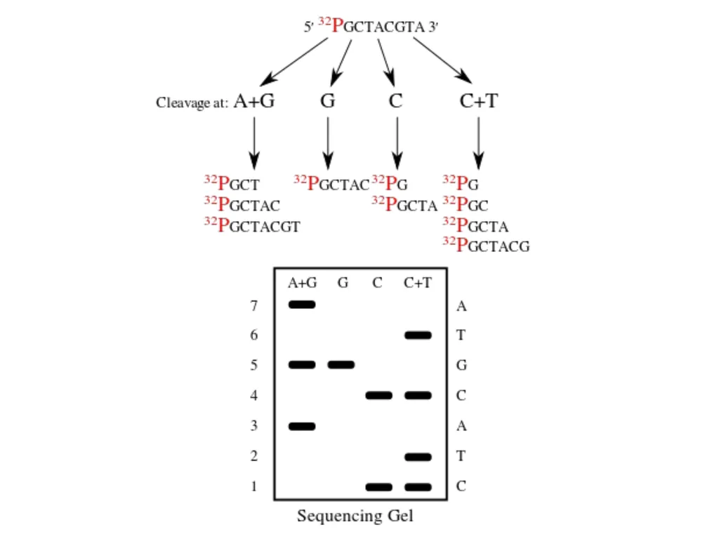
slide5. Maxam–Gilbert sequencing Maxam–Gilbert sequencing is a method of DNA sequencing developed by Allan Maxam and Walter Gilbert in 1976–1977. This method is based on nucleobase-specific partial chemical modification of DNA and subsequent cleavage of the DNA backbone at sites adjacent to the modified nucleotides. Maxam-Gilbert sequencing requires radioactive labeling at one 5' end of the DNA and purification of the DNA fragment to be sequenced. Chemical treatment then generates breaks at a small proportion of one or two of the four nucleotide bases in each of four reactions (G, A+G, C, C+T). The concentration of the modifying chemicals is controlled to introduce on average one modification per DNA molecule. Thus a series of labeled fragments is generated, from the radiolabeled end to the first "cut" site in each molecule. The fragments in the four reactions are electrophoresed side by side in denaturing acrylamide gels for size separation. To visualize the fragments, the gel is exposed to X-ray film for autoradiography, yielding a series of dark bands each corresponding to a radiolabeled DNA fragment, from which the sequence may be inferred.<br>
slide7. The first full DNA genome to be sequenced was that of bacteriophage φX174 in 1977. In 1995, Venter, Hamilton Smith, and colleagues at The Institute for Genomic Research (TIGR) published the first complete genome of a free-living organism, the bacterium Haemophilus influenzae.<br>
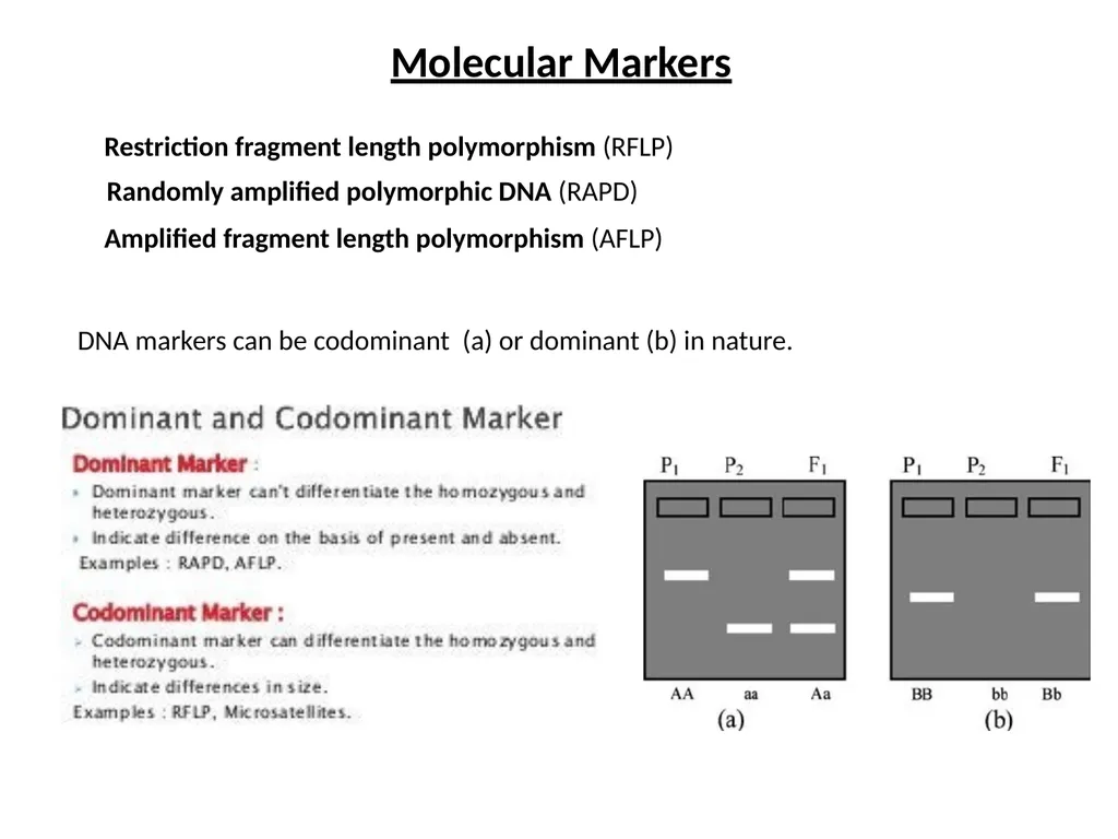
slide8. Molecular Markers Restriction fragment length polymorphism (RFLP) Randomly amplified polymorphic DNA (RAPD) Amplified fragment length polymorphism (AFLP) DNA markers can be codominant (a) or dominant (b) in nature.<br>
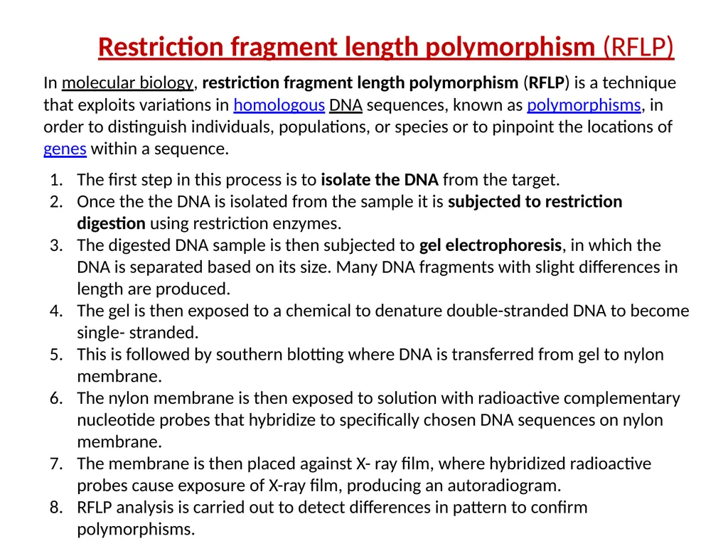
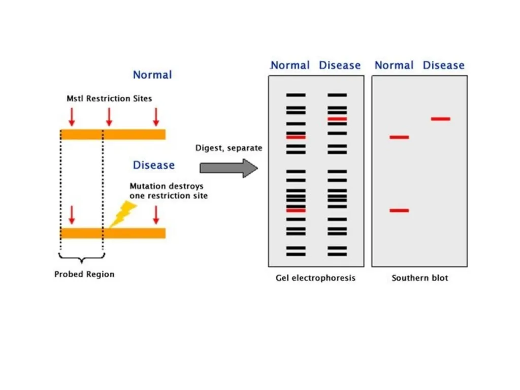
slide9. Restriction fragment length polymorphism (RFLP) The first step in this process is to isolate the DNA from the target.
Once the the DNA is isolated from the sample it is subjected to restriction digestion using restriction enzymes.
The digested DNA sample is then subjected to gel electrophoresis, in which the DNA is separated based on its size. Many DNA fragments with slight differences in length are produced.
The gel is then exposed to a chemical to denature double-stranded DNA to become single- stranded.
This is followed by southern blotting where DNA is transferred from gel to nylon membrane.
The nylon membrane is then exposed to solution with radioactive complementary nucleotide probes that hybridize to specifically chosen DNA sequences on nylon membrane.
The membrane is then placed against X- ray film, where hybridized radioactive probes cause exposure of X-ray film, producing an autoradiogram.
RFLP analysis is carried out to detect differences in pattern to confirm polymorphisms. In molecular biology, restriction fragment length polymorphism (RFLP) is a technique that exploits variations in homologous DNA sequences, known as polymorphisms, in order to distinguish individuals, populations, or species or to pinpoint the locations of genes within a sequence.<br>
slide11. Applications of Restriction Fragment Length Polymorphism (RFLP)
RFLP analysis was formerly an important tool in genome mapping, localization of genes for genetic disorders, determination of risk for disease, and paternity testing.
RFLP can be used in many different settings to accomplish different objectives:
In paternity cases or criminal cases to determine the source of a DNA sample. (i.e. it has forensic applications).
Determining the disease status of an individual. (e.g. it can be used in the detection of mutations)
To measure recombination rates which can lead to a genetic map with the distance between RFLP loci.
In the characterization of genetic diversity or breeding patterns in animal populations.
RFLP has been developed for chromosomes mapping of humans, mice, maize, tomato, rice, etc.<br>
slide12. 2. Randomly amplified polymorphic DNA (RAPD)
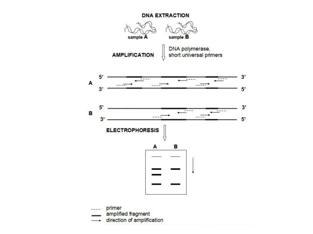
Randomly amplified polymorphic DNA (RAPD) is a PCR-based technique which uses arbitrary primers which bind to the nonspecific sites on the DNA and amplify the DNA. These amplified fragments are then migrated on agarose gel and difference in the band pattern is observed.
DOMINANT Marker<br>
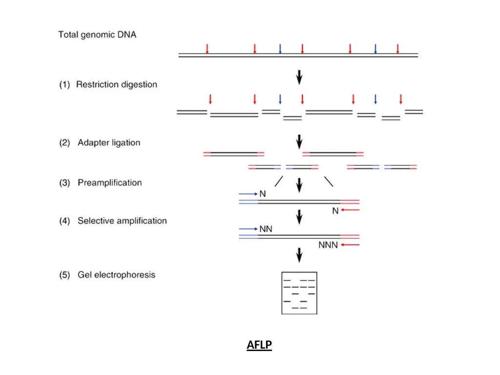
slide14. 3. Amplified fragment length polymorphism (AFLP) Amplified fragment length polymorphism (AFLP) is a PCR-based technique that uses selective amplification of a subset of digested DNA fragments to generate and compare unique fingerprints for genomes of interest.
Dominant Marker<br>
slide15. AFLP<br>
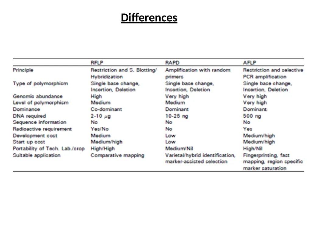
slide16. Differences<br>
slide2. Frederick Sanger, a pioneer of sequencing. Sanger is one of the few scientists who was awarded two Nobel prizes, one for the sequencing of proteins, and the other for the sequencing of DNA.<br>
slide3. Chain Termination Method
(Sanger sequencing or dideoxy method) Developed by Sanger and coworkers in 1977
This method is based on the DNA polymerase dependent synthesis of a complementary
DNA strand in the presence of dNTPs and dideoxynucleotides (ddNTPs) that serve
as terminator.
The DNA synthesis reaction is randomly terminated whenever a ddNTP is added in the
Growing oligonucleotide chain, resulting a truncated products of varying lengths with
An appropriate ddNTP at their 3’-terminus.
The products are separated by using capillary gel electrophoresis and its terminal ddNTPs
Are used to reveal the DNA sequence of the strand.<br>
slide5. Maxam–Gilbert sequencing Maxam–Gilbert sequencing is a method of DNA sequencing developed by Allan Maxam and Walter Gilbert in 1976–1977. This method is based on nucleobase-specific partial chemical modification of DNA and subsequent cleavage of the DNA backbone at sites adjacent to the modified nucleotides. Maxam-Gilbert sequencing requires radioactive labeling at one 5' end of the DNA and purification of the DNA fragment to be sequenced. Chemical treatment then generates breaks at a small proportion of one or two of the four nucleotide bases in each of four reactions (G, A+G, C, C+T). The concentration of the modifying chemicals is controlled to introduce on average one modification per DNA molecule. Thus a series of labeled fragments is generated, from the radiolabeled end to the first "cut" site in each molecule. The fragments in the four reactions are electrophoresed side by side in denaturing acrylamide gels for size separation. To visualize the fragments, the gel is exposed to X-ray film for autoradiography, yielding a series of dark bands each corresponding to a radiolabeled DNA fragment, from which the sequence may be inferred.<br>
slide7. The first full DNA genome to be sequenced was that of bacteriophage φX174 in 1977. In 1995, Venter, Hamilton Smith, and colleagues at The Institute for Genomic Research (TIGR) published the first complete genome of a free-living organism, the bacterium Haemophilus influenzae.<br>
slide8. Molecular Markers Restriction fragment length polymorphism (RFLP) Randomly amplified polymorphic DNA (RAPD) Amplified fragment length polymorphism (AFLP) DNA markers can be codominant (a) or dominant (b) in nature.<br>
slide9. Restriction fragment length polymorphism (RFLP) The first step in this process is to isolate the DNA from the target.
Once the the DNA is isolated from the sample it is subjected to restriction digestion using restriction enzymes.
The digested DNA sample is then subjected to gel electrophoresis, in which the DNA is separated based on its size. Many DNA fragments with slight differences in length are produced.
The gel is then exposed to a chemical to denature double-stranded DNA to become single- stranded.
This is followed by southern blotting where DNA is transferred from gel to nylon membrane.
The nylon membrane is then exposed to solution with radioactive complementary nucleotide probes that hybridize to specifically chosen DNA sequences on nylon membrane.
The membrane is then placed against X- ray film, where hybridized radioactive probes cause exposure of X-ray film, producing an autoradiogram.
RFLP analysis is carried out to detect differences in pattern to confirm polymorphisms. In molecular biology, restriction fragment length polymorphism (RFLP) is a technique that exploits variations in homologous DNA sequences, known as polymorphisms, in order to distinguish individuals, populations, or species or to pinpoint the locations of genes within a sequence.<br>
slide11. Applications of Restriction Fragment Length Polymorphism (RFLP)
RFLP analysis was formerly an important tool in genome mapping, localization of genes for genetic disorders, determination of risk for disease, and paternity testing.
RFLP can be used in many different settings to accomplish different objectives:
In paternity cases or criminal cases to determine the source of a DNA sample. (i.e. it has forensic applications).
Determining the disease status of an individual. (e.g. it can be used in the detection of mutations)
To measure recombination rates which can lead to a genetic map with the distance between RFLP loci.
In the characterization of genetic diversity or breeding patterns in animal populations.
RFLP has been developed for chromosomes mapping of humans, mice, maize, tomato, rice, etc.<br>
slide12. 2. Randomly amplified polymorphic DNA (RAPD)
Randomly amplified polymorphic DNA (RAPD) is a PCR-based technique which uses arbitrary primers which bind to the nonspecific sites on the DNA and amplify the DNA. These amplified fragments are then migrated on agarose gel and difference in the band pattern is observed.
DOMINANT Marker<br>
slide14. 3. Amplified fragment length polymorphism (AFLP) Amplified fragment length polymorphism (AFLP) is a PCR-based technique that uses selective amplification of a subset of digested DNA fragments to generate and compare unique fingerprints for genomes of interest.
Dominant Marker<br>
slide15. AFLP<br>
slide16. Differences<br>