Electron Microscopy Chelsea Aitken Peter Aspinall http:www.zyvexlabs.comEIPBNuGEIPBN20images05Ion.jpg Intro to Electron Microscopy Similar to optical microscopy except with electrons rather than photons Used to image samples with a
"Electron Microscopy Chelsea Aitken Peter Aspinall" is the property of its rightful owner. Permission is granted to
download and print the materials on this website for personal, non-commercial use only, and to display it
on your personal computer provided you do not modify the materials and that you retain all copyright
notices contained in the materials. By downloading content from our website, you accept the terms of this
agreement.
Presentation Transcript
01
Electron Microscopy Chelsea Aitken
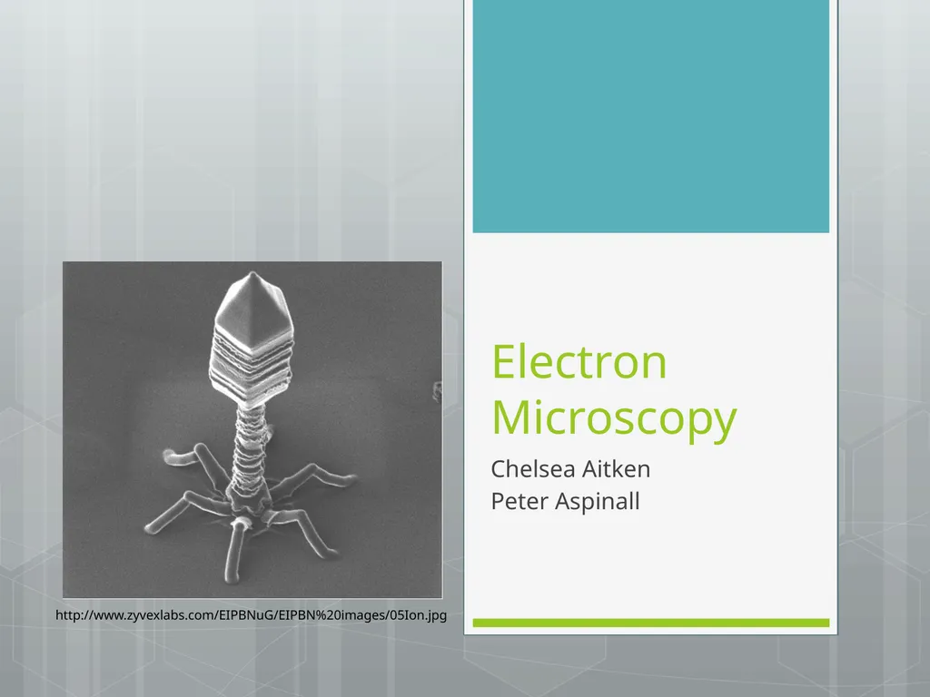
Peter Aspinall http://www.zyvexlabs.com/EIPBNuG/EIPBN%20images/05Ion.jpg<br>
02
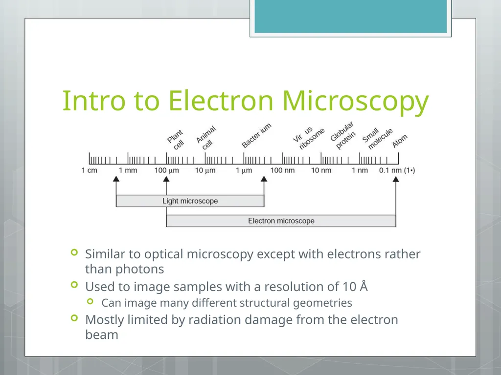
Intro to Electron Microscopy Similar to optical microscopy except with electrons rather than photons
Used to image samples with a resolution of 10 Å
Can image many different structural geometries
Mostly limited by radiation damage from the electron beam<br>
03
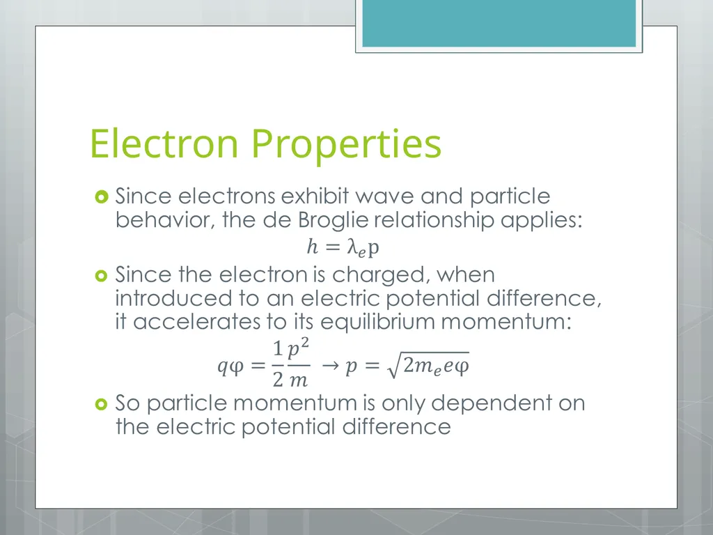
Electron Properties<br>
04
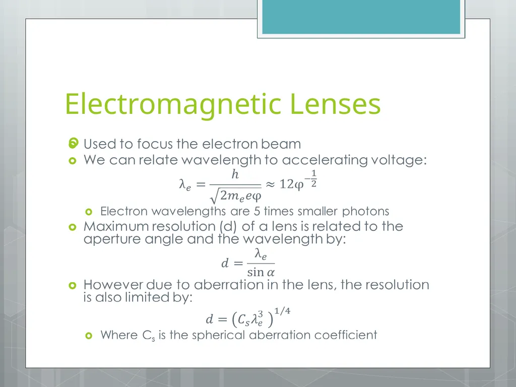
Electromagnetic Lenses<br>
05
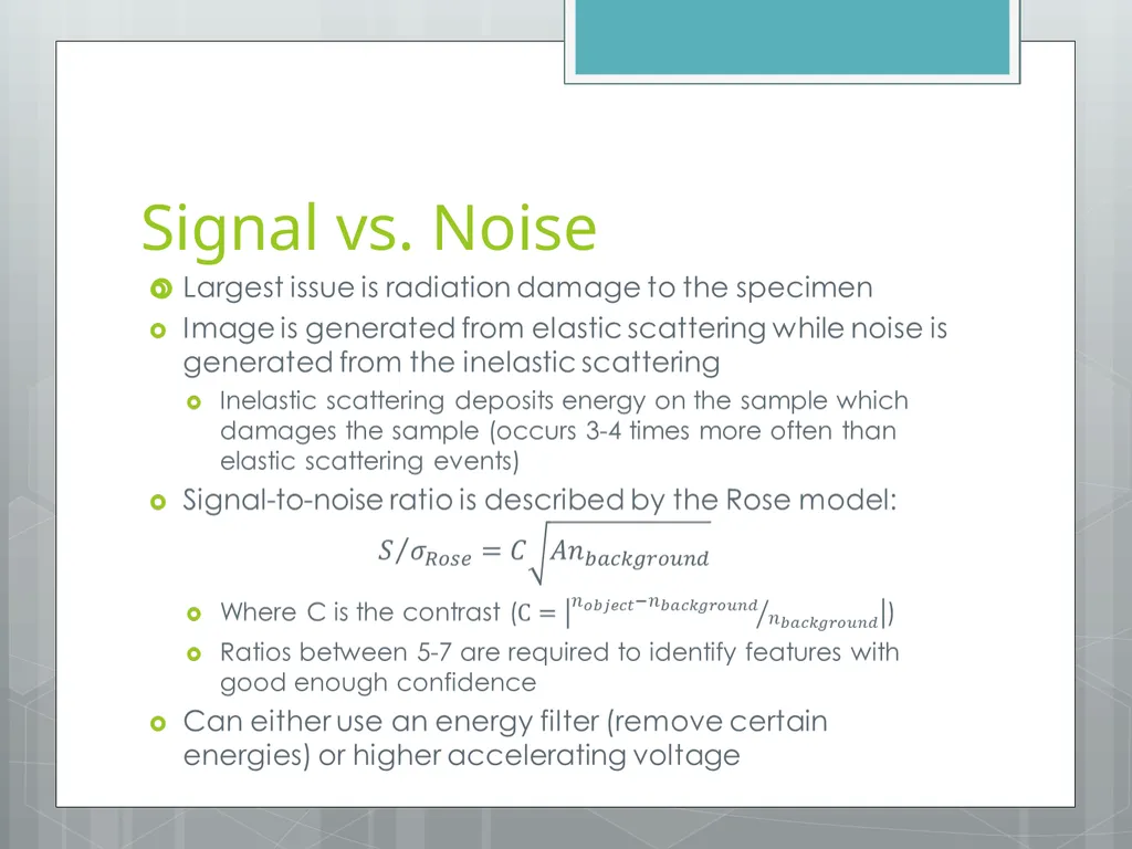
Signal vs. Noise<br>
06
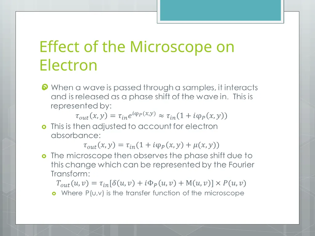
Effect of the Microscope on Electron<br>
07
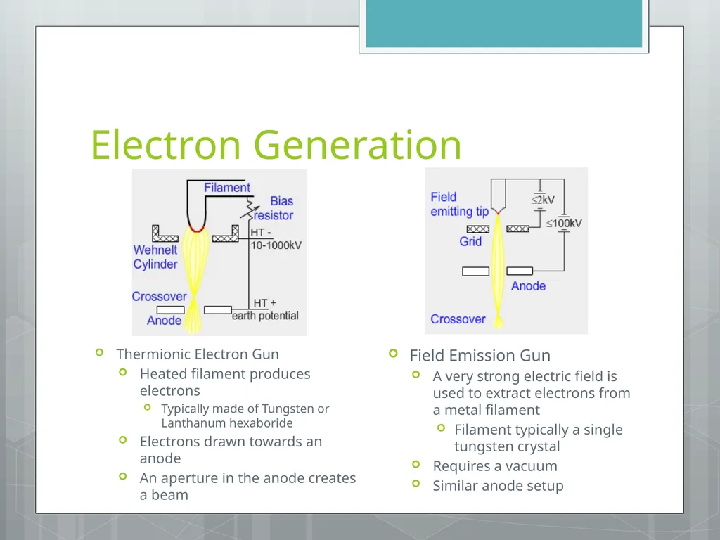
Electron Generation Thermionic Electron Gun
Heated filament produces electrons
Typically made of Tungsten or Lanthanum hexaboride
Electrons drawn towards an anode
An aperture in the anode creates a beam Field Emission Gun
A very strong electric field is used to extract electrons from a metal filament
Filament typically a single tungsten crystal
Requires a vacuum
Similar anode setup<br>
08
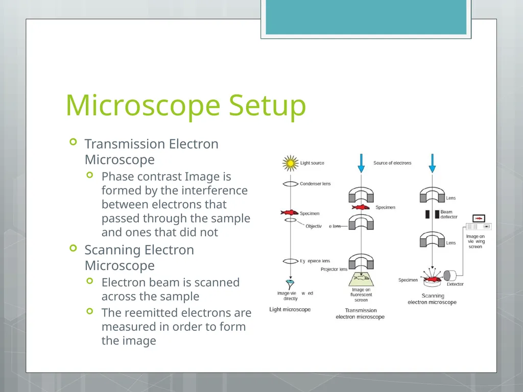
Microscope Setup Transmission Electron Microscope
Phase contrast Image is formed by the interference between electrons that passed through the sample and ones that did not
Scanning Electron Microscope
Electron beam is scanned across the sample
The reemitted electrons are measured in order to form the image<br>
09
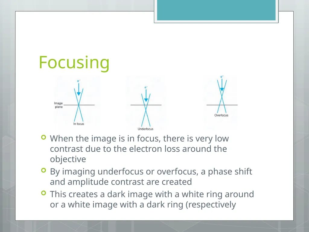
Focusing When the image is in focus, there is very low contrast due to the electron loss around the objective
By imaging underfocus or overfocus, a phase shift and amplitude contrast are created
This creates a dark image with a white ring around or a white image with a dark ring (respectively<br>
10
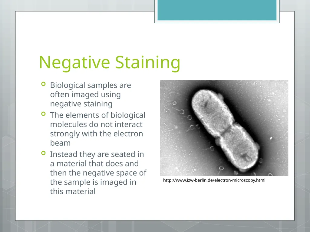
Negative Staining Biological samples are often imaged using negative staining
The elements of biological molecules do not interact strongly with the electron beam
Instead they are seated in a material that does and then the negative space of the sample is imaged in this material http://www.izw-berlin.de/electron-microscopy.html<br>
11
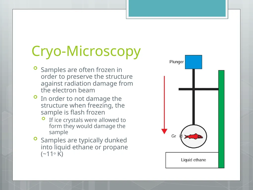
Cryo-Microscopy Samples are often frozen in order to preserve the structure against radiation damage from the electron beam
In order to not damage the structure when freezing, the sample is flash frozen
If ice crystals were allowed to form they would damage the sample
Samples are typically dunked into liquid ethane or propane (~11o K)<br>
12
Image Options EM can magnify a sample between 1000 and 200,000 times
However due to limitations, macromolecules are usually imaged between 40,000 and 60,000 for a resolution of 10 – 20 Å
Image intensity decreases as magnification goes up with 1/M2
Protein concentration is typically around 1 mg ml-1 to ensure sufficient particle density without being overcrowded
Biological samples can only be exposed to 10-15 electrons per Å2
Using a stain allows increased exposure
Lower temperatures can similarly protect the sample<br>
13
Data Collection Protocol Search/Focus/Exposure
Use low-dose/magnification to find area of interest to magnify
Specific defocus is picked and drift is checked
Sample is exposed to a high-does to image the sample<br>
14
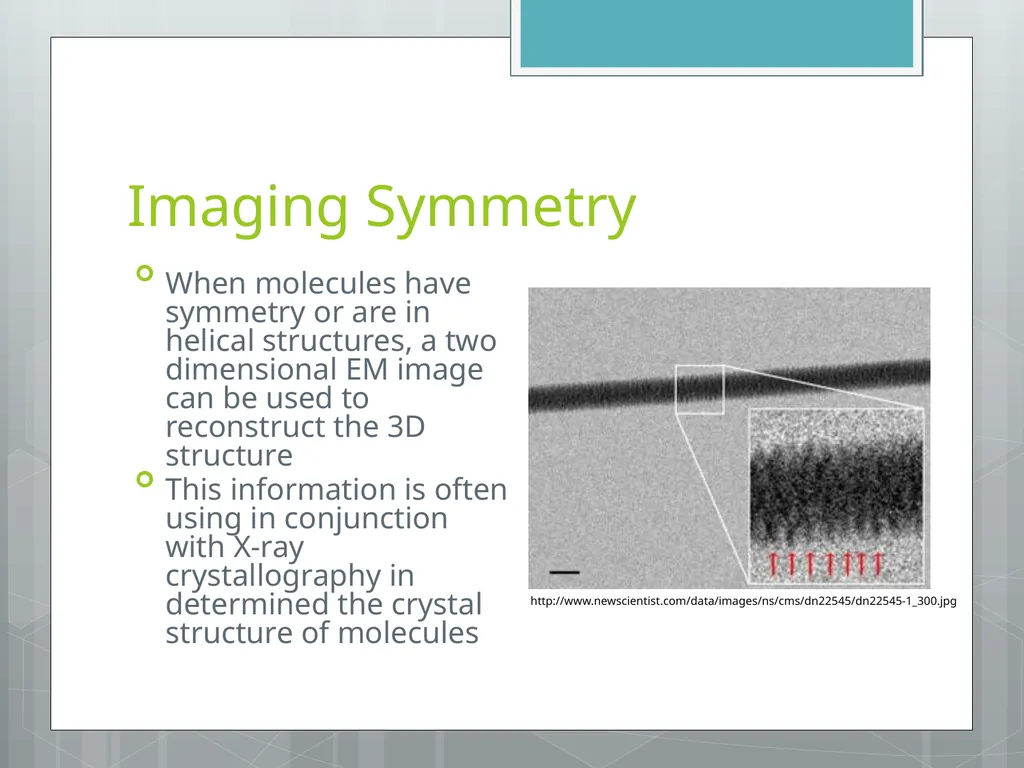
Imaging Symmetry When molecules have symmetry or are in helical structures, a two dimensional EM image can be used to reconstruct the 3D structure
This information is often using in conjunction with X-ray crystallography in determined the crystal structure of molecules http://www.newscientist.com/data/images/ns/cms/dn22545/dn22545-1_300.jpg<br>
15
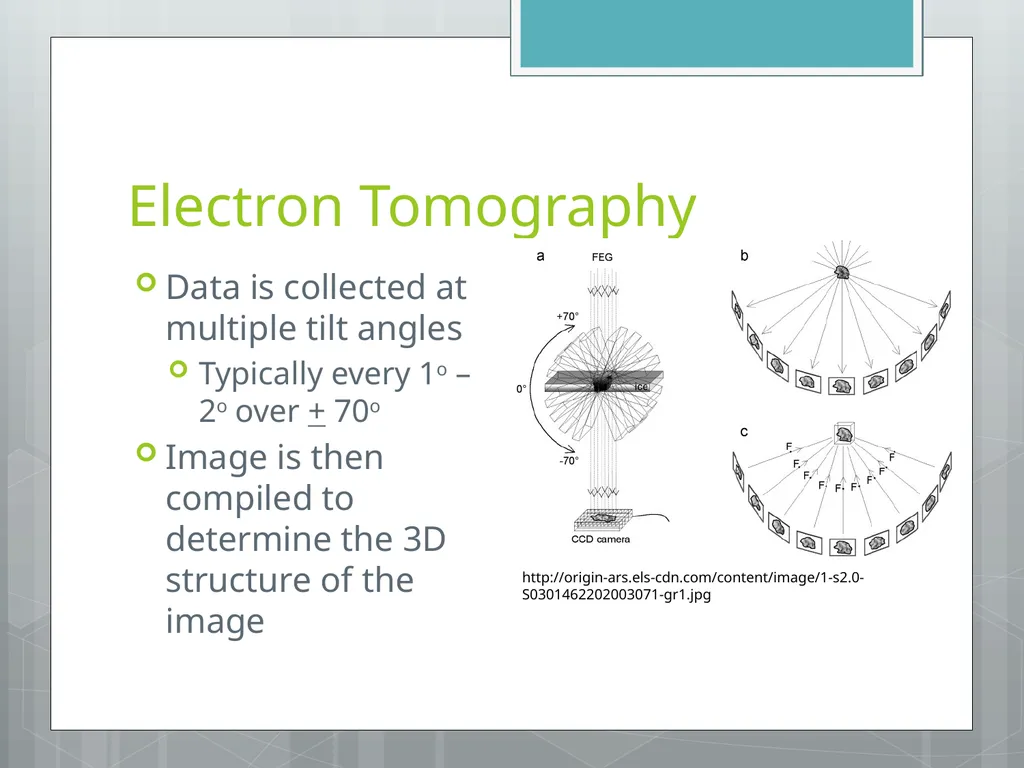
Electron Tomography Data is collected at multiple tilt angles
Typically every 1o – 2o over + 70o
Image is then compiled to determine the 3D structure of the image http://origin-ars.els-cdn.com/content/image/1-s2.0-S0301462202003071-gr1.jpg<br>
16
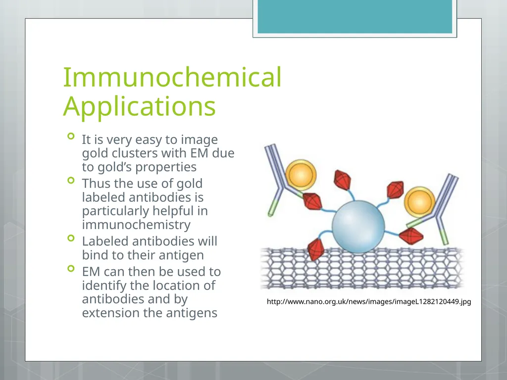
Immunochemical Applications It is very easy to image gold clusters with EM due to gold’s properties
Thus the use of gold labeled antibodies is particularly helpful in immunochemistry
Labeled antibodies will bind to their antigen
EM can then be used to identify the location of antibodies and by extension the antigens http://www.nano.org.uk/news/images/imageL1282120449.jpg<br>
17
Sources Serdyuk, Igor N., Nathan R. Zaccai, and Joseph Zaccai. Methods in Molecular Biophysics: Structure, Dynamics, Function. New York: Cambridge University Press, 2007. Print.
"Introduction to Electron Microscopes." Matter.org.uk. University of Liverpool, n.d. Web. 20 Oct 2013. <http://www.materials.ac.uk/elearning/matter/IntroductionToElectronMicroscopes/SEM/electron-gun.html>.<br>