
Figure 1. Optical coherence tomography imaging of
Description
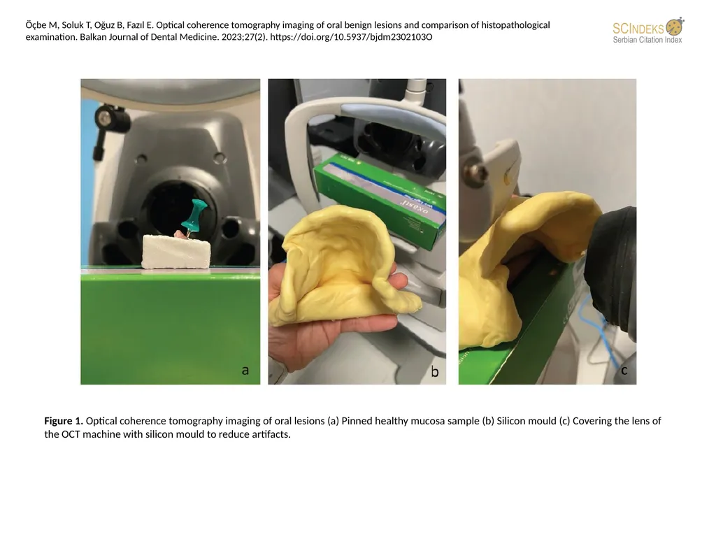
Figure 1. Optical coherence tomography imaging of oral lesions (a) Pinned healthy mucosa sample (b) Silicon mould (c) Covering the lens of the OCT machine with silicon mould to reduce artifacts. Öçbe M, Soluk T, Oğuz B, Fazıl E. Optical
Related Topics
Download this presentation From Below
"Figure 1. Optical coherence tomography imaging of" is the property of its rightful owner. Permission is granted to download and print the materials on this website for personal, non-commercial use only, and to display it on your personal computer provided you do not modify the materials and that you retain all copyright notices contained in the materials. By downloading content from our website, you accept the terms of this agreement.