
Human Anatomy and Physiology Eleventh Edition
YM
Published · 108 slides · 0 views

1 / 1







Description
Human Anatomy and Physiology Eleventh Edition Chapter 27 Part A The Reproductive System PowerPoint Lectures Slides prepared by Karen Dunbar Kareiva, Ivy Tech Community College Copyright 2019, 2016, 2013 Pearson Education, Inc. All Rights
Related Topics
Share
Embed code
Download this presentation From Below
"Human Anatomy and Physiology Eleventh Edition" is the property of its rightful owner. Permission is granted to download and print the materials on this website for personal, non-commercial use only, and to display it on your personal computer provided you do not modify the materials and that you retain all copyright notices contained in the materials. By downloading content from our website, you accept the terms of this agreement.
Presentation Transcript
01
Human Anatomy and Physiology Eleventh Edition Chapter 27 Part A The Reproductive System PowerPoint® Lectures Slides prepared by Karen Dunbar Kareiva, Ivy Tech Community College Copyright © 2019, 2016, 2013 Pearson Education, Inc. All Rights Reserved<br>
02
Why This Matters Understanding the anatomy and physiology of the reproductive system will help you recognize symptoms of disorders like STIs, give you the ability to treat your patients with reproductive diseases and help you educate your patients on how to have a healthy reproductive system.<br>
03
Video: Why This Matters (Career Connection) Click here to view ADA compliant video:
Why This Matters (Career Connection)
https://mediaplayer.pearsoncmg.com/assets/secs_wtm_ch_27_peter<br>
Why This Matters (Career Connection)
https://mediaplayer.pearsoncmg.com/assets/secs_wtm_ch_27_peter<br>
04
Reproductive System (1 of 2) Reproductive system does not function continuously like other organ systems
Does not become active until puberty
Male and female reproductive organs are quite different, but share same four tasks:
1. Form specialized cells for sexual reproduction call gametes
Sperm – gametes in males
Ova(eggs) – gametes in females<br>
Does not become active until puberty
Male and female reproductive organs are quite different, but share same four tasks:
1. Form specialized cells for sexual reproduction call gametes
Sperm – gametes in males
Ova(eggs) – gametes in females<br>
05
Reproductive System (2 of 2) 2. Bring gametes from male and female together through sexual intercourse (copulation)
3. Combine genetic information contained within gametes through fertilization
Zygote is formed when sperm and egg fuse
Zygote is first cell of new individual and all body cells arise from it
4. Support development of fetus (gestation) and birth of baby (parturition)<br>
3. Combine genetic information contained within gametes through fertilization
Zygote is formed when sperm and egg fuse
Zygote is first cell of new individual and all body cells arise from it
4. Support development of fetus (gestation) and birth of baby (parturition)<br>
06
27.1 Male and Female Reproductive System Share Common Features<br>
07
Gonads and Accessory Reproductive Organs (1 of 2) Male and female reproductive structures are homologous structures
Share a common origin during development
Example: male testes and female ovaries, or male penis and female clitoris<br>
Share a common origin during development
Example: male testes and female ovaries, or male penis and female clitoris<br>
08
Gonads and Accessory Reproductive Organs (2 of 2) Primary sex organs (gonads): testes (male) and ovaries (female) product two products:
1. Gametes: sperm (male) and ova (females)
Gamete formed by cell division called meiosis
2. Sex hormones (steroid hormones): Testosterone (males) and estrogens and progesterone (females)
Vital in development and function of reproductive organs and other organs/tissues, sexual behavior, sexual drives
Accessory reproductive organs: ducts, glands, and external genitalia<br>
1. Gametes: sperm (male) and ova (females)
Gamete formed by cell division called meiosis
2. Sex hormones (steroid hormones): Testosterone (males) and estrogens and progesterone (females)
Vital in development and function of reproductive organs and other organs/tissues, sexual behavior, sexual drives
Accessory reproductive organs: ducts, glands, and external genitalia<br>
09
Reproductive Hormone Secretion (1 of 5) Hypothalamic-Pituitary-Gonadal (HPG) Axis
Production of gametes and sex hormones is regulated by sequence of hormonal events involving hypothalamus, anterior pituitary gland, and testes
Referred to as hypothalamic-pituitary-gonadal (HPG) axis
Involves interacting hormones: GnRH, FSH, LH, testosterone, and inhibin<br>
Production of gametes and sex hormones is regulated by sequence of hormonal events involving hypothalamus, anterior pituitary gland, and testes
Referred to as hypothalamic-pituitary-gonadal (HPG) axis
Involves interacting hormones: GnRH, FSH, LH, testosterone, and inhibin<br>
10
Reproductive Hormone Secretion (2 of 5) HPG axis (cont.)
Hormones involved:
Gonadotropin-releasing hormone (GnRH)
Released from hypothalamus reaches anterior pituitary cells via hypophyseal portal system
Follicle-stimulating hormone (FSH) and Luteinizing hormone (LH) – gonadotropins released from anterior pituitary<br>
Hormones involved:
Gonadotropin-releasing hormone (GnRH)
Released from hypothalamus reaches anterior pituitary cells via hypophyseal portal system
Follicle-stimulating hormone (FSH) and Luteinizing hormone (LH) – gonadotropins released from anterior pituitary<br>
11
Reproductive Hormone Secretion (3 of 5) HPG axis (cont.)
Sex hormones: testosterone (males) and estrogen and progesterone (females) act at target tissues in body
Exert negative feedback on hypothalamus and anterior pituitary
Inhibin: released from gonads of both male and female
Exerts negative feedback on FSH release from anterior pituitary<br>
Sex hormones: testosterone (males) and estrogen and progesterone (females) act at target tissues in body
Exert negative feedback on hypothalamus and anterior pituitary
Inhibin: released from gonads of both male and female
Exerts negative feedback on FSH release from anterior pituitary<br>
12
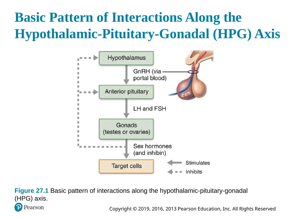
Basic Pattern of Interactions Along the
Hypothalamic-Pituitary-Gonadal (HPG) Axis Figure 27.1 Basic pattern of interactions along the hypothalamic-pituitary-gonadal (HPG) axis.<br>
13
Reproductive Hormone Secretion (4 of 5) Activation of the HPG Axis at Puberty
Puberty – period of life when reproductive organs grow to adult size and become functional
Earliest time that reproduction is possible
Before puberty, low levels of circulating levels of steroidal sex hormones are enough to suppress secretion of GnRH by hypothalamus
As puberty approaches, hypothalamus becomes less sensitive to inhibition by sex hormones
Hypothalamus releases GnRH in pulse like manner<br>
Puberty – period of life when reproductive organs grow to adult size and become functional
Earliest time that reproduction is possible
Before puberty, low levels of circulating levels of steroidal sex hormones are enough to suppress secretion of GnRH by hypothalamus
As puberty approaches, hypothalamus becomes less sensitive to inhibition by sex hormones
Hypothalamus releases GnRH in pulse like manner<br>
14
Reproductive Hormone Secretion (5 of 5) Activation of the HPG Axis at Puberty (cont.)
Anterior pituitary releases FSH and LH in response to increased GnRH levels
FSH and LH stimulate gonads to release more sex hormones from gonads
Threshold for inhibition of GnRH continues to rise resulting in more and more release of sex hormones
Sex hormones are released until adult pattern of hormone interaction is achieved<br>
Anterior pituitary releases FSH and LH in response to increased GnRH levels
FSH and LH stimulate gonads to release more sex hormones from gonads
Threshold for inhibition of GnRH continues to rise resulting in more and more release of sex hormones
Sex hormones are released until adult pattern of hormone interaction is achieved<br>
15
Meiosis (1 of 11) Meiosis – unique kind of nuclear division that occurs only in gonads
Same process for males and females
Sexual reproduction cannot take place without meiosis
Reduces number of chromosomes in gametes by half so zygote does not end up with twice as many chromosomes<br>
Same process for males and females
Sexual reproduction cannot take place without meiosis
Reduces number of chromosomes in gametes by half so zygote does not end up with twice as many chromosomes<br>
16
Meiosis (2 of 11) Terminology of Meiosis
Sister chromatids – cell’s DNA is replicated during interphase
Each chromosome has two sister chromatids that are identical and joined together by centromere
DNA coils and condenses, making chromosomes visible when viewed through light microscope
Homologous pairs of chromosomes – one member of each pair is from father (paternal chromosome) and other is from mother (material chromosome)
Look alike and carry same genes that code for same traits<br>
Sister chromatids – cell’s DNA is replicated during interphase
Each chromosome has two sister chromatids that are identical and joined together by centromere
DNA coils and condenses, making chromosomes visible when viewed through light microscope
Homologous pairs of chromosomes – one member of each pair is from father (paternal chromosome) and other is from mother (material chromosome)
Look alike and carry same genes that code for same traits<br>
17
Meiosis (3 of 11) Terminology of Meiosis (cont.)
Nonsister chromatids – within homologous pairs, chromatids of one chromosome are nonsister chromatids to other chromosome in pair
Nonsister chromatids exchange DNA with each other in process called crossing over
Process increases genetic variability
Diploid – diploid chromosomal number is normal number of chromosomes in most body cells
Consists of two sets of chromosomes (one from each parent) and is symbolized as 2n
Humans diploid number is 46; diploid cells contain 23 pairs of homologous chromosomes<br>
Nonsister chromatids – within homologous pairs, chromatids of one chromosome are nonsister chromatids to other chromosome in pair
Nonsister chromatids exchange DNA with each other in process called crossing over
Process increases genetic variability
Diploid – diploid chromosomal number is normal number of chromosomes in most body cells
Consists of two sets of chromosomes (one from each parent) and is symbolized as 2n
Humans diploid number is 46; diploid cells contain 23 pairs of homologous chromosomes<br>
18
Meiosis (4 of 11) Terminology of Meiosis (cont.)
Haploid – haploid chromosomal number, n, is the number of chromosomes in a gamete.
Gametes contain only one member of each homologous pair, either maternal and paternal
Humans haploid chromosomal number is 23
When sperm and egg fuse, they form a diploid, 2n, zygote<br>
Haploid – haploid chromosomal number, n, is the number of chromosomes in a gamete.
Gametes contain only one member of each homologous pair, either maternal and paternal
Humans haploid chromosomal number is 23
When sperm and egg fuse, they form a diploid, 2n, zygote<br>
19
Meiosis (5 of 11) Sequence of Events in Meiosis
Meiosis consists of two consecutive nuclear divisions referred to as meiosis I and meiosis II
DNA replication only occurs once
Product is four haploid daughter cells
Each nuclear division involves four steps:
Prophase
Metaphase
Anaphase
Telophase<br>
Meiosis consists of two consecutive nuclear divisions referred to as meiosis I and meiosis II
DNA replication only occurs once
Product is four haploid daughter cells
Each nuclear division involves four steps:
Prophase
Metaphase
Anaphase
Telophase<br>
20
Meiosis (6 of 11) Sequence of Events in Meiosis (cont.)
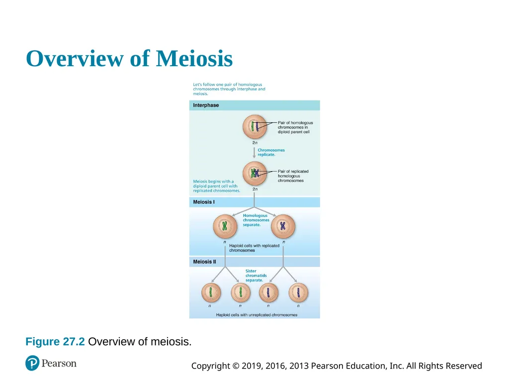
Overview of meiosis:
During interphase preceding meiosis, all chromosomes are replicated
During meiosis I, homologous pairs separate from each other, reducing number of chromosomes from 2n to n.
Referred to as reduction division of meiosis
During meiosis II, sister chromatids separate and each daughter cell gets equal share of chromosomes
Referred to as equational division of meiosis<br>
Overview of meiosis:
During interphase preceding meiosis, all chromosomes are replicated
During meiosis I, homologous pairs separate from each other, reducing number of chromosomes from 2n to n.
Referred to as reduction division of meiosis
During meiosis II, sister chromatids separate and each daughter cell gets equal share of chromosomes
Referred to as equational division of meiosis<br>
21
Overview of Meiosis Figure 27.2 Overview of meiosis.<br>
22
Meiosis (7 of 11) Sequence of Events in Meiosis (cont.)
Meiosis I
Chromosome number reduced from 2n → n
Prophase I has events not seen in mitosis or meiosis II
Synapsis: homologous chromosomes pair up forming tetrads consisting of 4 chromatids
Crossover (chiasmata): exchange of genetic material between male and female chromatids
Results in unique chromosomes that are mixtures of maternal and paternal chromosomes<br>
Meiosis I
Chromosome number reduced from 2n → n
Prophase I has events not seen in mitosis or meiosis II
Synapsis: homologous chromosomes pair up forming tetrads consisting of 4 chromatids
Crossover (chiasmata): exchange of genetic material between male and female chromatids
Results in unique chromosomes that are mixtures of maternal and paternal chromosomes<br>
23
Meiosis (8 of 11) Sequence of Events in Meiosis (cont.)
Meiosis I (cont.)
During metaphase I, tetrads line up randomly at spindle equator
Paternal or maternal homologous chromosome can be on either side of equator
During anaphase I, sister chromatids of one homologous chromosome are separated from sister chromatids of other homologous pair
Two joined sister chromatids of each homologous pair are distributed to opposite ends of cell<br>
Meiosis I (cont.)
During metaphase I, tetrads line up randomly at spindle equator
Paternal or maternal homologous chromosome can be on either side of equator
During anaphase I, sister chromatids of one homologous chromosome are separated from sister chromatids of other homologous pair
Two joined sister chromatids of each homologous pair are distributed to opposite ends of cell<br>
24
Meiosis (9 of 11) Sequence of Events in Meiosis (cont.)
Meiosis I (cont.)
At end of meiosis I, each daughter cell has:
Two copies (sister chromatids) of one member of each homologous pair (either maternal or paternal) and none of the other
Haploid chromosomal number because still-united sister chromatids are considered one chromosome (twice amount DNA in each chromosome)<br>
Meiosis I (cont.)
At end of meiosis I, each daughter cell has:
Two copies (sister chromatids) of one member of each homologous pair (either maternal or paternal) and none of the other
Haploid chromosomal number because still-united sister chromatids are considered one chromosome (twice amount DNA in each chromosome)<br>
25
Meiosis (1 of 2) Figure 27.3 Meiosis.<br>
26
Meiosis (10 of 11) Sequence of Events in Meiosis (cont.)
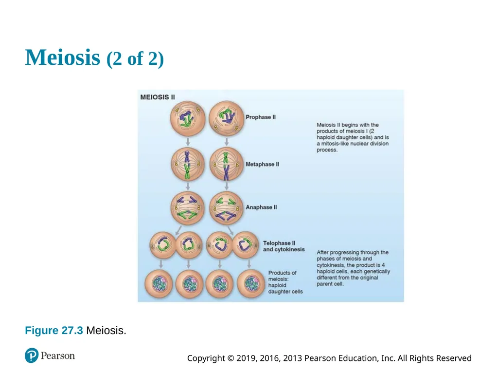
Meiosis II
Events mirrors mitosis in every way, except:
Chromosomes are not replicated before process begins
Cells that undergo meiosis II are haploid, not diploid<br>
Meiosis II
Events mirrors mitosis in every way, except:
Chromosomes are not replicated before process begins
Cells that undergo meiosis II are haploid, not diploid<br>
27
Meiosis (2 of 2) Figure 27.3 Meiosis.<br>
28
Meiosis (11 of 11) Meiosis Compared with Mitosis
Function of mitosis
Ensures that all cells in body have same complement of DNA for normal growth and repair
Functions of meiosis
Number of chromosomes are cut in half (2n to n)
Introduces genetic diversity, as all daughter cells are genetically different from original cell
Random alignment of homologous pairs provides variability
Crossing over increases variability<br>
Function of mitosis
Ensures that all cells in body have same complement of DNA for normal growth and repair
Functions of meiosis
Number of chromosomes are cut in half (2n to n)
Introduces genetic diversity, as all daughter cells are genetically different from original cell
Random alignment of homologous pairs provides variability
Crossing over increases variability<br>
29
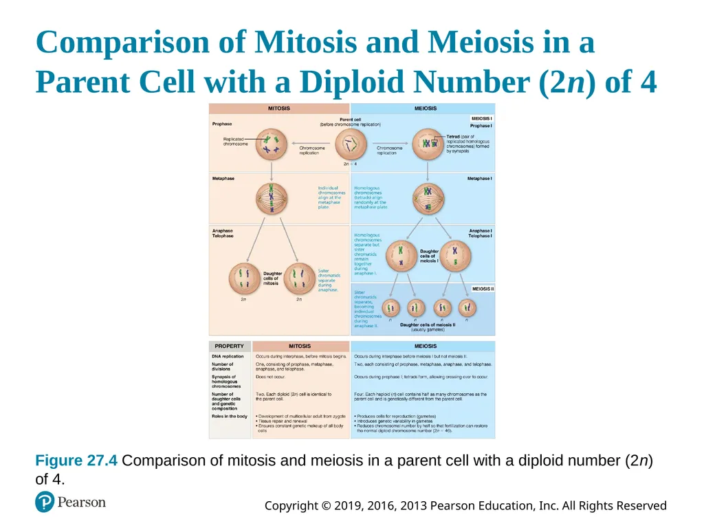
Comparison of Mitosis and Meiosis in a Parent Cell with a Diploid Number (2n) of 4 Figure 27.4 Comparison of mitosis and meiosis in a parent cell with a diploid number (2n) of 4.<br>
30
Animation: Interphase Click here to view ADA compliant Animation:
Interphase
https://mediaplayer.pearsoncmg.com/assets/interphase<br>
Interphase
https://mediaplayer.pearsoncmg.com/assets/interphase<br>
31
Animation: Prophase I Click here to view ADA compliant Animation:
Prophase I
https://mediaplayer.pearsoncmg.com/assets/meiosis-i-prophase-i<br>
Prophase I
https://mediaplayer.pearsoncmg.com/assets/meiosis-i-prophase-i<br>
32
Animation: Metaphase I Click here to view ADA compliant Animation:
Metaphase I
https://mediaplayer.pearsoncmg.com/assets/meiosis-i-metaphase-i<br>
Metaphase I
https://mediaplayer.pearsoncmg.com/assets/meiosis-i-metaphase-i<br>
33
Animation: Anaphase I Click here to view ADA compliant Animation:
Anaphase I
https://mediaplayer.pearsoncmg.com/assets/meiosis-i-anaphase-i<br>
Anaphase I
https://mediaplayer.pearsoncmg.com/assets/meiosis-i-anaphase-i<br>
34
Animation: Telophase I Click here to view ADA compliant Animation:
Telophase I
https://mediaplayer.pearsoncmg.com/assets/meiosis-i-telophase-i<br>
Telophase I
https://mediaplayer.pearsoncmg.com/assets/meiosis-i-telophase-i<br>
35
Animation: Meiosis II Click here to view ADA compliant Animation:
Meiosis II
https://mediaplayer.pearsoncmg.com/assets/meiosis-ii<br>
Meiosis II
https://mediaplayer.pearsoncmg.com/assets/meiosis-ii<br>
36
Bioflix: Meiosis Click here to view ADA compliant Animation:
Bioflix: Meiosis
https://mediaplayer.pearsoncmg.com/assets/_video.true/HKg3BIn5ihiaSoBwTDo_JpwVweCWk_FF<br>
Bioflix: Meiosis
https://mediaplayer.pearsoncmg.com/assets/_video.true/HKg3BIn5ihiaSoBwTDo_JpwVweCWk_FF<br>
37
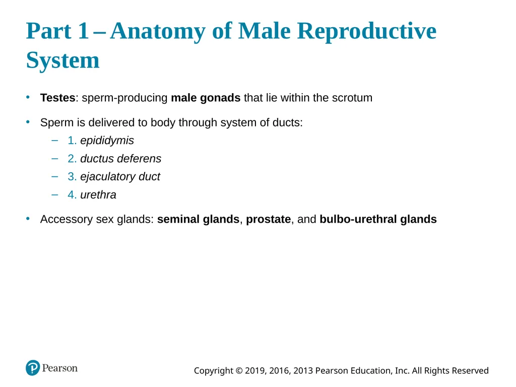
Part 1 – Anatomy of Male Reproductive System Testes: sperm-producing male gonads that lie within the scrotum
Sperm is delivered to body through system of ducts:
1. epididymis
2. ductus deferens
3. ejaculatory duct
4. urethra
Accessory sex glands: seminal glands, prostate, and bulbo-urethral glands<br>
Sperm is delivered to body through system of ducts:
1. epididymis
2. ductus deferens
3. ejaculatory duct
4. urethra
Accessory sex glands: seminal glands, prostate, and bulbo-urethral glands<br>
38
Animation: The Male Reproductive System Click here to view ADA compliant Animation:
The Male Reproductive System
https://mediaplayer.pearsoncmg.com/assets/_video.true/secs-campbell-reproductive-system-of-the-human-male<br>
The Male Reproductive System
https://mediaplayer.pearsoncmg.com/assets/_video.true/secs-campbell-reproductive-system-of-the-human-male<br>
39
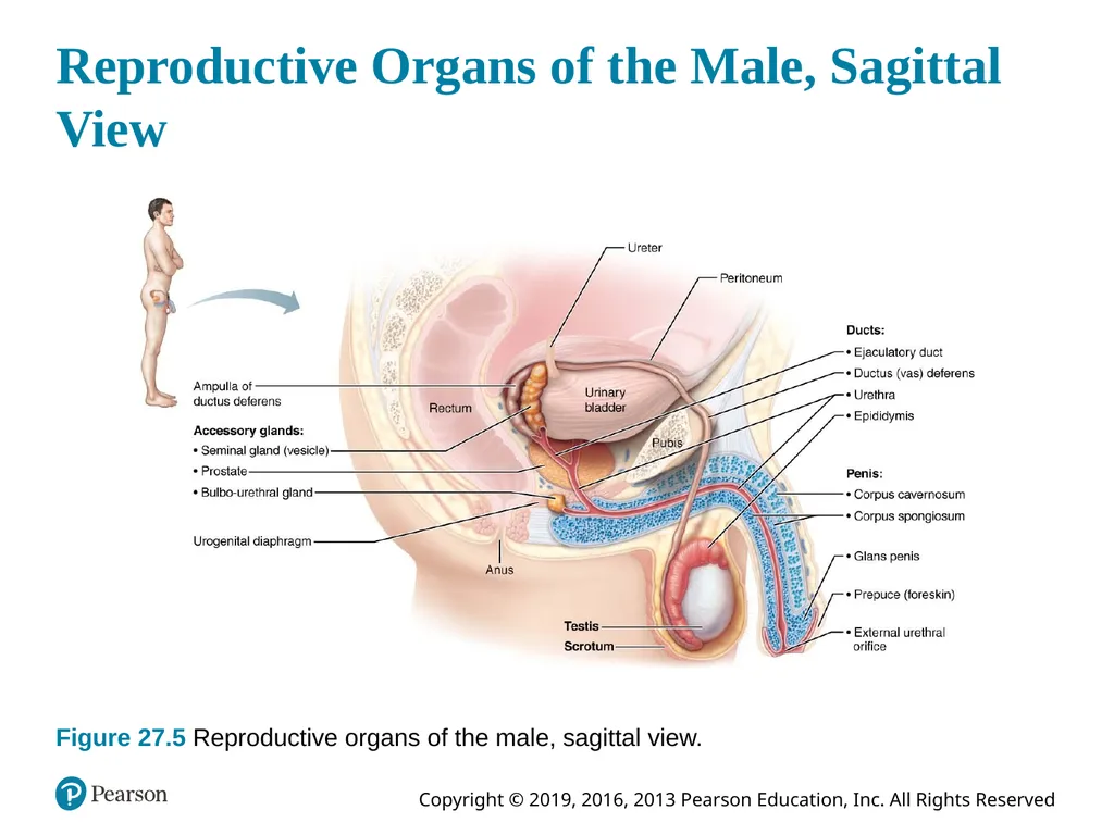
Reproductive Organs of the Male, Sagittal View Figure 27.5 Reproductive organs of the male, sagittal view.<br>
40
27.2 Scrotum and Testes<br>
41
The Scrotum (1 of 2) Sac of skin and superficial fascia
Hangs outside abdominopelvic cavity at root of penis
Contains paired testes
3°C lower than core body temperature
Lower temperature is necessary for sperm production
Midline septum divides scrotum into two compartments, one for each testis<br>
Hangs outside abdominopelvic cavity at root of penis
Contains paired testes
3°C lower than core body temperature
Lower temperature is necessary for sperm production
Midline septum divides scrotum into two compartments, one for each testis<br>
42
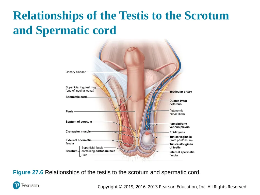
The Scrotum (2 of 2) Scrotum is affected by temperature changes
When cold, testes are pulled closer to warm body wall
Dartos muscle – layer of smooth muscle in superficial fascia that wrinkles skin
Cremaster muscles – bands of skeletal muscle elevate testes<br>
When cold, testes are pulled closer to warm body wall
Dartos muscle – layer of smooth muscle in superficial fascia that wrinkles skin
Cremaster muscles – bands of skeletal muscle elevate testes<br>
43
Relationships of the Testis to the Scrotum and Spermatic cord Figure 27.6 Relationships of the testis to the scrotum and spermatic cord.<br>
44
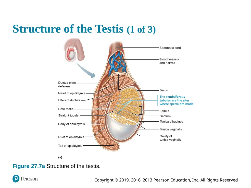
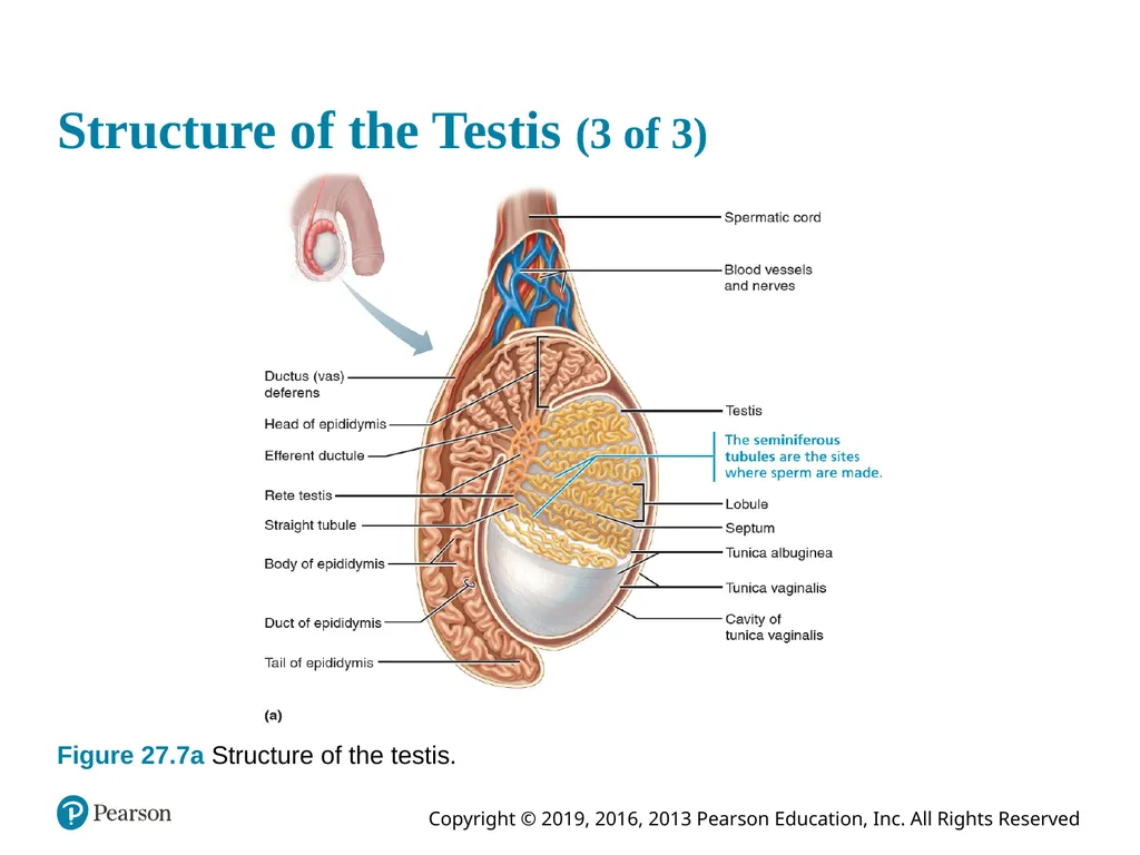
The Testes (1 of 3) Each testis is surrounded by two tunics
Tunica vaginalis: outer layer derived from peritoneum
Tunica albuginea: inner layer forms fibrous capsule
Septa divide testis into ~250 lobules, each containing one to four seminiferous tubules
Site of sperm production<br>
Tunica vaginalis: outer layer derived from peritoneum
Tunica albuginea: inner layer forms fibrous capsule
Septa divide testis into ~250 lobules, each containing one to four seminiferous tubules
Site of sperm production<br>
45
The Testes (2 of 3) Sperm is conveyed from seminiferous tubules to straight tubule → rete testis → efferent ductules → epididymis
Epididymis is made up of the head, the body, and the tail
Sperm are stored in the tail until ejaculation<br>
Epididymis is made up of the head, the body, and the tail
Sperm are stored in the tail until ejaculation<br>
46
The Testes (3 of 3) Blood supply
Testicular arteries arise from abdominal aorta
Testicular veins arise from pampiniform venous plexus surrounding each testicular artery
Cooler; absorb heat from testicular arteries
Keep testes cool
Spermatic cord encloses nerve fibers, blood vessels, and lymphatics that supply testes<br>
Testicular arteries arise from abdominal aorta
Testicular veins arise from pampiniform venous plexus surrounding each testicular artery
Cooler; absorb heat from testicular arteries
Keep testes cool
Spermatic cord encloses nerve fibers, blood vessels, and lymphatics that supply testes<br>
47
Structure of the Testis (1 of 3) Figure 27.7a Structure of the testis.<br>
48
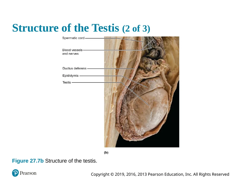
Structure of the Testis (2 of 3) Figure 27.7b Structure of the testis.<br>
49
Clinical – Homeostatic Imbalance 27.1 Testicular cancer
Rare, but most common cancer in men age 15–35
Having mumps that lead to orchitis (inflammation of testis) could be a risk factor
Cryptorchidism is most common risk factor
Nondescent of testes
Sign: painless, solid mass in testis
90% cured by surgical removal of testis and often radiation or chemotherapy<br>
Rare, but most common cancer in men age 15–35
Having mumps that lead to orchitis (inflammation of testis) could be a risk factor
Cryptorchidism is most common risk factor
Nondescent of testes
Sign: painless, solid mass in testis
90% cured by surgical removal of testis and often radiation or chemotherapy<br>
50
27.3 Male Duct System Accessory ducts carry sperm from testes to body exterior:
Epididymis
Ductus deferens
Ejaculatory duct
Urethra<br>
Epididymis
Ductus deferens
Ejaculatory duct
Urethra<br>
51
Epididymis (1 of 2) Sperm mature in epididymis
Head: contains efferent ductules that empty into highly coiled duct of the epididymis
Body and tail make up remainder of duct of the epididymis which is ~ 6 m (20 ft) in length
Pseudostratified epithelial cells of duct mucosa contain microvilli (stereocilia)
Large surface area allows absorption of excess testicular fluid and pass nutrients to stored sperm<br>
Head: contains efferent ductules that empty into highly coiled duct of the epididymis
Body and tail make up remainder of duct of the epididymis which is ~ 6 m (20 ft) in length
Pseudostratified epithelial cells of duct mucosa contain microvilli (stereocilia)
Large surface area allows absorption of excess testicular fluid and pass nutrients to stored sperm<br>
52
Epididymis (2 of 2) Nonmotile sperm enter, pass slowly through at rate that takes ~ 20 days, when they finally gain ability to swim
Can be stored several months
During ejaculation, epididymis contracts, expelling sperm into ductus deferens<br>
Can be stored several months
During ejaculation, epididymis contracts, expelling sperm into ductus deferens<br>
53
Ductus Deferens and Ejaculatory Duct Ductus deferens (vas deferens) ~ 45 cm long
Passes through inguinal canal to pelvic cavity
Expands to form ampulla
Joins duct of seminal vesicle to form ejaculatory duct
Smooth muscle in walls propels sperm from epididymis to urethra
Vasectomy: cutting and ligating ductus deferens
Nearly 100% effective form of birth control<br>
Passes through inguinal canal to pelvic cavity
Expands to form ampulla
Joins duct of seminal vesicle to form ejaculatory duct
Smooth muscle in walls propels sperm from epididymis to urethra
Vasectomy: cutting and ligating ductus deferens
Nearly 100% effective form of birth control<br>
54
Structure of the Testis (3 of 3) Figure 27.7a Structure of the testis.<br>
55
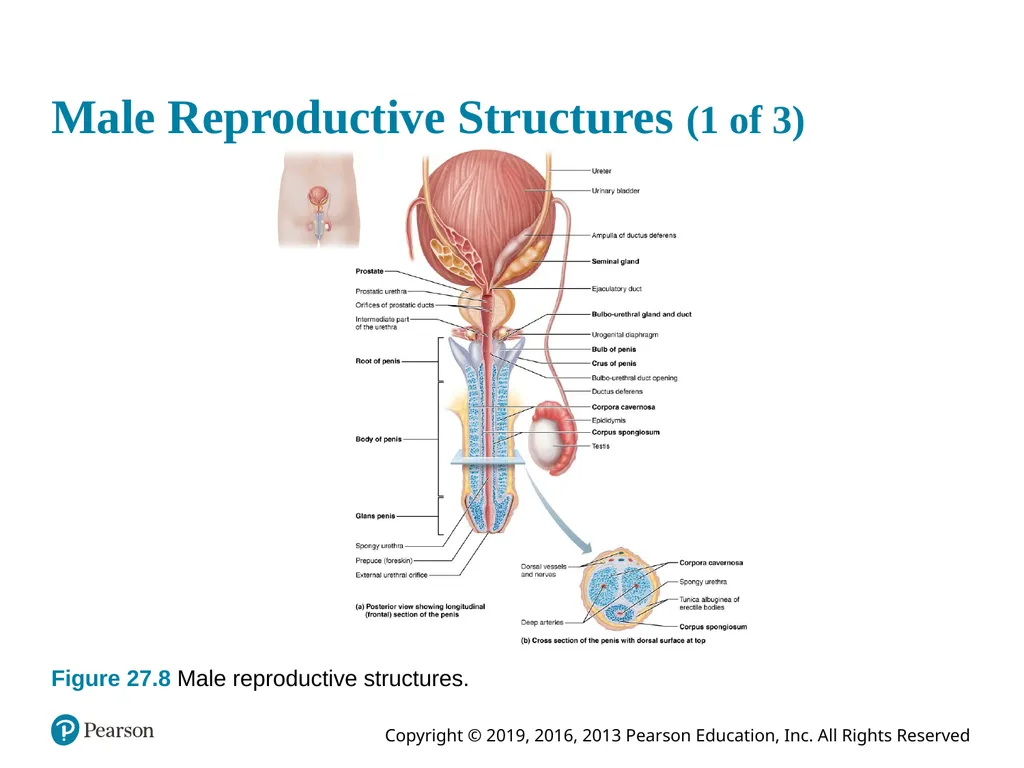
Urethra Conveys both urine and semen (at different times)
Has three regions
Prostatic urethra: surrounded by prostate
Intermediate part of the urethra (membranous urethra): in urogenital diaphragm
Spongy urethra: runs through penis; opens at external urethral orifice<br>
Has three regions
Prostatic urethra: surrounded by prostate
Intermediate part of the urethra (membranous urethra): in urogenital diaphragm
Spongy urethra: runs through penis; opens at external urethral orifice<br>
56
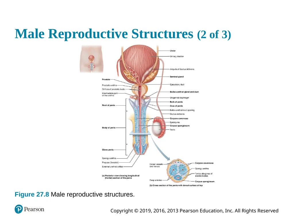
Male Reproductive Structures (1 of 3) Figure 27.8 Male reproductive structures.<br>
57
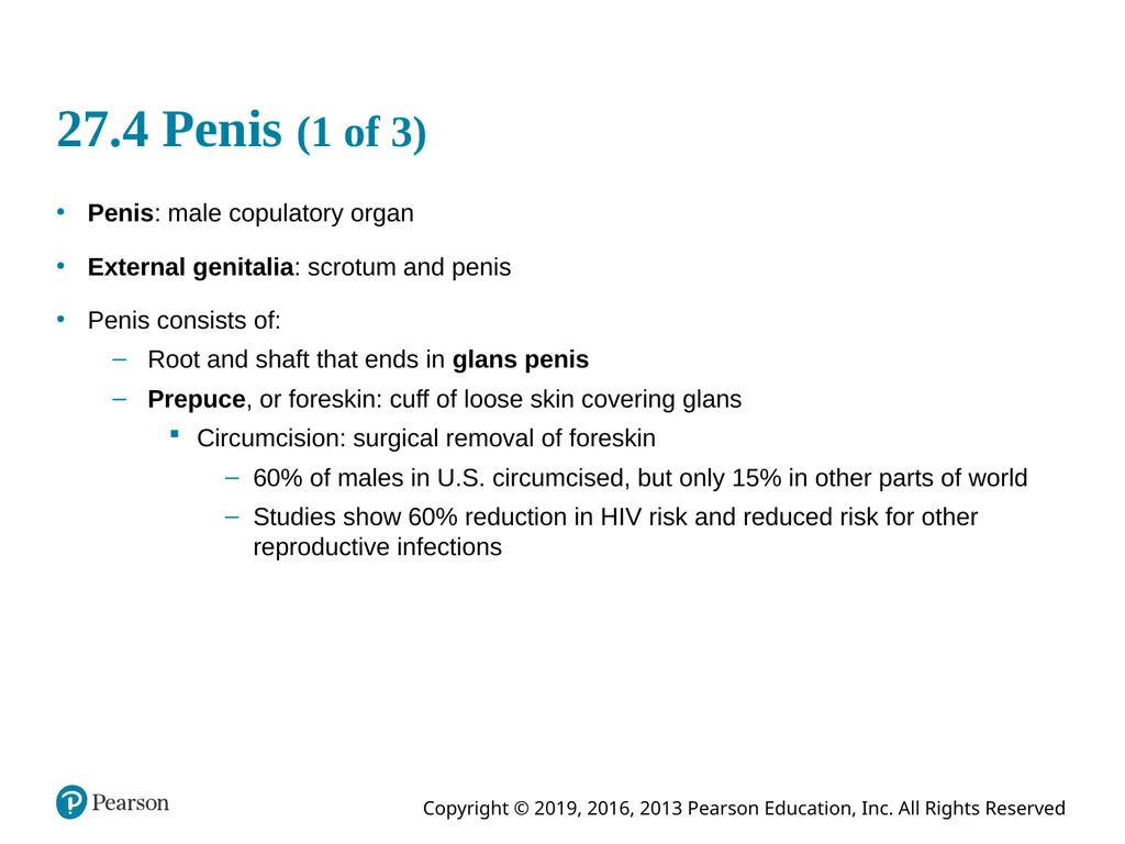
27.4 Penis (1 of 3) Penis: male copulatory organ
External genitalia: scrotum and penis
Penis consists of:
Root and shaft that ends in glans penis
Prepuce, or foreskin: cuff of loose skin covering glans
Circumcision: surgical removal of foreskin
60% of males in U.S. circumcised, but only 15% in other parts of world
Studies show 60% reduction in HIV risk and reduced risk for other reproductive infections<br>
External genitalia: scrotum and penis
Penis consists of:
Root and shaft that ends in glans penis
Prepuce, or foreskin: cuff of loose skin covering glans
Circumcision: surgical removal of foreskin
60% of males in U.S. circumcised, but only 15% in other parts of world
Studies show 60% reduction in HIV risk and reduced risk for other reproductive infections<br>
58
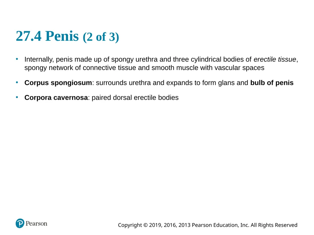
27.4 Penis (2 of 3) Internally, penis made up of spongy urethra and three cylindrical bodies of erectile tissue, spongy network of connective tissue and smooth muscle with vascular spaces
Corpus spongiosum: surrounds urethra and expands to form glans and bulb of penis
Corpora cavernosa: paired dorsal erectile bodies<br>
Corpus spongiosum: surrounds urethra and expands to form glans and bulb of penis
Corpora cavernosa: paired dorsal erectile bodies<br>
59
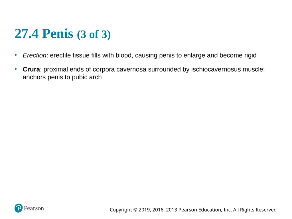
27.4 Penis (3 of 3) Erection: erectile tissue fills with blood, causing penis to enlarge and become rigid
Crura: proximal ends of corpora cavernosa surrounded by ischiocavernosus muscle; anchors penis to pubic arch<br>
Crura: proximal ends of corpora cavernosa surrounded by ischiocavernosus muscle; anchors penis to pubic arch<br>
60
Male Reproductive Structures (2 of 3) Figure 27.8 Male reproductive structures.<br>
61
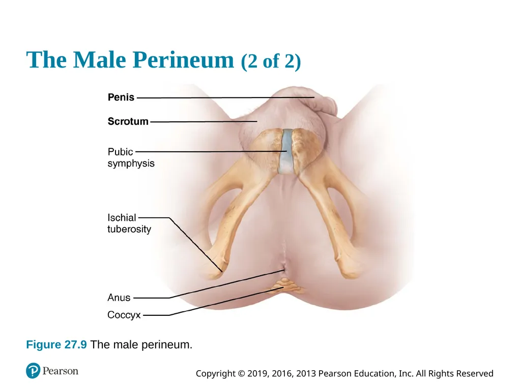
The Male Perineum (1 of 2) Diamond-shaped region between pubic symphysis, coccyx, and ischial tuberosities
Suspends scrotum
Contains root of penis and anus<br>
Suspends scrotum
Contains root of penis and anus<br>
62
The Male Perineum (2 of 2) Figure 27.9 The male perineum.<br>
63
27.5 Male Accessory Glands (1 of 3) Seminal glands (seminal vesicles)
On posterior bladder surface
Contains smooth muscle that contracts during ejaculation
Produces viscous alkaline seminal fluid
Fructose, citric acid, coagulating enzyme (vesiculase), and prostaglandins
Yellow pigment fluoresces with UV light
Comprises 70% volume of semen
Duct of seminal gland joins ductus deferens to form ejaculatory duct<br>
On posterior bladder surface
Contains smooth muscle that contracts during ejaculation
Produces viscous alkaline seminal fluid
Fructose, citric acid, coagulating enzyme (vesiculase), and prostaglandins
Yellow pigment fluoresces with UV light
Comprises 70% volume of semen
Duct of seminal gland joins ductus deferens to form ejaculatory duct<br>
64
27.5 Male Accessory Glands (2 of 3) Prostate
Encircles urethra inferior to bladder
Size of peach pit
Consists of smooth muscle that contracts during ejaculation
Secretes milky, slightly acid fluid
Contains citrate, enzymes, and prostate-specific antigen (PSA)
Plays a role in sperm activation
Enters prostatic urethra during ejaculation
Makes up one-third of semen volume<br>
Encircles urethra inferior to bladder
Size of peach pit
Consists of smooth muscle that contracts during ejaculation
Secretes milky, slightly acid fluid
Contains citrate, enzymes, and prostate-specific antigen (PSA)
Plays a role in sperm activation
Enters prostatic urethra during ejaculation
Makes up one-third of semen volume<br>
65
27.5 Male Accessory Glands (3 of 3) Bulbo-urethral glands
Pea-sized glands inferior to prostate
Produce thick, clear mucus during sexual arousal
Lubricate glans penis
Neutralize traces of acidic urine in urethra<br>
Pea-sized glands inferior to prostate
Produce thick, clear mucus during sexual arousal
Lubricate glans penis
Neutralize traces of acidic urine in urethra<br>
66
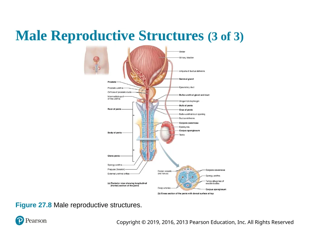
Male Reproductive Structures (3 of 3) Figure 27.8 Male reproductive structures.<br>
67
Clinical – Homeostatic Imbalance 27.2 (1 of 2) Benign prostatic hyperplasia
Common diseases that may be age-related
Affects ~50% of men age 50, 80% of men age 70
Benign growth of stroma and glands of prostate constricts urethra making urination difficult leading to incomplete emptying
Can lead to urinary tract infections or kidney damage
Treatment includes drugs to relax smooth muscle and inhibit growth; surgical transurethral resection widens prostatic urethra
Prostatitis: common reason for man to consult urologist – covered in Related Clinical Terms<br>
Common diseases that may be age-related
Affects ~50% of men age 50, 80% of men age 70
Benign growth of stroma and glands of prostate constricts urethra making urination difficult leading to incomplete emptying
Can lead to urinary tract infections or kidney damage
Treatment includes drugs to relax smooth muscle and inhibit growth; surgical transurethral resection widens prostatic urethra
Prostatitis: common reason for man to consult urologist – covered in Related Clinical Terms<br>
68
Clinical – Homeostatic Imbalance 27.2 (2 of 2) Prostate cancer
Third most common cause of cancer death in males that affects 1 in 6 men in U.S.
Digital exam screening, PSA levels can be checked by have high rate of false positives
Treated with surgery and sometimes radiation
Metastatic prostate cancer treated with drugs that block testosterone synthesis or action<br>
Third most common cause of cancer death in males that affects 1 in 6 men in U.S.
Digital exam screening, PSA levels can be checked by have high rate of false positives
Treated with surgery and sometimes radiation
Metastatic prostate cancer treated with drugs that block testosterone synthesis or action<br>
69
Semen (1 of 2) Milky-white mixture of sperm and accessory gland secretions
2–5 ml semen are ejaculated containing 20–150 million sperm/ml
Contains fructose for ATP production, protects and activates sperm, and facilitates sperm movement
Alkaline fluid neutralizes acidity of male urethra and female vagina and enhances motility<br>
2–5 ml semen are ejaculated containing 20–150 million sperm/ml
Contains fructose for ATP production, protects and activates sperm, and facilitates sperm movement
Alkaline fluid neutralizes acidity of male urethra and female vagina and enhances motility<br>
70
Semen (2 of 2) Characteristics of semen
Contain prostaglandins that decrease viscosity of mucus in cervix and stimulate reverse peristalsis in uterus
Contains the hormone relaxin and other enzymes that enhance sperm motility
Contains ATP for energy
Can suppress female immune response
Antibiotic chemicals destroy some bacteria
Clotting factors coagulate semen initially to prevent draining out, then liquefy it by fibrinolysin so sperm can finish journey<br>
Contain prostaglandins that decrease viscosity of mucus in cervix and stimulate reverse peristalsis in uterus
Contains the hormone relaxin and other enzymes that enhance sperm motility
Contains ATP for energy
Can suppress female immune response
Antibiotic chemicals destroy some bacteria
Clotting factors coagulate semen initially to prevent draining out, then liquefy it by fibrinolysin so sperm can finish journey<br>
71
Part 2 – Male Reproductive Physiology<br>
72
27.6 Male Sexual Response<br>
73
Erection (1 of 2) Erection: enlargement and stiffening of penis
Arterioles are normally constricted
Sexual excitement causes CNS activation of parasympathetic neurons innervating internal pudendal arteries, causing local release of nitric oxide (NO)
NO release causes relaxation of local vascular smooth muscle<br>
Arterioles are normally constricted
Sexual excitement causes CNS activation of parasympathetic neurons innervating internal pudendal arteries, causing local release of nitric oxide (NO)
NO release causes relaxation of local vascular smooth muscle<br>
74
Erection (2 of 2) When smooth muscles relax, arterioles dilate
Corpora cavernosa expands and retards venous drainage, leading to engorgement of erectile tissues with blood and enlargement and stiffening of penis
Corpus spongiosum keeps urethra open
Initiated by sexual stimuli such as touch; mechanical stimulation of penis; erotic sights, sounds, and smells
Can be induced or inhibited by emotions or higher mental activity<br>
Corpora cavernosa expands and retards venous drainage, leading to engorgement of erectile tissues with blood and enlargement and stiffening of penis
Corpus spongiosum keeps urethra open
Initiated by sexual stimuli such as touch; mechanical stimulation of penis; erotic sights, sounds, and smells
Can be induced or inhibited by emotions or higher mental activity<br>
75
Ejaculation (1 of 3) Propulsion of semen from male duct system
Spinal reflex is initiated over sympathetic nerves serving genital organs, resulting in:
1. Ductus deferens, prostate, and seminal glands contract and empty contents into prostatic urethra; bladder internal sphincter muscle constricts, preventing expulsion of urine or reflux of semen into bladder<br>
Spinal reflex is initiated over sympathetic nerves serving genital organs, resulting in:
1. Ductus deferens, prostate, and seminal glands contract and empty contents into prostatic urethra; bladder internal sphincter muscle constricts, preventing expulsion of urine or reflux of semen into bladder<br>
76
Ejaculation (2 of 3) 2. Semen in urethra triggers spinal reflex through somatic neurons
Bulbospongiosus muscles undergo rapid series of contractions that cause expulsion of semen at ~ 500 cm/s (close to 11 mph)
Ejaculatory event accompanied by intense pleasure referred to as climax (orgasm), and increased heart rate and blood pressure<br>
Bulbospongiosus muscles undergo rapid series of contractions that cause expulsion of semen at ~ 500 cm/s (close to 11 mph)
Ejaculatory event accompanied by intense pleasure referred to as climax (orgasm), and increased heart rate and blood pressure<br>
77
Ejaculation (3 of 3) Ejaculation followed by resolution, period of muscular and psychological relaxation
Arteries and arterioles constricted by signals from sympathetic nerves reducing blood flow to penis
Latent (refractory) period – time during which man is unable to achieve another orgasm
Can last minutes to hours
Latent period lengthens with age<br>
Arteries and arterioles constricted by signals from sympathetic nerves reducing blood flow to penis
Latent (refractory) period – time during which man is unable to achieve another orgasm
Can last minutes to hours
Latent period lengthens with age<br>
78
Clinical – Homeostatic Imbalance 27.3 (1 of 2) Erectile dysfunction
Parasympathetic nerves of penis release too little NO leading to inability to attain erection
Affects ~50% of American men over 40
Temporary ED caused by psychological factors, alcohol or drugs (antihypertensives, antidepressants, etc.)<br>
Parasympathetic nerves of penis release too little NO leading to inability to attain erection
Affects ~50% of American men over 40
Temporary ED caused by psychological factors, alcohol or drugs (antihypertensives, antidepressants, etc.)<br>
79
Clinical – Homeostatic Imbalance 27.3 (2 of 2) Chronic ED result of problems with blood vessels (atherosclerosis, varicose veins) or nervous system (stroke, penile nerve damage, multiple sclerosis)
Diabetes mellitus is often underlying cause
New drugs, sildenafil (Viagra) and others enhance effect of existing NO<br>
Diabetes mellitus is often underlying cause
New drugs, sildenafil (Viagra) and others enhance effect of existing NO<br>
80
27.7 Spermatogenesis Spermatogenesis: process of forming male gametes
Occurs in seminiferous tubules
Begins at puberty, around 14 years of age
Adult males make ~90 million sperm daily<br>
Occurs in seminiferous tubules
Begins at puberty, around 14 years of age
Adult males make ~90 million sperm daily<br>
81
Histology of the Seminiferous Tubules (1 of 3) Tubules consists of a thick stratified epithelium surrounding a central fluid-filled lumen containing four important types of cells:
Sustentocytes: large columnar cells act as supporting cells and play role in sperm formation
Spermatogenic cells: cells that are surrounded by sustentocytes and give rise to sperm
Myoid cells: smooth muscle-like cells surrounding seminiferous tubule that contract to squeeze sperm and testicular fluid through tubules
Interstitial endocrine cells: (Leydig cells) produce androgens and some estrogen<br>
Sustentocytes: large columnar cells act as supporting cells and play role in sperm formation
Spermatogenic cells: cells that are surrounded by sustentocytes and give rise to sperm
Myoid cells: smooth muscle-like cells surrounding seminiferous tubule that contract to squeeze sperm and testicular fluid through tubules
Interstitial endocrine cells: (Leydig cells) produce androgens and some estrogen<br>
82
Histology of the Seminiferous Tubules (2 of 3) Figure 27.10a Histology of the seminiferous tubule.<br>
83
Histology of the Seminiferous Tubules (3 of 3) Figure 27.10b Histology of the seminiferous tubule.<br>
84
Spermatogenesis: Summary of Events
in the Seminiferous Tubules (1 of 9) Occurs in seminiferous tubules of testis
Spermatogenic cells give rise to sperm
Overview of three steps of spermatogenesis:
Mitosis of spermatogonia (stem cell) forms two spermatocytes
Meiosis
Spermatocytes form secondary spermatocytes, which form spermatids
Spermiogenesis
Spermatids become sperm<br>
Spermatogenic cells give rise to sperm
Overview of three steps of spermatogenesis:
Mitosis of spermatogonia (stem cell) forms two spermatocytes
Meiosis
Spermatocytes form secondary spermatocytes, which form spermatids
Spermiogenesis
Spermatids become sperm<br>
85
Spermatogenesis: Summary of Events
in the Seminiferous Tubules (2 of 9) Mitosis of spermatogonia: forming
spermatocytes
Spermatogenesis begins at puberty
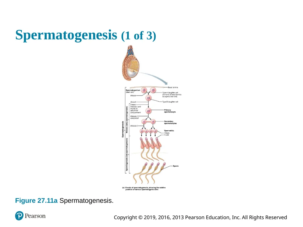
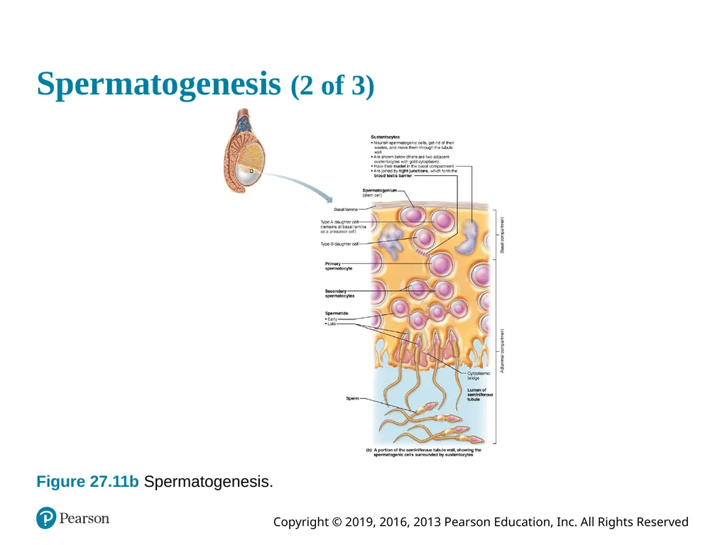
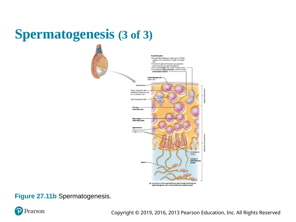
Begins with spermatogonia - stem cells that are in direct contact with epithelial basal lamina that divide more or less continuously by mitosis
After puberty, each division produces:
Type A daughter cells remain at basal lamina to maintain pool of dividing germ cells
Type B daughter cells move toward lumen and develop into primary spermatocytes<br>
Spermatogenesis begins at puberty
Begins with spermatogonia - stem cells that are in direct contact with epithelial basal lamina that divide more or less continuously by mitosis
After puberty, each division produces:
Type A daughter cells remain at basal lamina to maintain pool of dividing germ cells
Type B daughter cells move toward lumen and develop into primary spermatocytes<br>
86
Spermatogenesis (1 of 3) Figure 27.11a Spermatogenesis.<br>
87
Spermatogenesis: Summary of Events
in the Seminiferous Tubules (3 of 9) Meiosis: spermatocytes to spermatids
Meiosis I:
Primary spermatocyte (2n) undergoes meiosis I, forming two secondary spermatocytes (n)
Meiosis II
Secondary spermatocyte (n) rapidly undergoes meiosis II to become two spermatids (n)
Spermatids: small, round cells with large nuclei found close to lumen of tubule
Nearly all genes are turned off and DNA compacted into dense pellets<br>
Meiosis I:
Primary spermatocyte (2n) undergoes meiosis I, forming two secondary spermatocytes (n)
Meiosis II
Secondary spermatocyte (n) rapidly undergoes meiosis II to become two spermatids (n)
Spermatids: small, round cells with large nuclei found close to lumen of tubule
Nearly all genes are turned off and DNA compacted into dense pellets<br>
88
Spermatogenesis (2 of 3) Figure 27.11b Spermatogenesis.<br>
89
Spermatogenesis: Summary of Events
in the Seminiferous Tubules (4 of 9) Spermiogenesis: spermatids to sperm
Spermatids contain correct haploid chromosome number needed for fertilization (n)
Are still nonmotile
Spermiogenesis
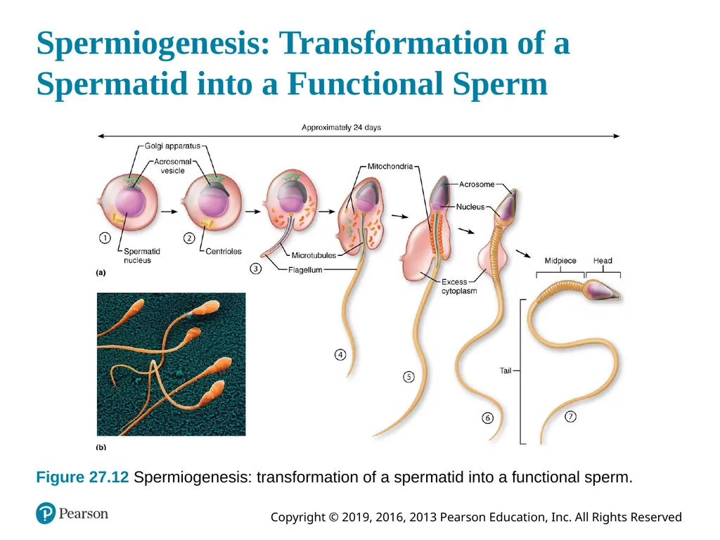
Streamlining process where spermatid elongates, loses excess cytoplasm, and forms a tail.<br>
Spermatids contain correct haploid chromosome number needed for fertilization (n)
Are still nonmotile
Spermiogenesis
Streamlining process where spermatid elongates, loses excess cytoplasm, and forms a tail.<br>
90
Spermatogenesis: Summary of Events
in the Seminiferous Tubules (5 of 9) Spermiogenesis: spermatids to sperm (cont.)
Major regions of sperm:
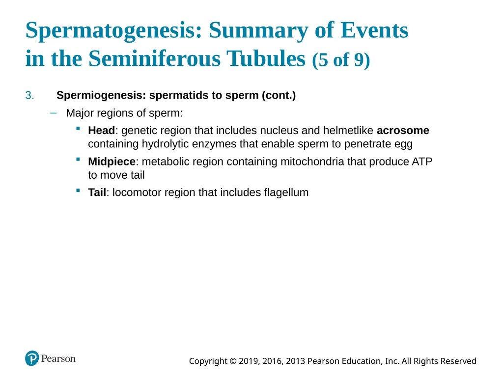
Head: genetic region that includes nucleus and helmetlike acrosome containing hydrolytic enzymes that enable sperm to penetrate egg
Midpiece: metabolic region containing mitochondria that produce ATP to move tail
Tail: locomotor region that includes flagellum<br>
Major regions of sperm:
Head: genetic region that includes nucleus and helmetlike acrosome containing hydrolytic enzymes that enable sperm to penetrate egg
Midpiece: metabolic region containing mitochondria that produce ATP to move tail
Tail: locomotor region that includes flagellum<br>
91
Spermiogenesis: Transformation of a Spermatid into a Functional Sperm Figure 27.12 Spermiogenesis: transformation of a spermatid into a functional sperm.<br>
92
Spermatogenesis: Summary of Events
in the Seminiferous Tubules (6 of 9) Spermatogenesis takes 64–72 days if conditions are hospitable
Sperm are unable to swim, but pressure of testicular fluid pushes immotile sperm into epididymis, where they gain motility and fertilizing power<br>
Sperm are unable to swim, but pressure of testicular fluid pushes immotile sperm into epididymis, where they gain motility and fertilizing power<br>
93
Spermatogenesis: Summary of Events
in the Seminiferous Tubules (7 of 9) Role of sustentocytes
Large supporting cells (also called Sertoli cells) extend from basal lamina to tubule lumen and surround developing spermatogonium
Sustentocytes contain tight junctions that divide tubule into two compartments
Basal compartment: basal lamina to tight junctions; spermatogonia and primary spermatocytes located here
Adluminal compartment: internal to tight junction; area where meiotically active cells and tubule lumen are located<br>
Large supporting cells (also called Sertoli cells) extend from basal lamina to tubule lumen and surround developing spermatogonium
Sustentocytes contain tight junctions that divide tubule into two compartments
Basal compartment: basal lamina to tight junctions; spermatogonia and primary spermatocytes located here
Adluminal compartment: internal to tight junction; area where meiotically active cells and tubule lumen are located<br>
94
Spermatogenesis: Summary of Events
in the Seminiferous Tubules (8 of 9) Role of sustentocytes (cont.)
Tight junctions form blood testis barrier
Prevents sperm antigens from escaping into blood and causing activation of immune system
Sperm is not formed until puberty, so it is absent during immune system development
Results in sperm not being recognized as “self”
Therefore, sperm needs to be kept separated from rest of body to avoid being attacked by immune system<br>
Tight junctions form blood testis barrier
Prevents sperm antigens from escaping into blood and causing activation of immune system
Sperm is not formed until puberty, so it is absent during immune system development
Results in sperm not being recognized as “self”
Therefore, sperm needs to be kept separated from rest of body to avoid being attacked by immune system<br>
95
Spermatogenesis: Summary of Events
in the Seminiferous Tubules (9 of 9) Role of sustentocytes (cont.)
Sustentocytes also:
Provide nutrients and signals to dividing cells
Move spermatocytes and spermatids along to lumen
Secrete testicular fluid (rich in androgens and metabolic acid) into lumen for sperm transport
Phagocytize faulty germ cells and excess cytoplasm
Produce 2 mediators to regulate spermatogenesis:
Androgen-binding protein (ABP): keeps testosterone levels high to stimulate spermatogenesis
Inhibin: inhibits spermatogenesis by inhibiting FSH release by anterior pituitary<br>
Sustentocytes also:
Provide nutrients and signals to dividing cells
Move spermatocytes and spermatids along to lumen
Secrete testicular fluid (rich in androgens and metabolic acid) into lumen for sperm transport
Phagocytize faulty germ cells and excess cytoplasm
Produce 2 mediators to regulate spermatogenesis:
Androgen-binding protein (ABP): keeps testosterone levels high to stimulate spermatogenesis
Inhibin: inhibits spermatogenesis by inhibiting FSH release by anterior pituitary<br>
96
Spermatogenesis (3 of 3) Figure 27.11b Spermatogenesis.<br>
97
Clinical – Homeostatic Imbalance 27.4 (1 of 2) Infertility affects roughly 1 in 7 couples in America
Often caused by problems with sperm quality or quantity
Gradual decline in male fertility has been occurring in past 50 years
Some believe main culprit is foreign molecules we ingest such as environmental toxins, phthalates (oily solvents from plastics), pesticides, herbicides, etc.<br>
Often caused by problems with sperm quality or quantity
Gradual decline in male fertility has been occurring in past 50 years
Some believe main culprit is foreign molecules we ingest such as environmental toxins, phthalates (oily solvents from plastics), pesticides, herbicides, etc.<br>
98
Clinical – Homeostatic Imbalance 27.4 (2 of 2) Other possibilities include:
Estrogen-like compounds block action of male sex hormones
Antibiotics such a tetracycline may suppress sperm formation
Radiation, lead, marijuana, and excessive alcohol can cause abnormal sperm to be produced (two-headed, multiple-tailed)
Defects in calcium channels, hormonal imbalances and oxidative stress
Thermal related events, such as sperm or overuse of hot tubs may inhibit sperm maturation<br>
Estrogen-like compounds block action of male sex hormones
Antibiotics such a tetracycline may suppress sperm formation
Radiation, lead, marijuana, and excessive alcohol can cause abnormal sperm to be produced (two-headed, multiple-tailed)
Defects in calcium channels, hormonal imbalances and oxidative stress
Thermal related events, such as sperm or overuse of hot tubs may inhibit sperm maturation<br>
99
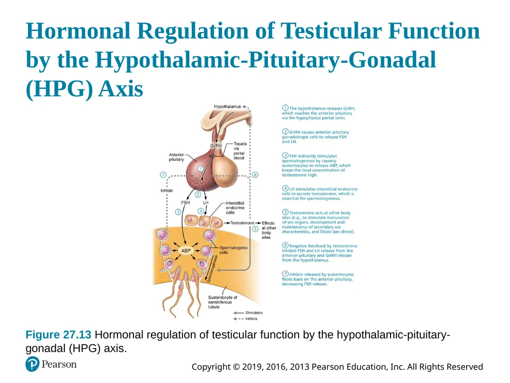
27.8 Regulation of Male Reproductive System (1 of 5) Production of gametes and sex hormones is regulated by sequence of hormonal events involving hypothalamus, anterior pituitary gland, and testes
Referred to as hypothalamic-pituitary-gonadal (HPG) axis
Involves interacting hormones: GnRH, FSH, LH, testosterone, and inhibin<br>
Referred to as hypothalamic-pituitary-gonadal (HPG) axis
Involves interacting hormones: GnRH, FSH, LH, testosterone, and inhibin<br>
100
Hormonal Regulation of Testicular Function by the Hypothalamic-Pituitary-Gonadal (HPG) Axis Figure 27.13 Hormonal regulation of testicular function by the hypothalamic-pituitary- gonadal (HPG) axis.<br>
101
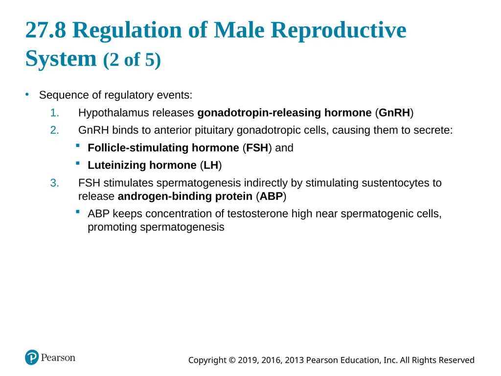
27.8 Regulation of Male Reproductive System (2 of 5) Sequence of regulatory events:
Hypothalamus releases gonadotropin-releasing hormone (GnRH)
GnRH binds to anterior pituitary gonadotropic cells, causing them to secrete:
Follicle-stimulating hormone (FSH) and
Luteinizing hormone (LH)
FSH stimulates spermatogenesis indirectly by stimulating sustentocytes to release androgen-binding protein (ABP)
ABP keeps concentration of testosterone high near spermatogenic cells, promoting spermatogenesis<br>
Hypothalamus releases gonadotropin-releasing hormone (GnRH)
GnRH binds to anterior pituitary gonadotropic cells, causing them to secrete:
Follicle-stimulating hormone (FSH) and
Luteinizing hormone (LH)
FSH stimulates spermatogenesis indirectly by stimulating sustentocytes to release androgen-binding protein (ABP)
ABP keeps concentration of testosterone high near spermatogenic cells, promoting spermatogenesis<br>
102
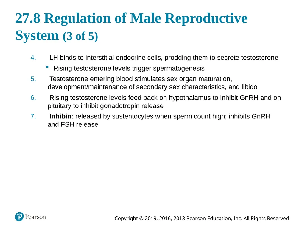
27.8 Regulation of Male Reproductive System (3 of 5) LH binds to interstitial endocrine cells, prodding them to secrete testosterone
Rising testosterone levels trigger spermatogenesis
Testosterone entering blood stimulates sex organ maturation, development/maintenance of secondary sex characteristics, and libido
Rising testosterone levels feed back on hypothalamus to inhibit GnRH and on pituitary to inhibit gonadotropin release
Inhibin: released by sustentocytes when sperm count high; inhibits GnRH and FSH release<br>
Rising testosterone levels trigger spermatogenesis
Testosterone entering blood stimulates sex organ maturation, development/maintenance of secondary sex characteristics, and libido
Rising testosterone levels feed back on hypothalamus to inhibit GnRH and on pituitary to inhibit gonadotropin release
Inhibin: released by sustentocytes when sperm count high; inhibits GnRH and FSH release<br>
103
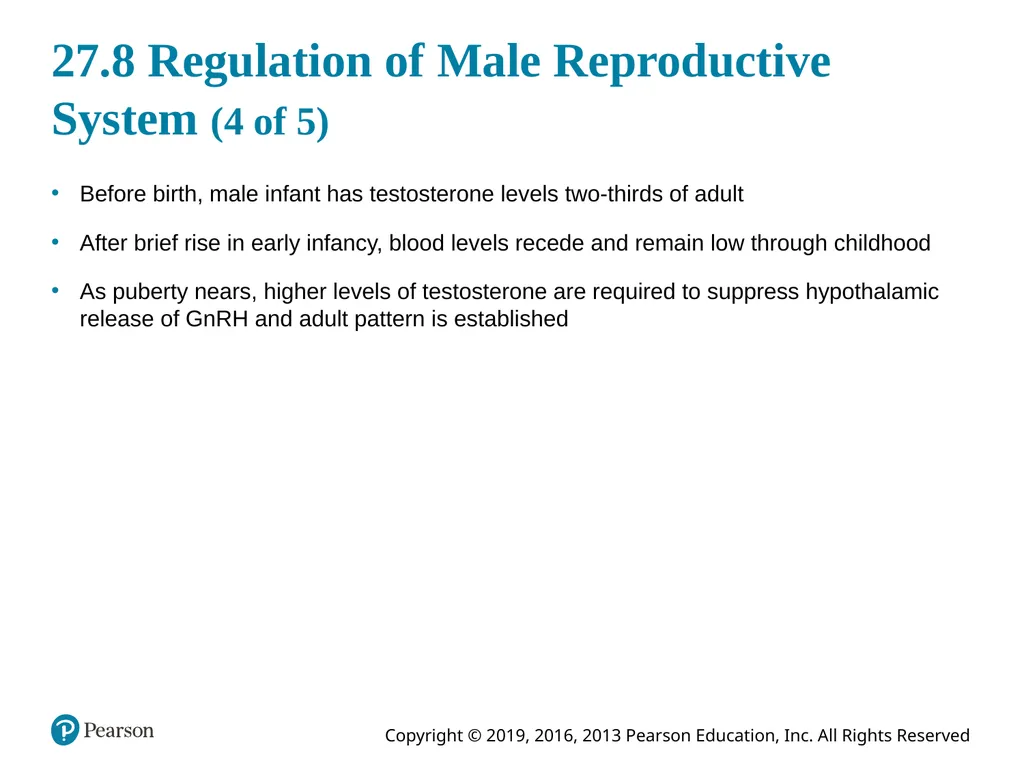
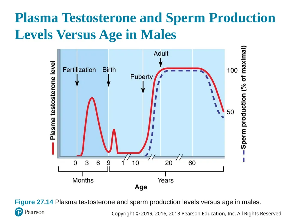
27.8 Regulation of Male Reproductive System (4 of 5) Before birth, male infant has testosterone levels two-thirds of adult
After brief rise in early infancy, blood levels recede and remain low through childhood
As puberty nears, higher levels of testosterone are required to suppress hypothalamic release of GnRH and adult pattern is established<br>
After brief rise in early infancy, blood levels recede and remain low through childhood
As puberty nears, higher levels of testosterone are required to suppress hypothalamic release of GnRH and adult pattern is established<br>
104
Plasma Testosterone and Sperm Production
Levels Versus Age in Males Figure 27.14 Plasma testosterone and sperm production levels versus age in males.<br>
105
27.8 Regulation of Male Reproductive System (5 of 5) Amount of testosterone and sperm produced by testes reflects balance among interacting hormones of HPG axis.
Balance takes 3 years to achieve, after which testosterone and sperm production are fairly stable throughout life
Without GnRH and gonadotropins, testes atrophy, and sperm and testosterone production ceases<br>
Balance takes 3 years to achieve, after which testosterone and sperm production are fairly stable throughout life
Without GnRH and gonadotropins, testes atrophy, and sperm and testosterone production ceases<br>
106
Mechanism and Effects of Testosterone Activity (1 of 2) Testosterone, synthesized from cholesterol, is transformed at some target cells
Converted to dihydrotestosterone (DHT) in prostate and estradiol in some brain neurons
Prompts spermatogenesis and targets all accessory organs
Has multiple anabolic effects throughout body
Deficiency leads to atrophy of accessory organs, semen volume declines, and erection/ejaculation are impaired; treatment: testosterone replacement<br>
Converted to dihydrotestosterone (DHT) in prostate and estradiol in some brain neurons
Prompts spermatogenesis and targets all accessory organs
Has multiple anabolic effects throughout body
Deficiency leads to atrophy of accessory organs, semen volume declines, and erection/ejaculation are impaired; treatment: testosterone replacement<br>
107
Mechanism and Effects of Testosterone Activity (2 of 2) Male secondary sex characteristics: features induced in nonreproductive organs by male sex hormones (mainly testosterone)
Appearance of pubic, axillary, and facial hair
Enhanced growth of hair on chest or other areas
Larynx enlargements causes deepening of voice
Skin thickens and becomes oily
Bones grow, increase in density
Skeletal muscles increase in size and mass
Boosts basal metabolic rate
Basis of sex drive (libido) in males<br>
Appearance of pubic, axillary, and facial hair
Enhanced growth of hair on chest or other areas
Larynx enlargements causes deepening of voice
Skin thickens and becomes oily
Bones grow, increase in density
Skeletal muscles increase in size and mass
Boosts basal metabolic rate
Basis of sex drive (libido) in males<br>
108
Copyright<br>