
Paramedic Care: Principles & Practice Sixth
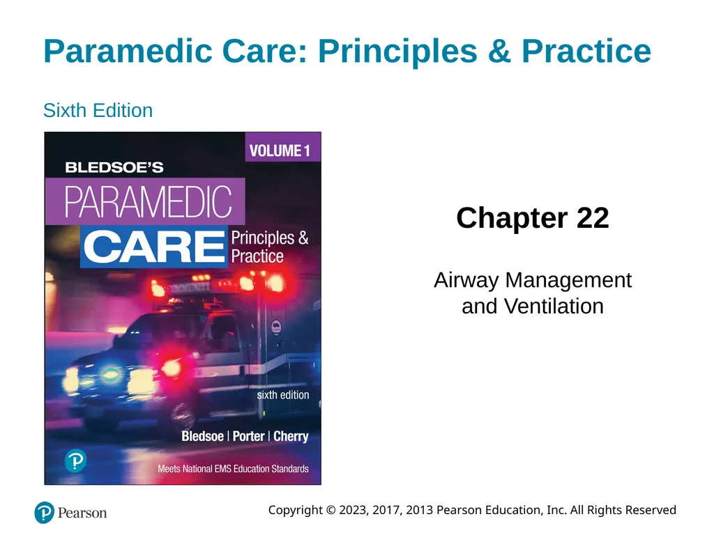


Description: Paramedic Care: Principles Practice Sixth Edition Chapter 22 Airway Management and Ventilation Copyright 2023, 2017, 2013 Pearson Education, Inc. All Rights Reserved Standard Airway Management, Respiration, and Artificial Ventilation
Related Topics
Download Presentation
"Paramedic Care: Principles & Practice Sixth" is the property of its rightful owner. Permission is granted to download and print the materials on this website for personal, non-commercial use only, and to display it on your personal computer provided you do not modify the materials and that you retain all copyright notices contained in the materials. By downloading content from our website, you accept the terms of this agreement.
Presentation Transcript
slide1. Paramedic Care: Principles & Practice Sixth Edition Chapter 22 Airway Management and Ventilation Copyright © 2023, 2017, 2013 Pearson Education, Inc. All Rights Reserved<br>
slide2. Standard Airway Management, Respiration, and Artificial Ventilation<br>
slide3. Competency Integrates comprehensive knowledge of anatomy, physiology, and pathophysiology into the assessment to develop and implement a treatment plan with the goal of ensuring a patent airway, adequate mechanical ventilation, and respiration for patients of all ages.<br>
slide4. Introduction (1 of 3) Airway management and ventilation are the first and most critical steps during the primary assessment of every patient
Establish and maintain airway patency while providing adequate oxygen
Without adequate airway maintenance and ventilation, patient will succumb to brain injury or death in as little as 4 minutes<br>
slide5. Introduction (2 of 3) The primary assessment typically follows the A B C ((airway, breathing, circulation) approach to the patient
If however, the patient is believed to be in cardiac arrest, then the sequence becomes C A B (compressions, airway, breathing)
Airway and ventilation often assessed and managed together<br>
slide6. Introduction (3 of 3) Airway and ventilation problems are at times, approached in a nonlinear fashion
Simple, basic airway skills are key to successful airway management
The inability to properly assess and manage an airway will doom all other treatments to failure<br>
slide7. Part 1: Respiratory Anatomy, Physiology, and Assessment<br>
slide8. Respiratory Anatomy, Physiology, and Assessment (1 of 39) Anatomy of the Respiratory System
Provides passage for oxygen and carbon dioxide
Movement of gases from atmosphere to body cells is called ventilation
Respiration is the gas exchange that occurs at the cellular level<br>
slide9. Respiratory Anatomy, Physiology, and Assessment (2 of 39) Anatomy of the Respiratory System (continued)
Upper Airway Anatomy
Upper airway:
from mouth and nose to larynx
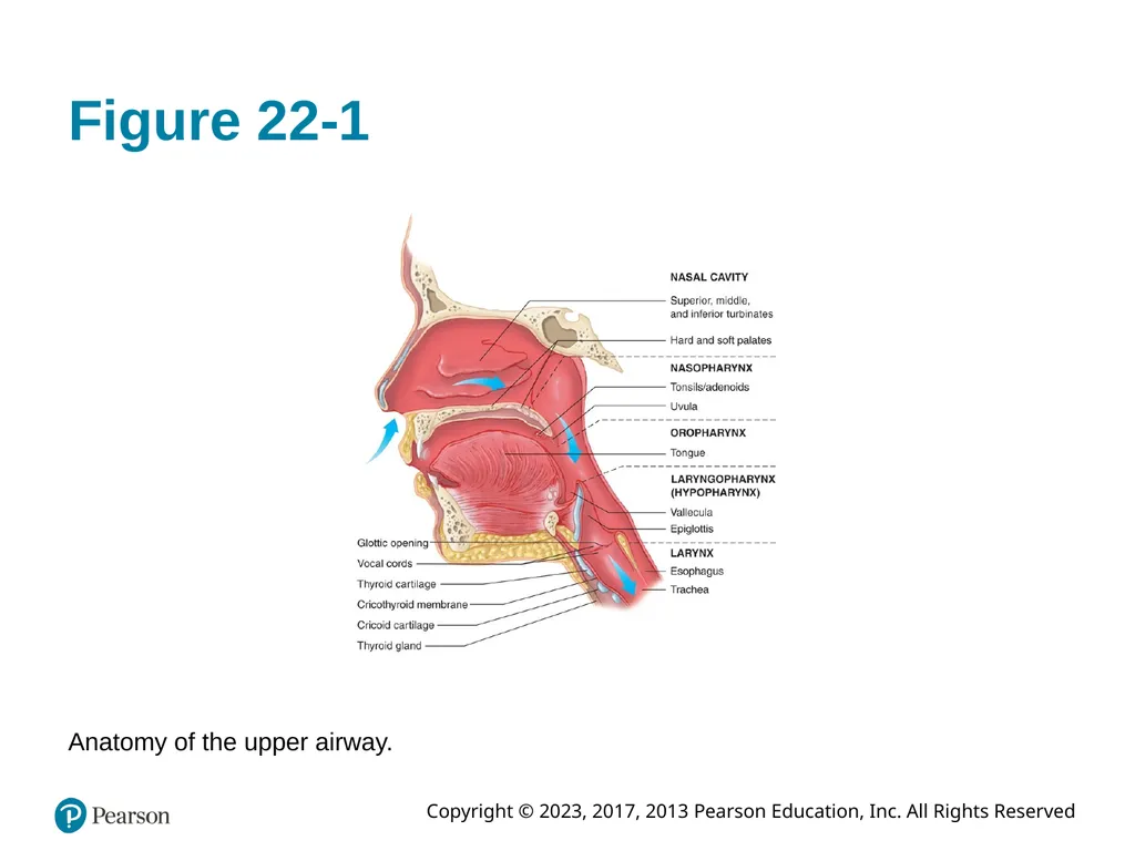
Includes nasal cavity, oral cavity, pharynx
Larynx joins upper and lower airways<br>
slide10. Figure 22-1 Anatomy of the upper airway.<br>
slide11. Respiratory Anatomy, Physiology, and Assessment (3 of 39) Anatomy of the Respiratory System (continued)
Upper Airway Anatomy (continued)
Nasal cavity
Oral cavity
U-shaped hyoid bone
Pharynx
Nasopharynx, Oropharynx, Hypopharynx
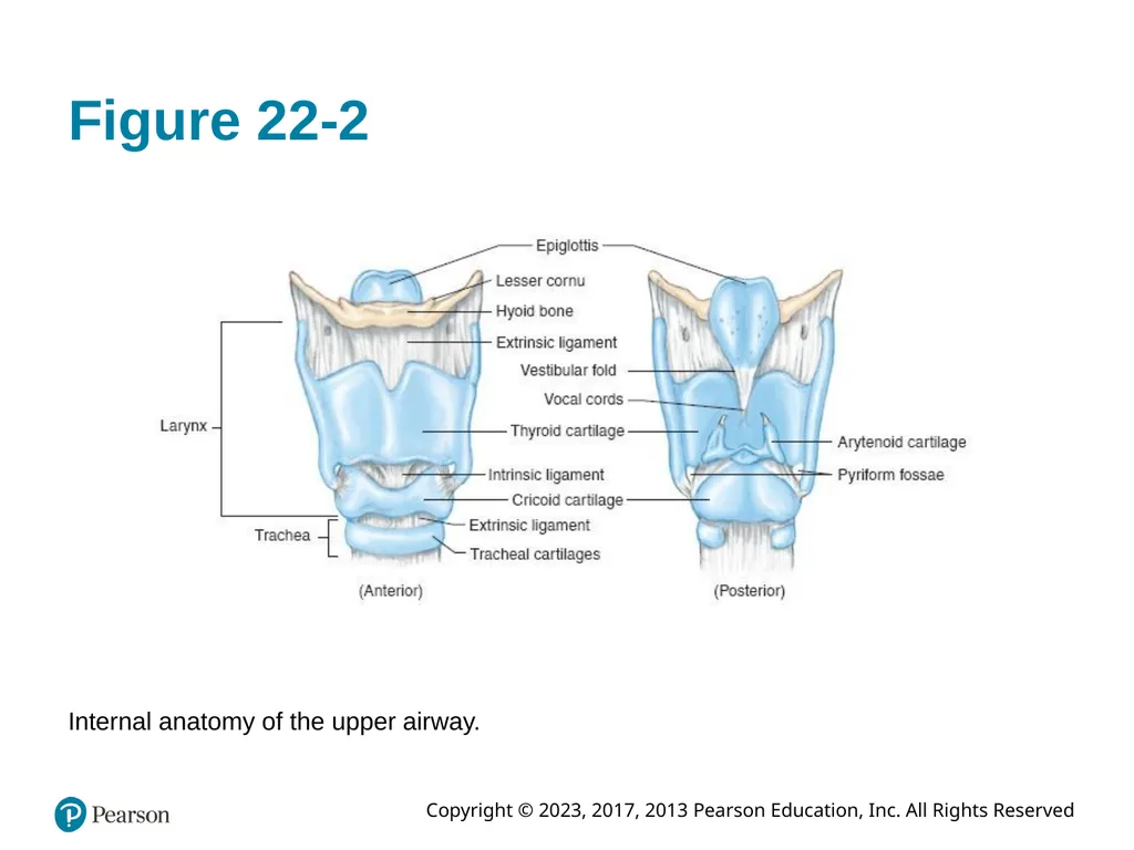
Larynx
Glottic opening (beneath epiglottis), thyroid and cricoid cartilages. Houses vocal cords<br>
slide12. Figure 22-2 Internal anatomy of the upper airway.<br>
slide13. Respiratory Anatomy, Physiology, and Assessment (4 of 39) Anatomy of the Respiratory System (continued)
Lower Airway Anatomy
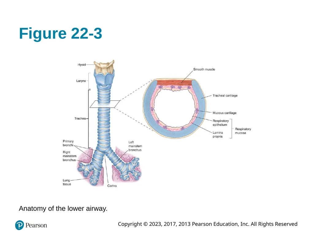
Trachea
Tube that connects larynx to two mainstem bronchi
Trachea divides into right and left mainstem bronchi<br>
slide14. Figure 22-3 Anatomy of the lower airway.<br>
slide15. Respiratory Anatomy, Physiology, and Assessment (5 of 39) Anatomy of the Respiratory System (continued)
Lower Airway Anatomy (continued)
Bronchi
Trachea bifurcates at carina into two mainstem bronchi
Right mainstem often site of aspirated foreign bodies
Bronchi (supported by cartilage) gives rise to bronchiole structures supported by smooth muscle
After about 22 divisions, bronchioles give rise to respiratory bronchioles that have limited capacity for gas exchange<br>
slide16. Respiratory Anatomy, Physiology, and Assessment (6 of 39) Anatomy of the Respiratory System (continued)
Lower Airway Anatomy (continued)
Alveoli
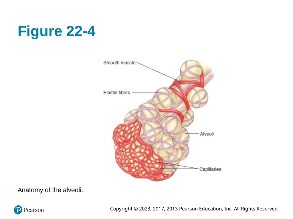
Respiratory bronchioles divide into alveolar ducts
Alveolar ducts terminate into alveoli sacs
Individual alveoli are sites of primary gas exchange
Surfactant layer to prevent atelectasis<br>
slide17. Figure 22-4 Anatomy of the alveoli.<br>
slide18. Respiratory Anatomy, Physiology, and Assessment (7 of 39) Anatomy of the Respiratory System (continued)
Lower Airway Anatomy (continued)
Lung Parenchyma
Two pulmonary lobules; anatomic division of lungs. Further divided into lobes
Pleura:
Membranous connective tissue that covers lungs
Parietal pleura
Visceral pleura<br>
slide19. Respiratory Anatomy, Physiology, and Assessment (8 of 39) Anatomy of the Respiratory System (continued)
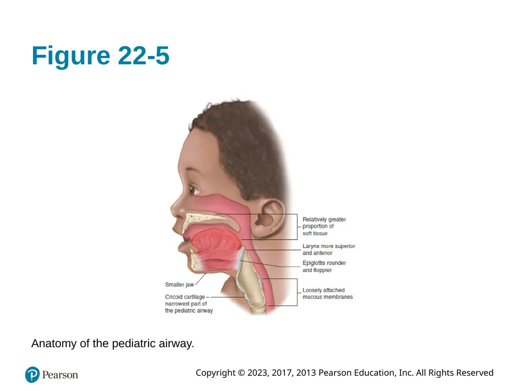
Pediatric Airway
Pediatric airway smaller in all aspects; softer and more fragile
Epiglottis for floppier and rounder
Larynx more anterior and funnel shaped
Ribs and cartilage of pediatric thoracic cage softer and more pliable
Primarily diaphragmatic breathers<br>
slide20. Figure 22-5 Anatomy of the pediatric airway.<br>
slide21. Respiratory Anatomy, Physiology, and Assessment (9 of 39) Physiology of the Respiratory System
Respiration and Ventilation
Pulmonary respiration occurs in lungs
Cellular respiration occurs in peripheral capillaries
Ventilation: mechanical process that moves air into and out of lungs; necessary for respiration to occur
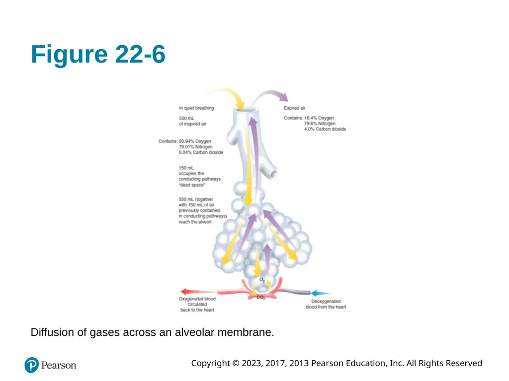
Gases exchanged between red blood cells and alveoli through capillary membranes<br>
slide22. Figure 22-6 Diffusion of gases across an alveolar membrane.<br>
slide23. Respiratory Anatomy, Physiology, and Assessment (10 of 39) Physiology of the Respiratory System (continued)
Respiration and Ventilation (continued)
Respiratory Cycle
Normal expiration: passive process
Inspiration: active process, using energy
Musculature to change intrathoracic size
Diaphragm, intercostal, accessory muscles<br>
slide24. Respiratory Anatomy, Physiology, and Assessment (11 of 39) Physiology of the Respiratory System (continued)
Respiration and Ventilation (continued)
Respiratory Cycle (continued)
Pulmonary ventilation: depends on changes in pressure within thoracic cavity
Respiratory cycle: coordinated interaction among respiratory system, central nervous system, musculoskeletal system<br>
slide25. Respiratory Anatomy, Physiology, and Assessment (12 of 39) Physiology of the Respiratory System (continued)
Respiration and Ventilation (continued)
Pulmonary Circulation
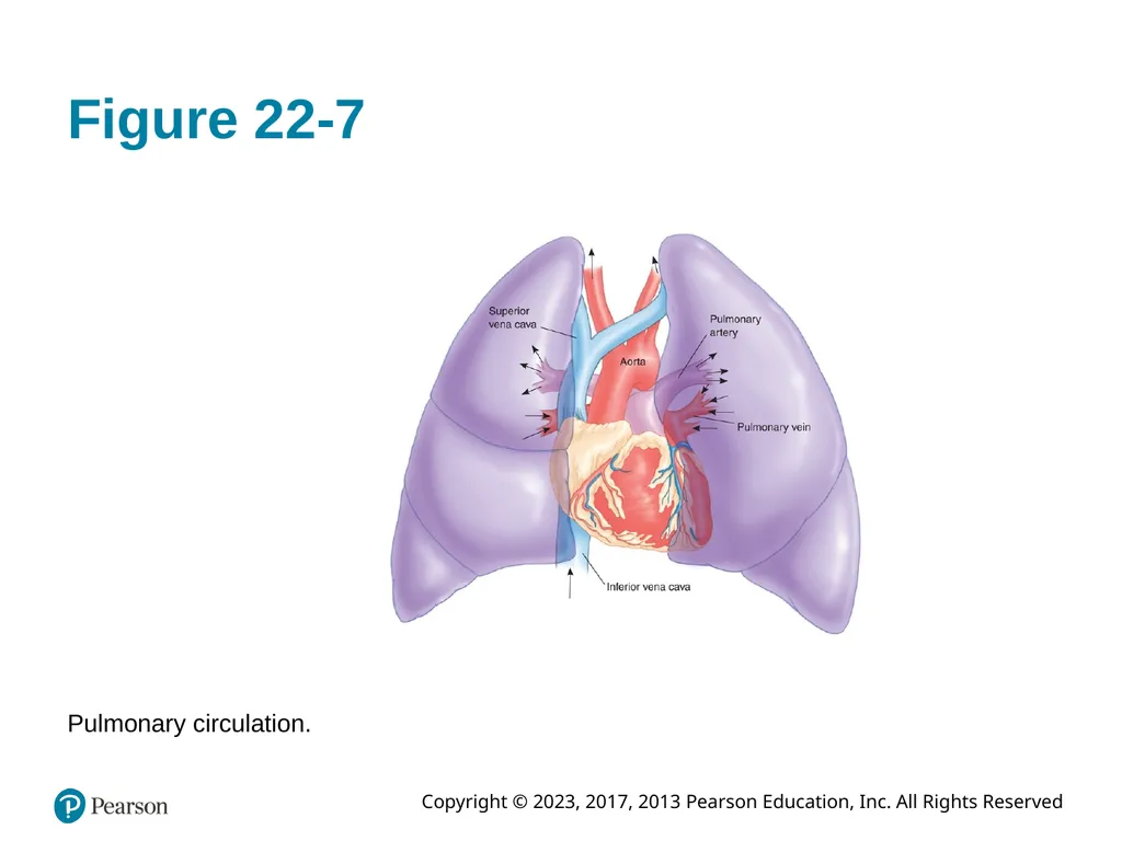
Respiration requires intact circulatory system
Heart pumps as much blood to lungs as it pumps to peripheral tissues
Right side of heart pumps to lungs via pulmonary artery to lungs for oxygenation and offloading of carbon dioxide waste from tissues<br>
slide26. Figure 22-7 Pulmonary circulation.<br>
slide27. Respiratory Anatomy, Physiology, and Assessment (13 of 39) Measurement of Oxygen and Carbon Dioxide Levels
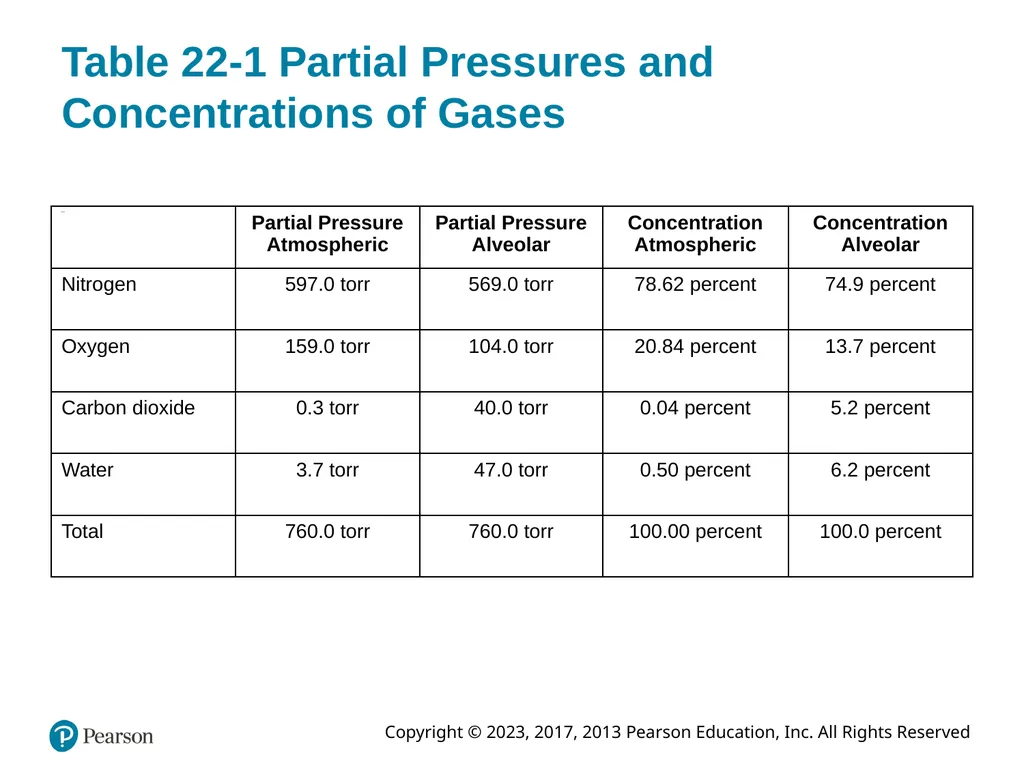
Partial pressure: pressure exerted by each component of gas mixture
Major respiratory gases Nitrogen Oxygen Carbon dioxide Water<br>
slide28. Table 22-1 Partial Pressures and Concentrations of Gases<br>
slide29. Respiratory Anatomy, Physiology, and Assessment (14 of 39) Measurement of Oxygen and Carbon Dioxide Levels (continued)
Diffusion
Movement of gas from area of higher concentration to area of lower concentration
Transfers gases between lungs and blood; blood and peripheral tissues<br>
slide30. Respiratory Anatomy, Physiology, and Assessment (15 of 39) Measurement of Oxygen and Carbon Dioxide Levels (continued)
Oxygen Concentrations in the Blood
Oxygen diffuses into blood plasma; combines with hemoglobin (97% of oxygen)
Remainder dissolved in the plasma (3%)
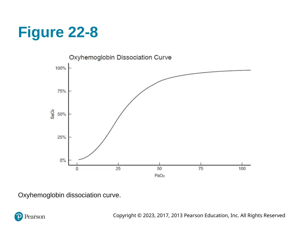
The characteristics of oxygen binding to hemoglobin graphically represented by oxy-hemoglobin dissociation curve<br>
slide31. Figure 22-8 Oxyhemoglobin dissociation curve.<br>
slide32. Respiratory Anatomy, Physiology, and Assessment (16 of 39) Measurement of Oxygen and Carbon Dioxide Levels (continued)
Oxygen Concentrations in the Blood (continued)
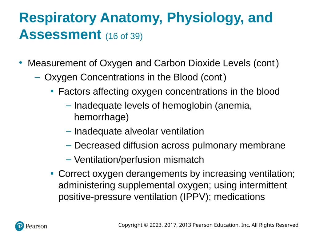
Factors affecting oxygen concentrations in the blood
Inadequate levels of hemoglobin (anemia, hemorrhage)
Inadequate alveolar ventilation
Decreased diffusion across pulmonary membrane
Ventilation/perfusion mismatch
Correct oxygen derangements by increasing ventilation; administering supplemental oxygen; using intermittent positive-pressure ventilation (I P P V); medications<br>
slide33. Respiratory Anatomy, Physiology, and Assessment (17 of 39) Measurement of Oxygen and Carbon Dioxide Levels (continued)
Carbon Dioxide Concentrations in the Blood
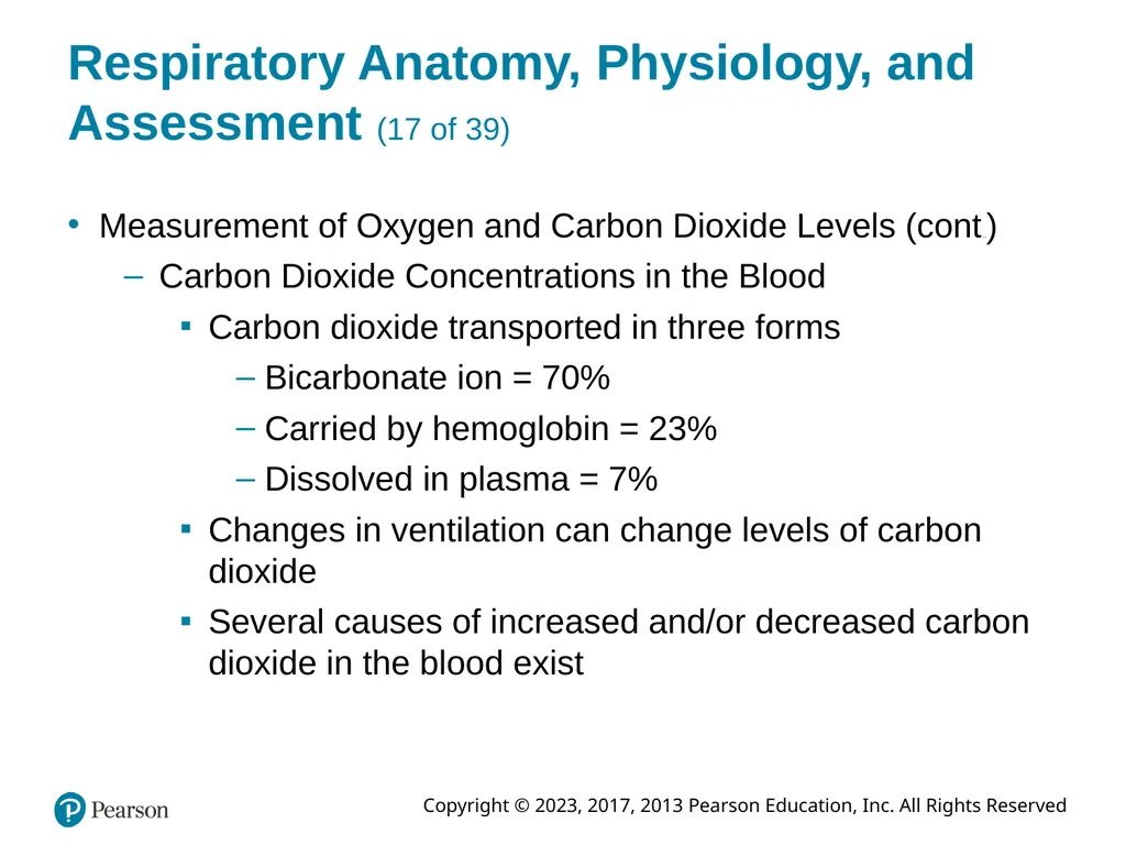
Carbon dioxide transported in three forms
Bicarbonate ion = 70%
Carried by hemoglobin = 23%
Dissolved in plasma = 7%
Changes in ventilation can change levels of carbon dioxide
Several causes of increased and/or decreased carbon dioxide in the blood exist<br>
slide34. Respiratory Anatomy, Physiology, and Assessment (18 of 39) Regulation of Respiration
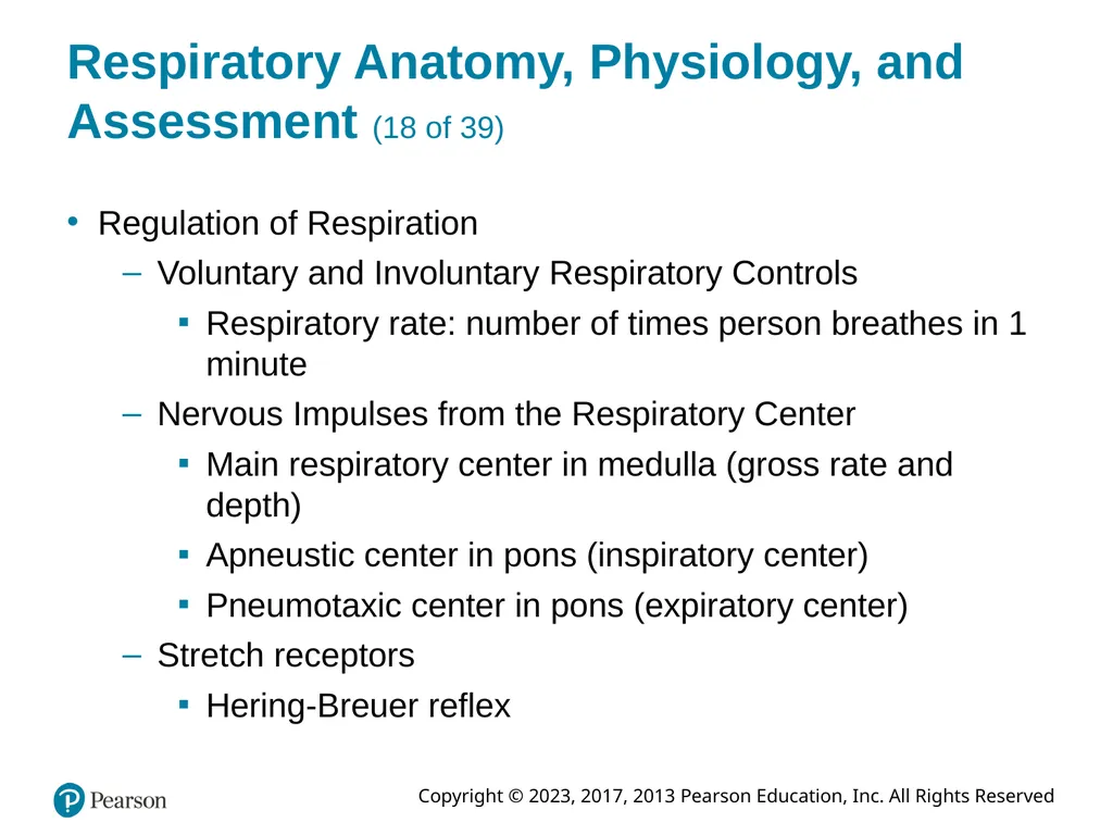
Voluntary and Involuntary Respiratory Controls
Respiratory rate: number of times person breathes in 1 minute
Nervous Impulses from the Respiratory Center
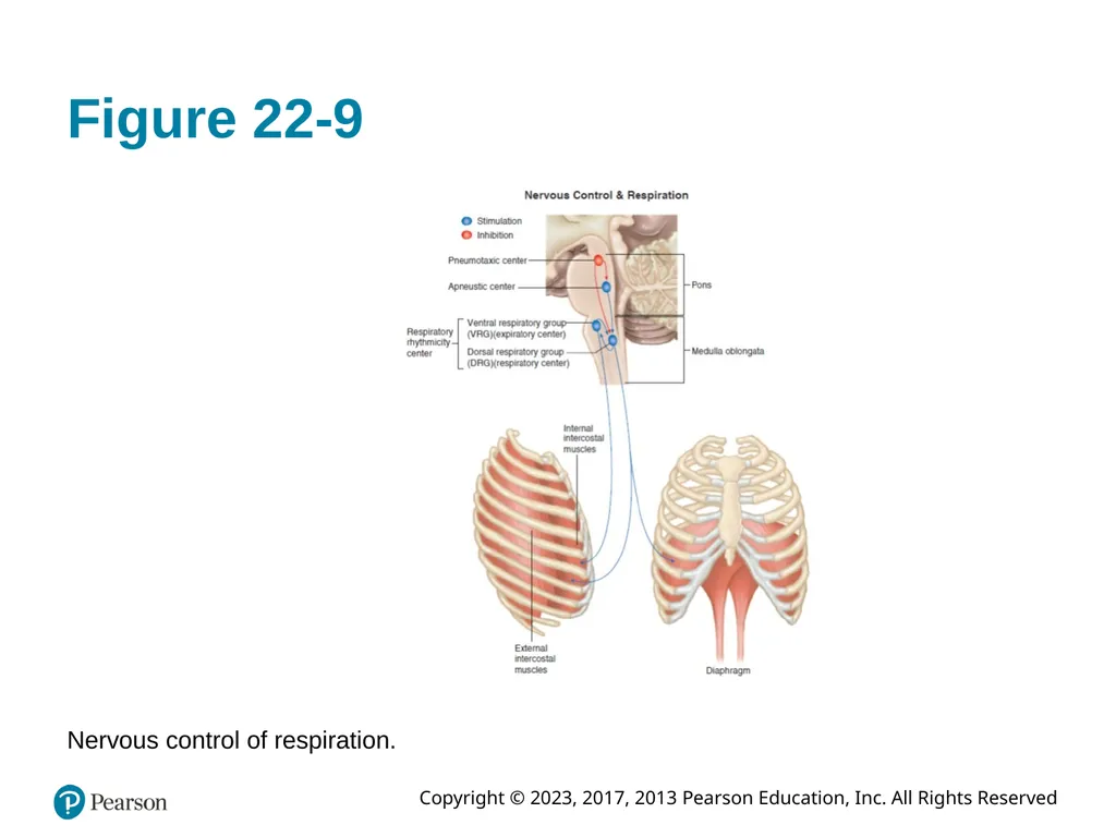
Main respiratory center in medulla (gross rate and depth)
Apneustic center in pons (inspiratory center)
Pneumotaxic center in pons (expiratory center)
Stretch receptors
Hering-Breuer reflex<br>
slide35. Respiratory Anatomy, Physiology, and Assessment (19 of 39) Regulation of Respiration (continued)
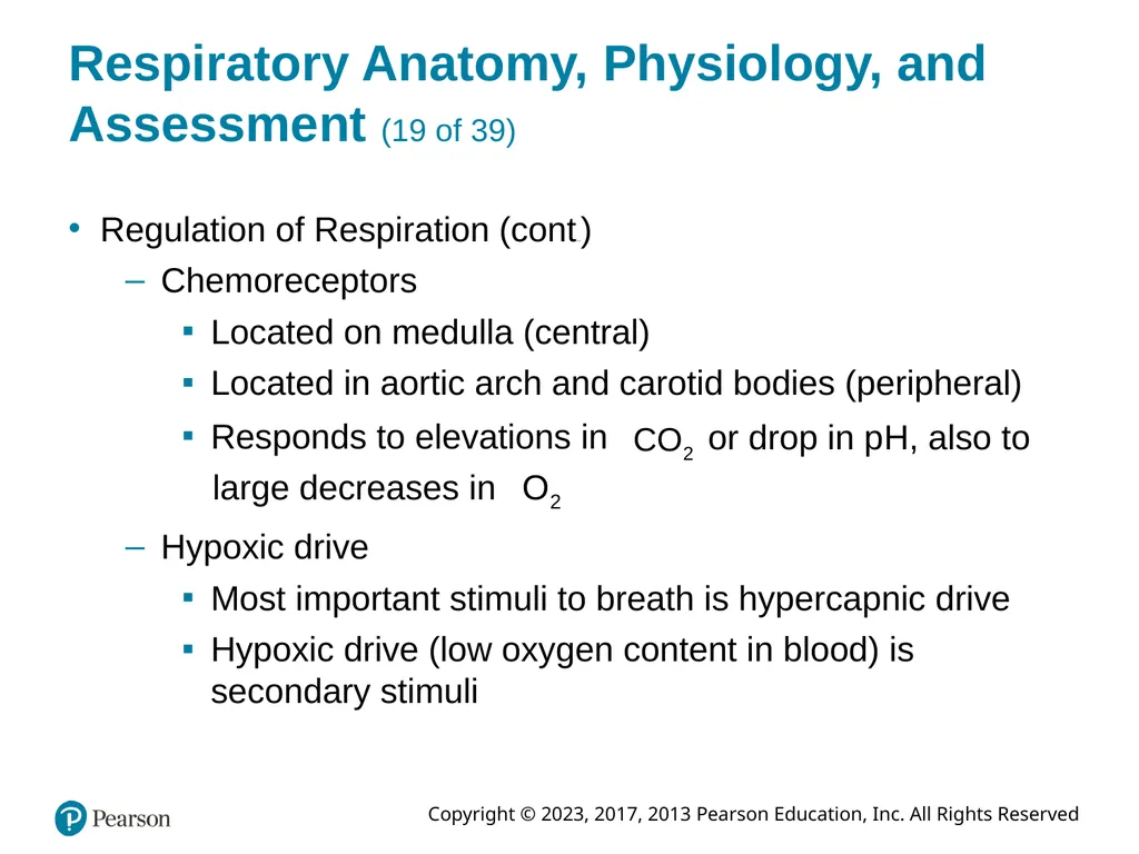
Chemoreceptors
Located on medulla (central)
Located in aortic arch and carotid bodies (peripheral) Responds to elevations in or drop in p H, also to large decreases in Hypoxic drive
Most important stimuli to breath is hypercapnic drive
Hypoxic drive (low oxygen content in blood) is secondary stimuli<br>
slide36. Figure 22-9 Nervous control of respiration.<br>
slide37. Respiratory Anatomy, Physiology, and Assessment (20 of 39) Regulation of Respiration (continued)
Measure of Respiratory Function
Factors that affect respiratory rate:
Fever
Emotion
Pain
Hypoxia
Acidosis
Stimulant or depressant drugs
Sleep<br>
slide38. Respiratory Anatomy, Physiology, and Assessment (21 of 39) Regulation of Respiration (continued)
Measure of Respiratory Function (continued)
Respiratory capacities and measurements:
Total lung capacity (T L C) Tidal volume Dead space volume Alveolar volume Minute volume Alveolar minute volume<br>
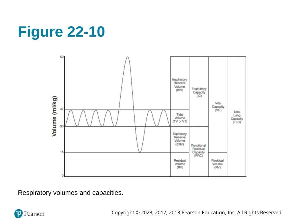
slide39. Respiratory Anatomy, Physiology, and Assessment (22 of 39) Regulation of Respiration (continued)
Measure of Respiratory Function (continued)
Respiratory capacities and measurements: (continued)
Inspiratory reserve volume (I R V)
Expiratory reserve volume (E R V)
Residual volume (R V)
Functional residual capacity (F R C)
Forced expiratory volume (F E V)<br>
slide40. Figure 22-10 Respiratory volumes and capacities.<br>
slide41. Respiratory Anatomy, Physiology, and Assessment (23 of 39) Respiratory Problems
Airway Obstruction
Blockage of airway immediate threat to patient's life; true emergency
Causes of airway obstruction:
Tongue
Foreign bodies
Trauma
Laryngeal spasm and edema
Aspiration; vomitus or blood<br>
slide42. Respiratory Anatomy, Physiology, and Assessment (24 of 39) Respiratory Problems (continued)
Airway Obstruction (continued)
Airway obstruction partial or complete
Tongue most common cause of airway obstruction<br>
slide43. Respiratory Anatomy, Physiology, and Assessment (25 of 39) Respiratory Problems (continued)
Inadequate Ventilation
Reduction of rate or volume of inhalation leads to reduction in minute volume
If there are no alveolar breath sounds when auscultating, then there is no alveolar ventilation This means no gas exchange!<br>
slide44. Respiratory Anatomy, Physiology, and Assessment (26 of 39) Respiratory System Assessment
Primary Assessment
Vigilance key to airway management in every patient
Primary assessment: identify any immediate threats to patient's life; airway, breathing, circulation (A B C s)
For patients in cardiac arrest, compressions come before airway and breathing (C A B)<br>
slide45. Respiratory Anatomy, Physiology, and Assessment (27 of 39) Respiratory System Assessment (continued)
Primary Assessment (continued)
Patients with altered mental status warrant further evaluation
Assess airway to ensure it is patent
Look and listen for air exchange through mouth/nose
If airway partially or totally occluded, establish patency immediately with progressive airway interventions
Determine adequacy of breathing after airway assured
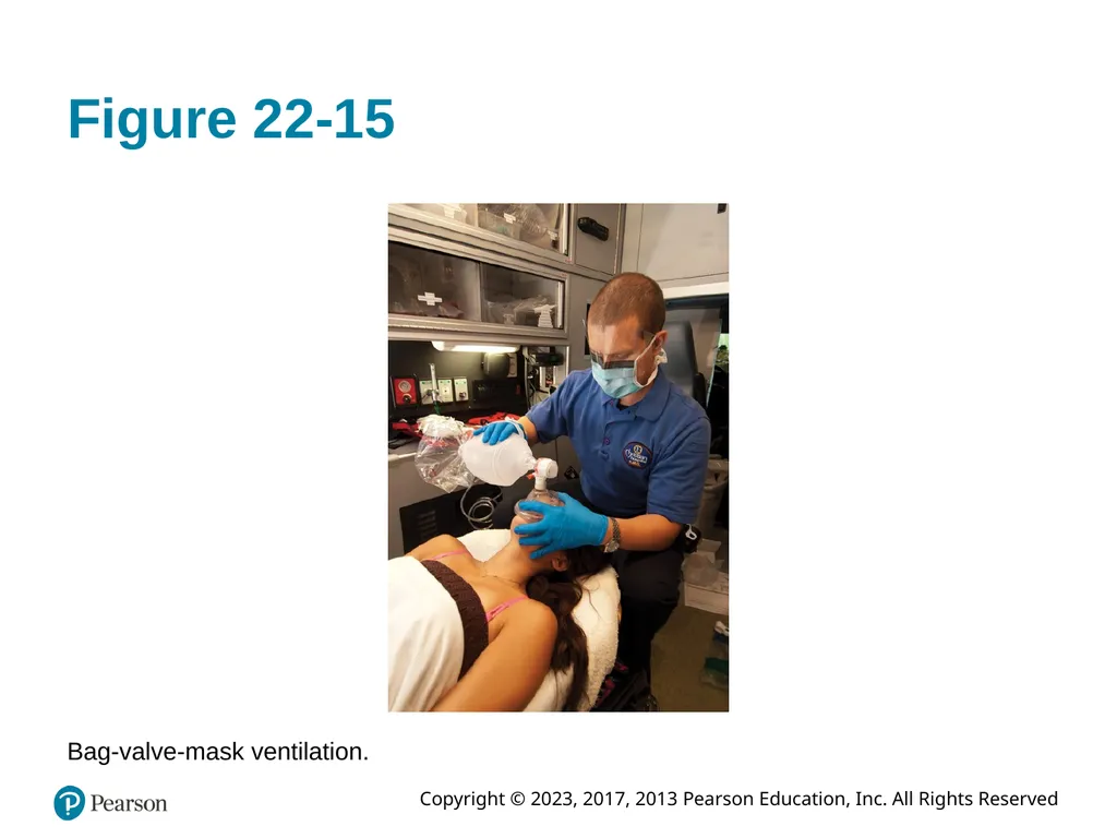
If breathing inadequately, establish good ventilations immediately with B V M<br>
slide46. Figure 22-15 Bag-valve-mask ventilation.<br>
slide47. Respiratory Anatomy, Physiology, and Assessment (28 of 39) Respiratory System Assessment (continued)
Primary Assessment (continued)
With airway and breathing assured or being supported, move on with assessment
Assess circulatory status
Check peripheral pulses, if none, check central pulses
Initiate C P R as needed
Stop any acute external hemorrhaging
Evaluate skin findings
Make priority determination of patient’s status<br>
slide48. Respiratory Anatomy, Physiology, and Assessment (29 of 39) Secondary Assessment
Complete after primary assessment and correction of any immediate life threats
Determine History
Recent history leading to onset of symptoms; identify possible triggers
Past medical history
Begin physical examination of patient<br>
slide49. Respiratory Anatomy, Physiology, and Assessment (30 of 39) Secondary Assessment (continued)
Physical Examination
Physical examination techniques
Inspection
Auscultation
Palpation
Percussion
Begin physical assessment by inspecting patient<br>
slide50. Respiratory Anatomy, Physiology, and Assessment (31 of 39) Secondary Assessment (continued)
Noninvasive Respiratory Monitoring
Pulse oximetry: "fifth vital sign"
Noninvasive measure of hemoglobin oxygen saturation in peripheral tissues
Pulse C O Oximetry
Monitors for abnormal hemoglobin states
Carboxyhemoglobin
Methemoglobin
Total hemoglobin<br>
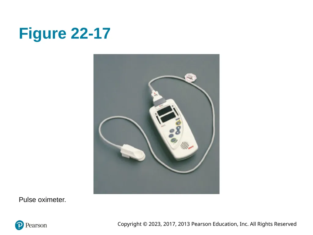
slide51. Figure 22-17 Pulse oximeter.<br>
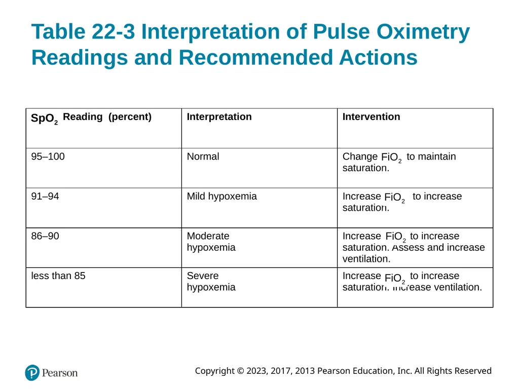
slide52. Table 22-3 Interpretation of Pulse Oximetry Readings and Recommended Actions<br>
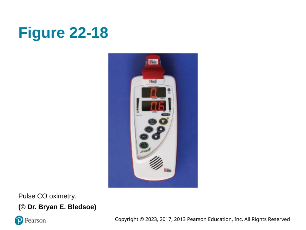
slide53. Figure 22-18 Pulse CO oximetry.
(© Dr. Bryan E. Bledsoe)<br>
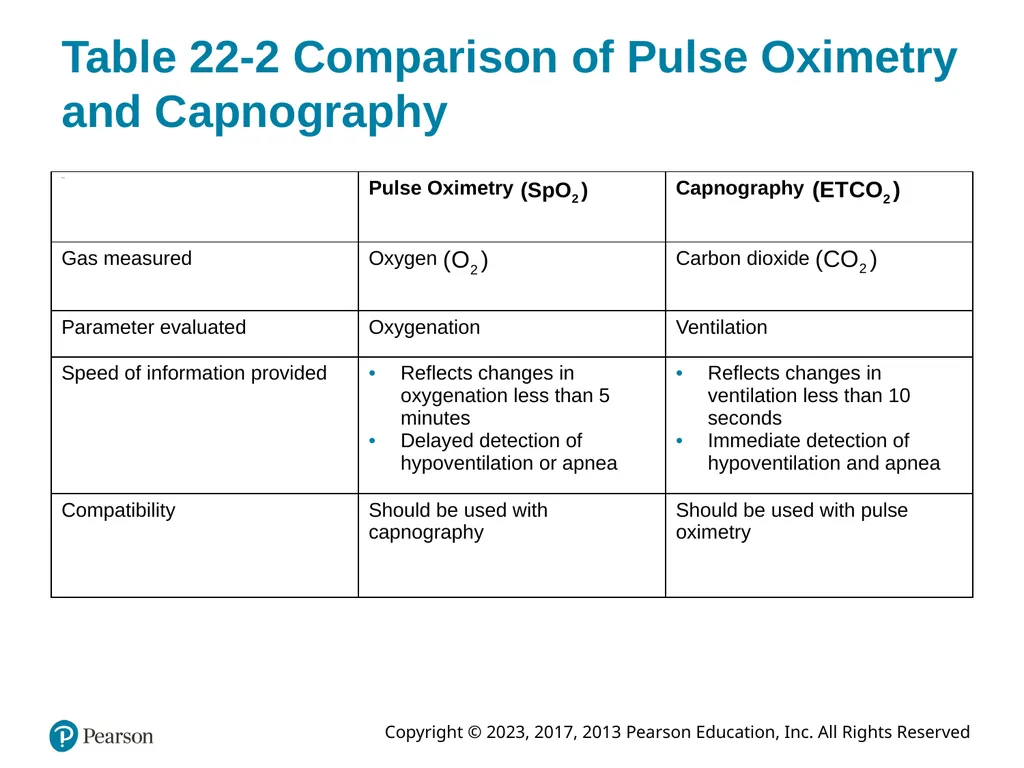
slide54. Table 22-2 Comparison of Pulse Oximetry and Capnography<br>
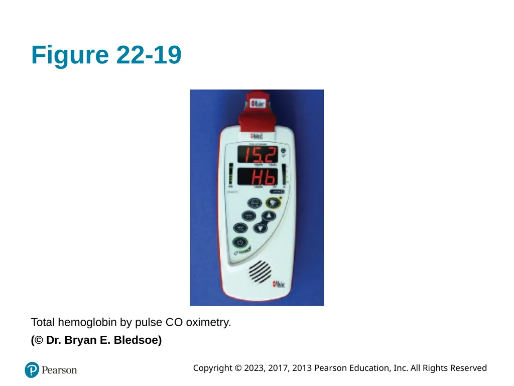
slide55. Figure 22-19 Total hemoglobin by pulse C O oximetry.
(© Dr. Bryan E. Bledsoe)<br>
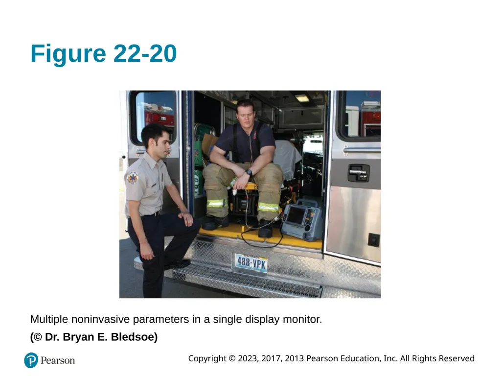
slide56. Figure 22-20 Multiple noninvasive parameters in a single display monitor.
(© Dr. Bryan E. Bledsoe)<br>
slide57. Respiratory Anatomy, Physiology, and Assessment (32 of 39) Secondary Assessment (continued)
Noninvasive Respiratory Monitoring (continued)
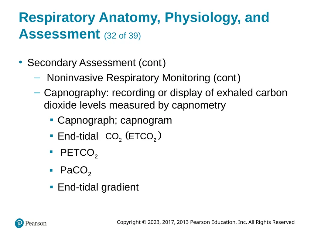
Capnography: recording or display of exhaled carbon dioxide levels measured by capnometry
Capnograph; capnogram End-tidal End-tidal gradient<br>
slide58. Respiratory Anatomy, Physiology, and Assessment (33 of 39) Secondary Assessment (continued)
Noninvasive Respiratory Monitoring (continued)
Capnography
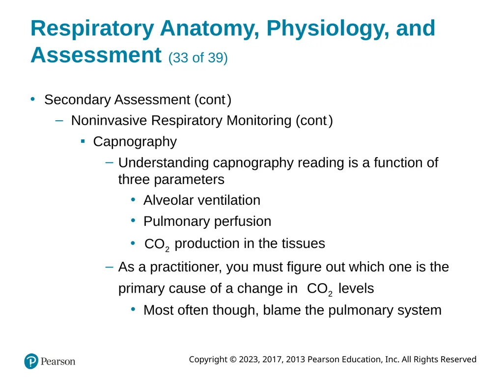
Understanding capnography reading is a function of three parameters
Alveolar ventilation
Pulmonary perfusion production in the tissues As a practitioner, you must figure out which one is the primary cause of a change in levels Most often though, blame the pulmonary system<br>
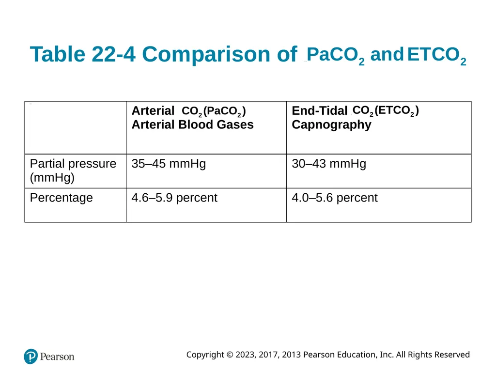
slide59. Table 22-4 Comparison of P a C O sub 2 and E T C O sub 2<br>
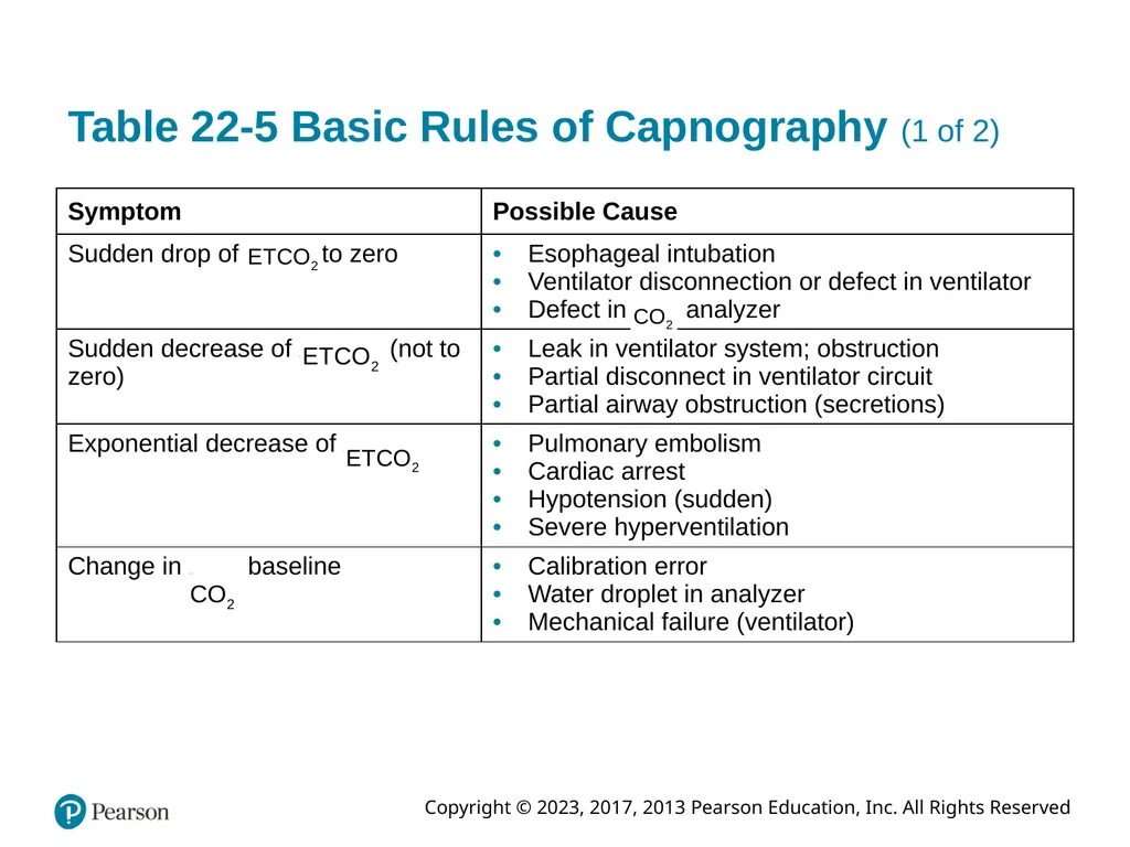
slide60. Table 22-5 Basic Rules of Capnography (1 of 2)<br>
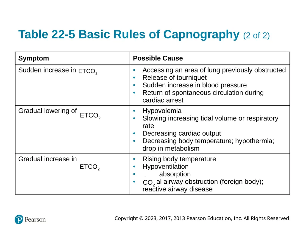
slide61. Table 22-5 Basic Rules of Capnography (2 of 2)<br>
slide62. Respiratory Anatomy, Physiology, and Assessment (34 of 39) Secondary Assessment (continued)
Noninvasive Respiratory Monitoring (continued)
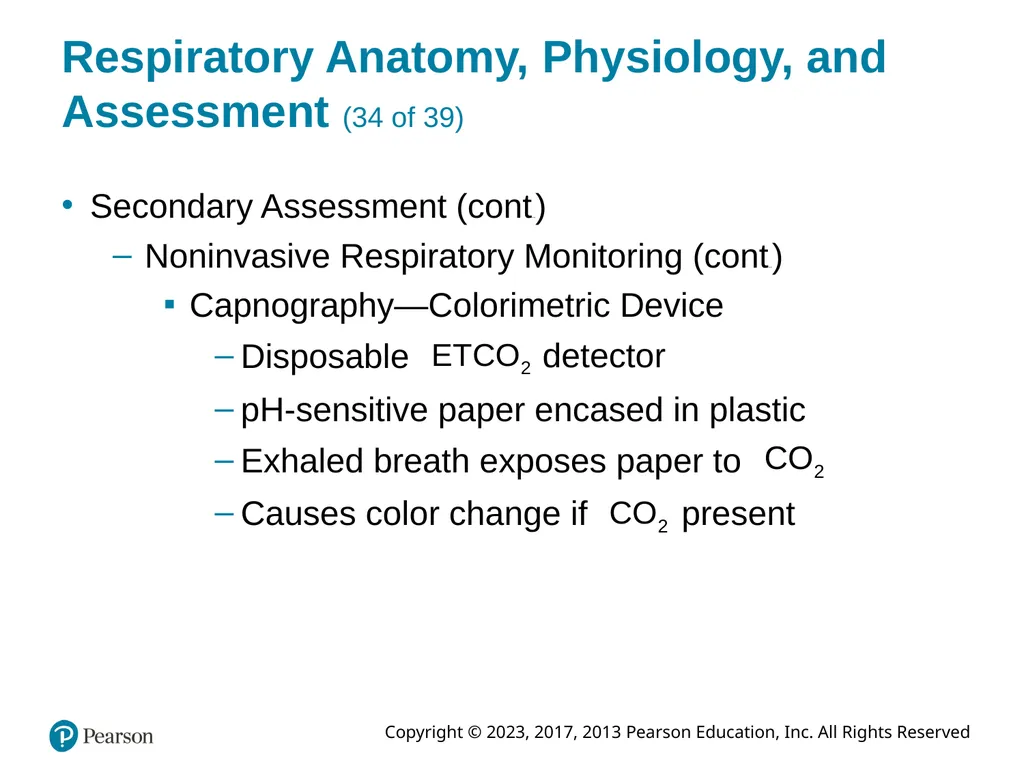
Capnography—Colorimetric Device Disposable detector p H-sensitive paper encased in plastic Exhaled breath exposes paper to Causes color change if present<br>
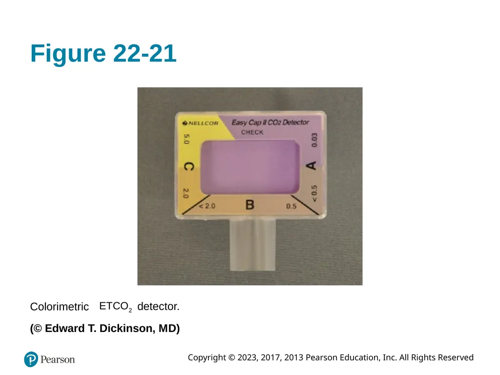
slide63. Figure 22-21 Colorimetric detector. (© Edward T. Dickinson, M D)<br>
slide64. Respiratory Anatomy, Physiology, and Assessment (35 of 39) Secondary Assessment (continued)
Noninvasive Respiratory Monitoring (continued)
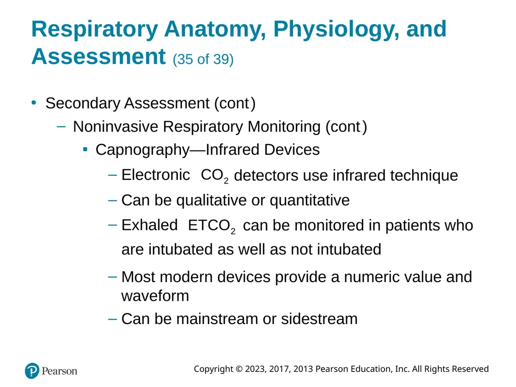
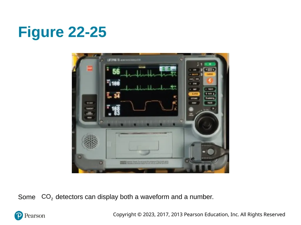
Capnography—Infrared Devices Electronic detectors use infrared technique Can be qualitative or quantitative Exhaled can be monitored in patients who are intubated as well as not intubated Most modern devices provide a numeric value and waveform
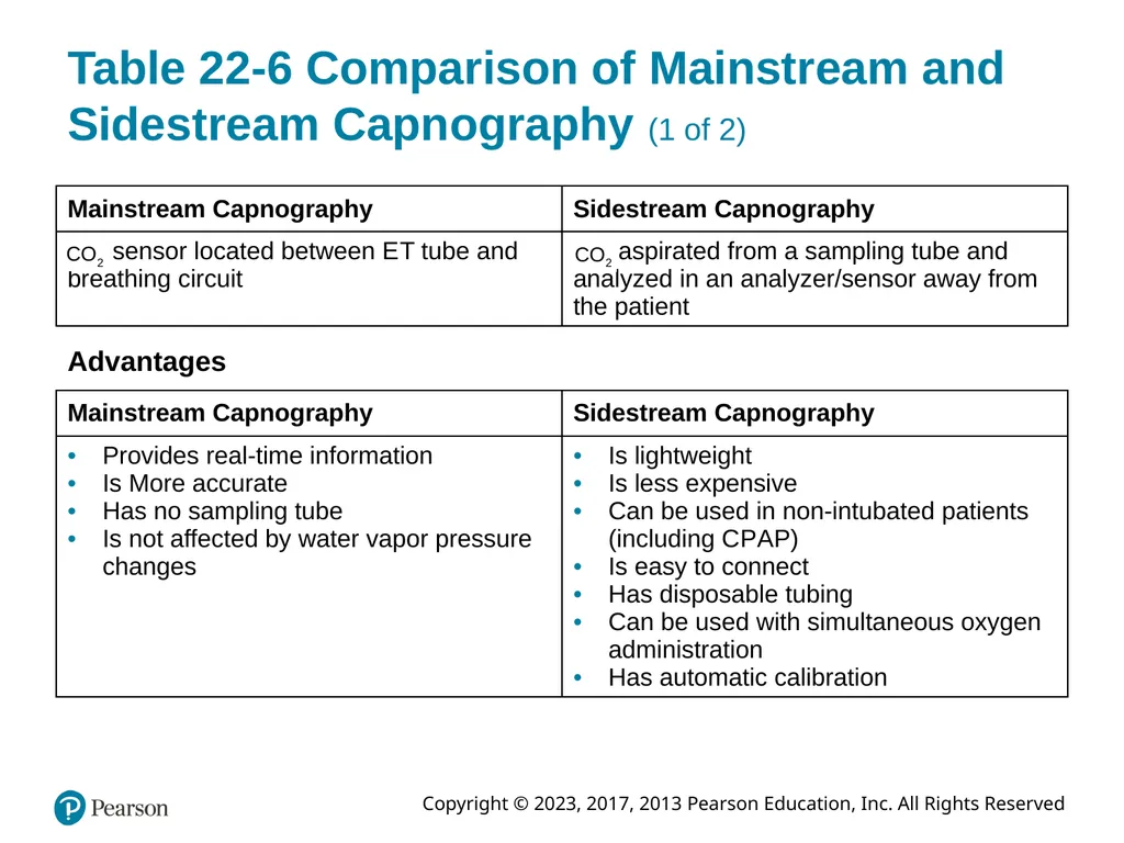
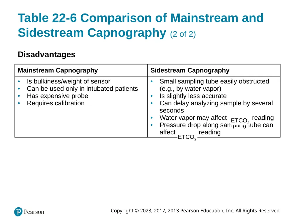
Can be mainstream or sidestream<br>
slide65. Table 22-6 Comparison of Mainstream and Sidestream Capnography (1 of 2) Advantages<br>
slide66. Table 22-6 Comparison of Mainstream and Sidestream Capnography (2 of 2) Disadvantages<br>
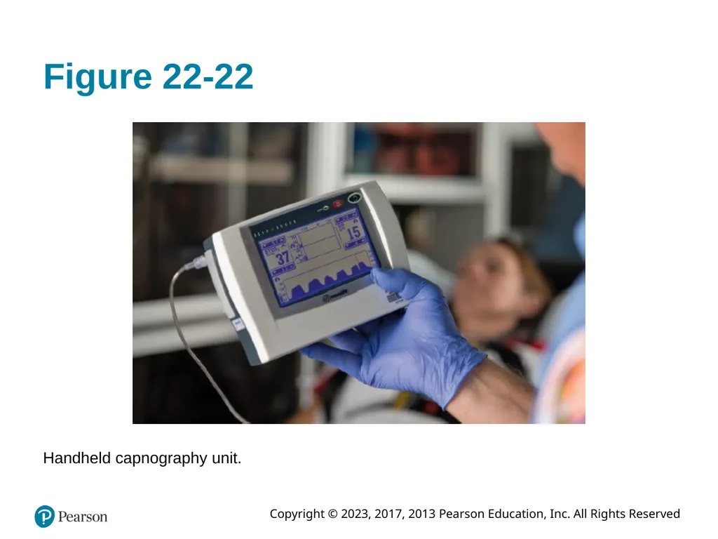
slide67. Figure 22-22 Handheld capnography unit.<br>
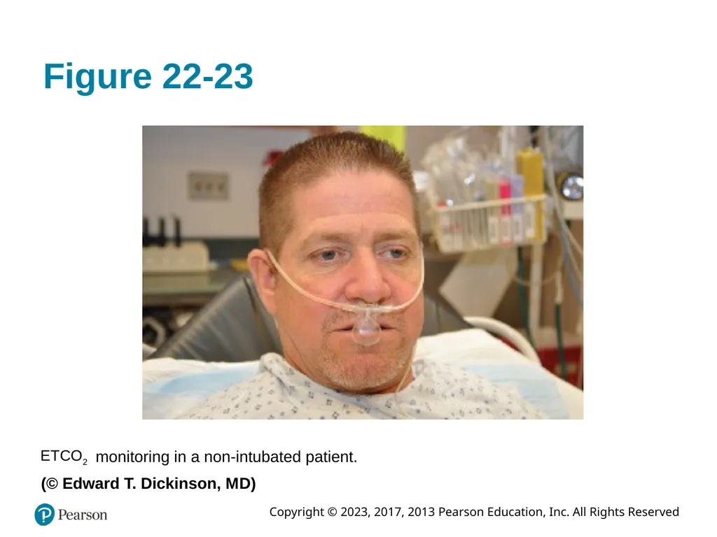
slide68. Figure 22-23 monitoring in a non-intubated patient. (© Edward T. Dickinson, M D)<br>
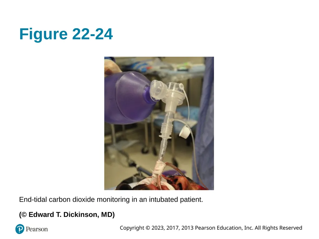
slide69. Figure 22-24 End-tidal carbon dioxide monitoring in an intubated patient.
(© Edward T. Dickinson, M D)<br>
slide70. Figure 22-25 Some detectors can display both a waveform and a number.<br>
slide71. Respiratory Anatomy, Physiology, and Assessment (36 of 39) Secondary Assessment (continued)
Noninvasive Respiratory Monitoring (continued)
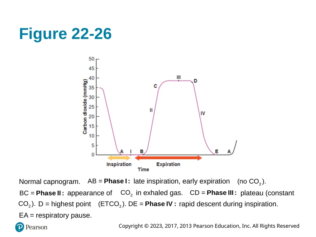
Capnography—Capnogram Capnogram reflects exhaled concentrations over time
Its shape is reflective of the expiratory phase and elimination from the body Four phases of the waveform
Changes to the waveform provide clinical information<br>
slide72. Figure 22-26 Normal capnogram. late inspiration, early expiration appearance of in exhaled gas. plateau (constant D = highest point rapid descent during inspiration. E A = respiratory pause.<br>
slide73. Respiratory Anatomy, Physiology, and Assessment (37 of 39) Secondary Assessment (continued)
Noninvasive Respiratory Monitoring (continued)
Capnography—Clinical Applications
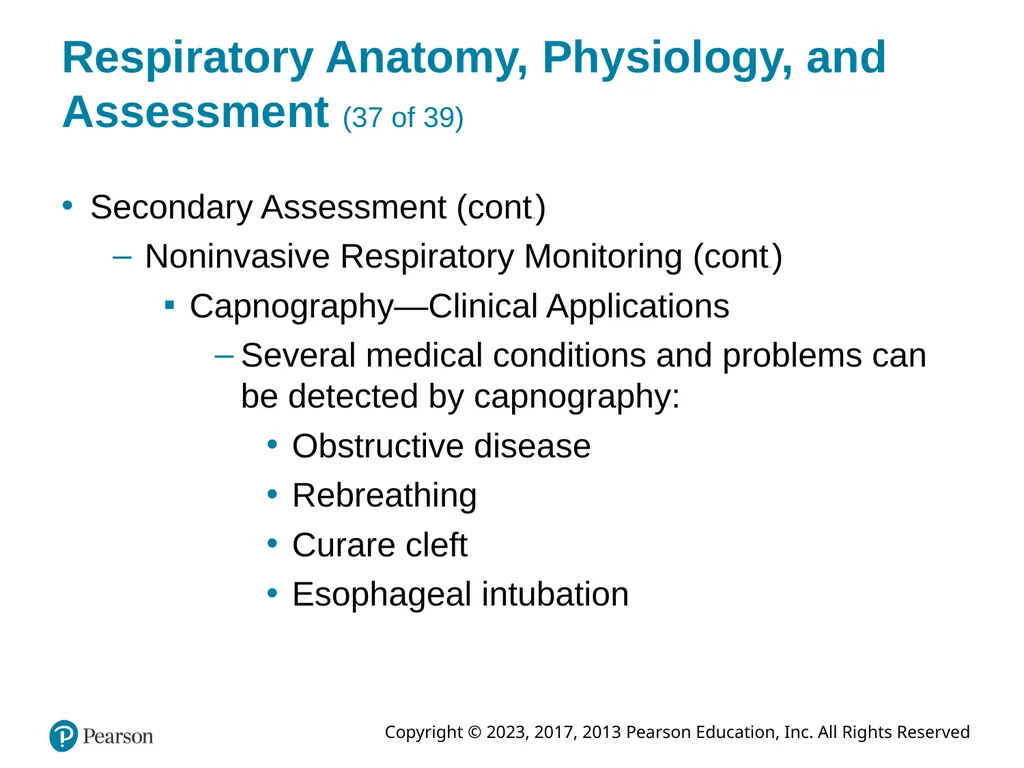
Several medical conditions and problems can be detected by capnography:
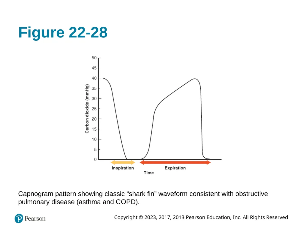
Obstructive disease
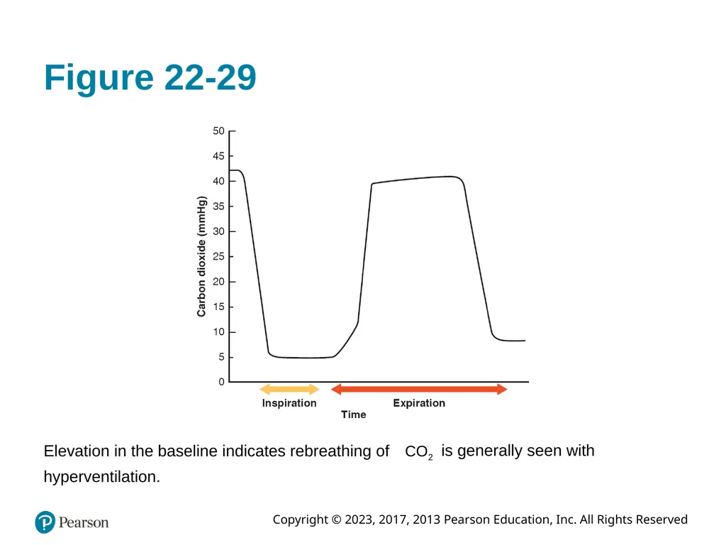
Rebreathing
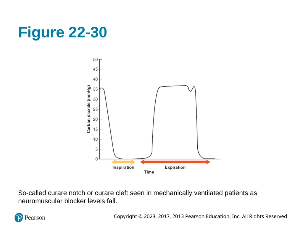
Curare cleft
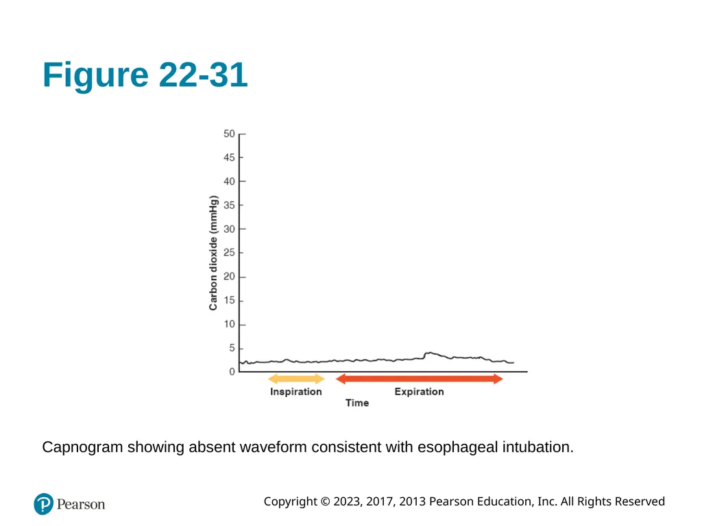
Esophageal intubation<br>
slide74. Respiratory Anatomy, Physiology, and Assessment (38 of 39) Secondary Assessment (continued)
Noninvasive Respiratory Monitoring (continued)
Capnography—Clinical Applications (continued)
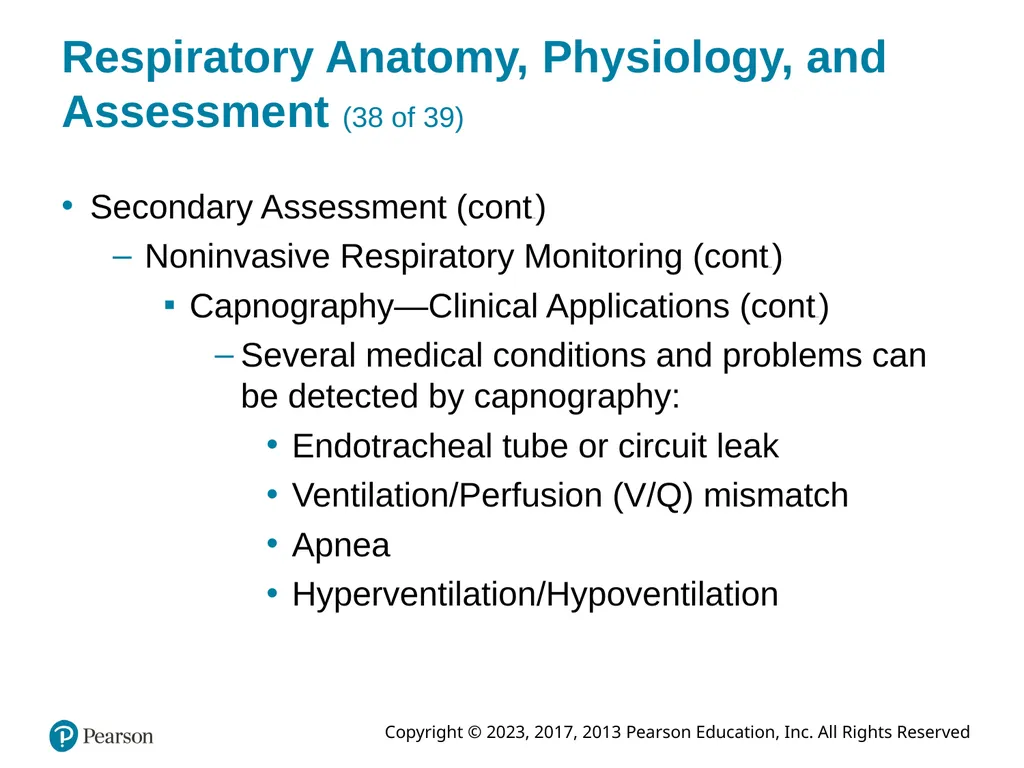
Several medical conditions and problems can be detected by capnography:
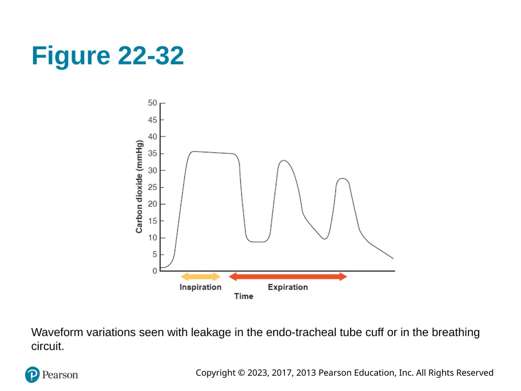
Endotracheal tube or circuit leak
Ventilation/Perfusion (V/Q) mismatch
Apnea
Hyperventilation/Hypoventilation<br>
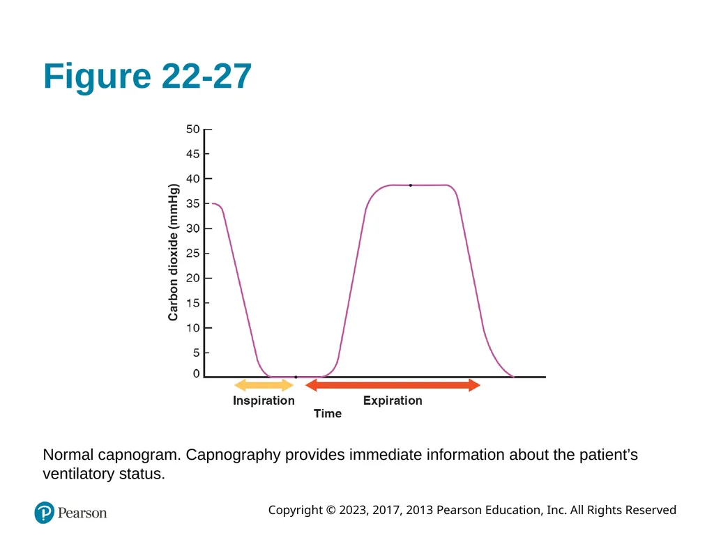
slide75. Figure 22-27 Normal capnogram. Capnography provides immediate information about the patient’s ventilatory status.<br>
slide76. Figure 22-28 Capnogram pattern showing classic “shark fin” waveform consistent with obstructive pulmonary disease (asthma and C O P D).<br>
slide77. Figure 22-29 Elevation in the baseline indicates rebreathing of is generally seen with hyperventilation.<br>
slide78. Figure 22-30 So-called curare notch or curare cleft seen in mechanically ventilated patients as neuromuscular blocker levels fall.<br>
slide79. Figure 22-31 Capnogram showing absent waveform consistent with esophageal intubation.<br>
slide80. Figure 22-32 Waveform variations seen with leakage in the endo-tracheal tube cuff or in the breathing circuit.<br>
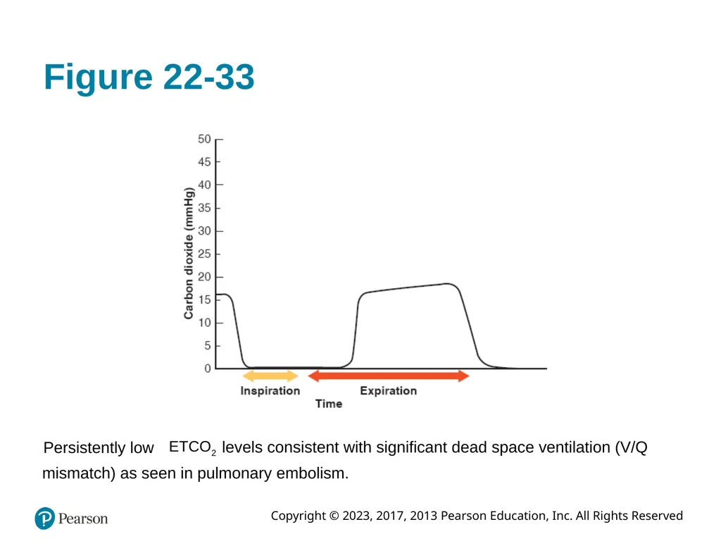
slide81. Figure 22-33 Persistently low levels consistent with significant dead space ventilation (V/Q mismatch) as seen in pulmonary embolism.<br>
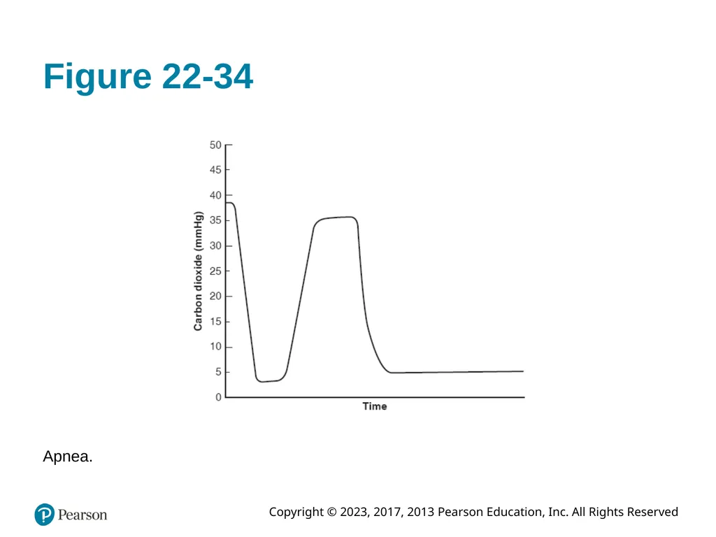
slide82. Figure 22-34 Apnea.<br>
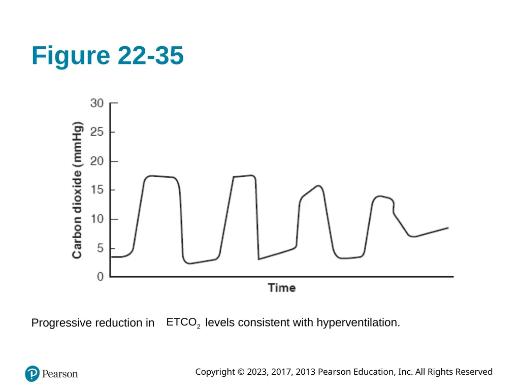
slide83. Figure 22-35 Progressive reduction in levels consistent with hyperventilation.<br>
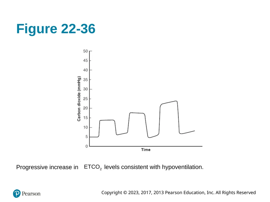
slide84. Figure 22-36 Progressive increase in levels consistent with hypoventilation.<br>
slide85. Respiratory Anatomy, Physiology, and Assessment (39 of 39) Secondary Assessment (continued)
Noninvasive Respiratory Monitoring (continued)
Peak Expiratory Flow Testing
Disposable plastic chamber into which patient exhales forcefully after maximal inhalation
Crude measure of respiratory efficacy
Improving measurements can indicate a good response to treatment of a pulmonary condition<br>
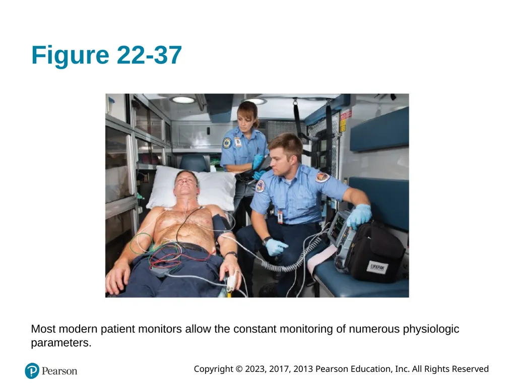
slide86. Figure 22-37 Most modern patient monitors allow the constant monitoring of numerous physiologic parameters.<br>
slide87. Copyright This work is protected by United States copyright laws and is provided solely for the use of instructors in teaching their courses and assessing student learning. Dissemination or sale of any part of this work (including on the World Wide Web) will destroy the integrity of the work and is not permitted. The work and materials from it should never be made available to students except by instructors using the accompanying text in their classes. All recipients of this work are expected to abide by these restrictions and to honor the intended pedagogical purposes and the needs of other instructors who rely on these materials.<br>
slide2. Standard Airway Management, Respiration, and Artificial Ventilation<br>
slide3. Competency Integrates comprehensive knowledge of anatomy, physiology, and pathophysiology into the assessment to develop and implement a treatment plan with the goal of ensuring a patent airway, adequate mechanical ventilation, and respiration for patients of all ages.<br>
slide4. Introduction (1 of 3) Airway management and ventilation are the first and most critical steps during the primary assessment of every patient
Establish and maintain airway patency while providing adequate oxygen
Without adequate airway maintenance and ventilation, patient will succumb to brain injury or death in as little as 4 minutes<br>
slide5. Introduction (2 of 3) The primary assessment typically follows the A B C ((airway, breathing, circulation) approach to the patient
If however, the patient is believed to be in cardiac arrest, then the sequence becomes C A B (compressions, airway, breathing)
Airway and ventilation often assessed and managed together<br>
slide6. Introduction (3 of 3) Airway and ventilation problems are at times, approached in a nonlinear fashion
Simple, basic airway skills are key to successful airway management
The inability to properly assess and manage an airway will doom all other treatments to failure<br>
slide7. Part 1: Respiratory Anatomy, Physiology, and Assessment<br>
slide8. Respiratory Anatomy, Physiology, and Assessment (1 of 39) Anatomy of the Respiratory System
Provides passage for oxygen and carbon dioxide
Movement of gases from atmosphere to body cells is called ventilation
Respiration is the gas exchange that occurs at the cellular level<br>
slide9. Respiratory Anatomy, Physiology, and Assessment (2 of 39) Anatomy of the Respiratory System (continued)
Upper Airway Anatomy
Upper airway:
from mouth and nose to larynx
Includes nasal cavity, oral cavity, pharynx
Larynx joins upper and lower airways<br>
slide10. Figure 22-1 Anatomy of the upper airway.<br>
slide11. Respiratory Anatomy, Physiology, and Assessment (3 of 39) Anatomy of the Respiratory System (continued)
Upper Airway Anatomy (continued)
Nasal cavity
Oral cavity
U-shaped hyoid bone
Pharynx
Nasopharynx, Oropharynx, Hypopharynx
Larynx
Glottic opening (beneath epiglottis), thyroid and cricoid cartilages. Houses vocal cords<br>
slide12. Figure 22-2 Internal anatomy of the upper airway.<br>
slide13. Respiratory Anatomy, Physiology, and Assessment (4 of 39) Anatomy of the Respiratory System (continued)
Lower Airway Anatomy
Trachea
Tube that connects larynx to two mainstem bronchi
Trachea divides into right and left mainstem bronchi<br>
slide14. Figure 22-3 Anatomy of the lower airway.<br>
slide15. Respiratory Anatomy, Physiology, and Assessment (5 of 39) Anatomy of the Respiratory System (continued)
Lower Airway Anatomy (continued)
Bronchi
Trachea bifurcates at carina into two mainstem bronchi
Right mainstem often site of aspirated foreign bodies
Bronchi (supported by cartilage) gives rise to bronchiole structures supported by smooth muscle
After about 22 divisions, bronchioles give rise to respiratory bronchioles that have limited capacity for gas exchange<br>
slide16. Respiratory Anatomy, Physiology, and Assessment (6 of 39) Anatomy of the Respiratory System (continued)
Lower Airway Anatomy (continued)
Alveoli
Respiratory bronchioles divide into alveolar ducts
Alveolar ducts terminate into alveoli sacs
Individual alveoli are sites of primary gas exchange
Surfactant layer to prevent atelectasis<br>
slide17. Figure 22-4 Anatomy of the alveoli.<br>
slide18. Respiratory Anatomy, Physiology, and Assessment (7 of 39) Anatomy of the Respiratory System (continued)
Lower Airway Anatomy (continued)
Lung Parenchyma
Two pulmonary lobules; anatomic division of lungs. Further divided into lobes
Pleura:
Membranous connective tissue that covers lungs
Parietal pleura
Visceral pleura<br>
slide19. Respiratory Anatomy, Physiology, and Assessment (8 of 39) Anatomy of the Respiratory System (continued)
Pediatric Airway
Pediatric airway smaller in all aspects; softer and more fragile
Epiglottis for floppier and rounder
Larynx more anterior and funnel shaped
Ribs and cartilage of pediatric thoracic cage softer and more pliable
Primarily diaphragmatic breathers<br>
slide20. Figure 22-5 Anatomy of the pediatric airway.<br>
slide21. Respiratory Anatomy, Physiology, and Assessment (9 of 39) Physiology of the Respiratory System
Respiration and Ventilation
Pulmonary respiration occurs in lungs
Cellular respiration occurs in peripheral capillaries
Ventilation: mechanical process that moves air into and out of lungs; necessary for respiration to occur
Gases exchanged between red blood cells and alveoli through capillary membranes<br>
slide22. Figure 22-6 Diffusion of gases across an alveolar membrane.<br>
slide23. Respiratory Anatomy, Physiology, and Assessment (10 of 39) Physiology of the Respiratory System (continued)
Respiration and Ventilation (continued)
Respiratory Cycle
Normal expiration: passive process
Inspiration: active process, using energy
Musculature to change intrathoracic size
Diaphragm, intercostal, accessory muscles<br>
slide24. Respiratory Anatomy, Physiology, and Assessment (11 of 39) Physiology of the Respiratory System (continued)
Respiration and Ventilation (continued)
Respiratory Cycle (continued)
Pulmonary ventilation: depends on changes in pressure within thoracic cavity
Respiratory cycle: coordinated interaction among respiratory system, central nervous system, musculoskeletal system<br>
slide25. Respiratory Anatomy, Physiology, and Assessment (12 of 39) Physiology of the Respiratory System (continued)
Respiration and Ventilation (continued)
Pulmonary Circulation
Respiration requires intact circulatory system
Heart pumps as much blood to lungs as it pumps to peripheral tissues
Right side of heart pumps to lungs via pulmonary artery to lungs for oxygenation and offloading of carbon dioxide waste from tissues<br>
slide26. Figure 22-7 Pulmonary circulation.<br>
slide27. Respiratory Anatomy, Physiology, and Assessment (13 of 39) Measurement of Oxygen and Carbon Dioxide Levels
Partial pressure: pressure exerted by each component of gas mixture
Major respiratory gases Nitrogen Oxygen Carbon dioxide Water<br>
slide28. Table 22-1 Partial Pressures and Concentrations of Gases<br>
slide29. Respiratory Anatomy, Physiology, and Assessment (14 of 39) Measurement of Oxygen and Carbon Dioxide Levels (continued)
Diffusion
Movement of gas from area of higher concentration to area of lower concentration
Transfers gases between lungs and blood; blood and peripheral tissues<br>
slide30. Respiratory Anatomy, Physiology, and Assessment (15 of 39) Measurement of Oxygen and Carbon Dioxide Levels (continued)
Oxygen Concentrations in the Blood
Oxygen diffuses into blood plasma; combines with hemoglobin (97% of oxygen)
Remainder dissolved in the plasma (3%)
The characteristics of oxygen binding to hemoglobin graphically represented by oxy-hemoglobin dissociation curve<br>
slide31. Figure 22-8 Oxyhemoglobin dissociation curve.<br>
slide32. Respiratory Anatomy, Physiology, and Assessment (16 of 39) Measurement of Oxygen and Carbon Dioxide Levels (continued)
Oxygen Concentrations in the Blood (continued)
Factors affecting oxygen concentrations in the blood
Inadequate levels of hemoglobin (anemia, hemorrhage)
Inadequate alveolar ventilation
Decreased diffusion across pulmonary membrane
Ventilation/perfusion mismatch
Correct oxygen derangements by increasing ventilation; administering supplemental oxygen; using intermittent positive-pressure ventilation (I P P V); medications<br>
slide33. Respiratory Anatomy, Physiology, and Assessment (17 of 39) Measurement of Oxygen and Carbon Dioxide Levels (continued)
Carbon Dioxide Concentrations in the Blood
Carbon dioxide transported in three forms
Bicarbonate ion = 70%
Carried by hemoglobin = 23%
Dissolved in plasma = 7%
Changes in ventilation can change levels of carbon dioxide
Several causes of increased and/or decreased carbon dioxide in the blood exist<br>
slide34. Respiratory Anatomy, Physiology, and Assessment (18 of 39) Regulation of Respiration
Voluntary and Involuntary Respiratory Controls
Respiratory rate: number of times person breathes in 1 minute
Nervous Impulses from the Respiratory Center
Main respiratory center in medulla (gross rate and depth)
Apneustic center in pons (inspiratory center)
Pneumotaxic center in pons (expiratory center)
Stretch receptors
Hering-Breuer reflex<br>
slide35. Respiratory Anatomy, Physiology, and Assessment (19 of 39) Regulation of Respiration (continued)
Chemoreceptors
Located on medulla (central)
Located in aortic arch and carotid bodies (peripheral) Responds to elevations in or drop in p H, also to large decreases in Hypoxic drive
Most important stimuli to breath is hypercapnic drive
Hypoxic drive (low oxygen content in blood) is secondary stimuli<br>
slide36. Figure 22-9 Nervous control of respiration.<br>
slide37. Respiratory Anatomy, Physiology, and Assessment (20 of 39) Regulation of Respiration (continued)
Measure of Respiratory Function
Factors that affect respiratory rate:
Fever
Emotion
Pain
Hypoxia
Acidosis
Stimulant or depressant drugs
Sleep<br>
slide38. Respiratory Anatomy, Physiology, and Assessment (21 of 39) Regulation of Respiration (continued)
Measure of Respiratory Function (continued)
Respiratory capacities and measurements:
Total lung capacity (T L C) Tidal volume Dead space volume Alveolar volume Minute volume Alveolar minute volume<br>
slide39. Respiratory Anatomy, Physiology, and Assessment (22 of 39) Regulation of Respiration (continued)
Measure of Respiratory Function (continued)
Respiratory capacities and measurements: (continued)
Inspiratory reserve volume (I R V)
Expiratory reserve volume (E R V)
Residual volume (R V)
Functional residual capacity (F R C)
Forced expiratory volume (F E V)<br>
slide40. Figure 22-10 Respiratory volumes and capacities.<br>
slide41. Respiratory Anatomy, Physiology, and Assessment (23 of 39) Respiratory Problems
Airway Obstruction
Blockage of airway immediate threat to patient's life; true emergency
Causes of airway obstruction:
Tongue
Foreign bodies
Trauma
Laryngeal spasm and edema
Aspiration; vomitus or blood<br>
slide42. Respiratory Anatomy, Physiology, and Assessment (24 of 39) Respiratory Problems (continued)
Airway Obstruction (continued)
Airway obstruction partial or complete
Tongue most common cause of airway obstruction<br>
slide43. Respiratory Anatomy, Physiology, and Assessment (25 of 39) Respiratory Problems (continued)
Inadequate Ventilation
Reduction of rate or volume of inhalation leads to reduction in minute volume
If there are no alveolar breath sounds when auscultating, then there is no alveolar ventilation This means no gas exchange!<br>
slide44. Respiratory Anatomy, Physiology, and Assessment (26 of 39) Respiratory System Assessment
Primary Assessment
Vigilance key to airway management in every patient
Primary assessment: identify any immediate threats to patient's life; airway, breathing, circulation (A B C s)
For patients in cardiac arrest, compressions come before airway and breathing (C A B)<br>
slide45. Respiratory Anatomy, Physiology, and Assessment (27 of 39) Respiratory System Assessment (continued)
Primary Assessment (continued)
Patients with altered mental status warrant further evaluation
Assess airway to ensure it is patent
Look and listen for air exchange through mouth/nose
If airway partially or totally occluded, establish patency immediately with progressive airway interventions
Determine adequacy of breathing after airway assured
If breathing inadequately, establish good ventilations immediately with B V M<br>
slide46. Figure 22-15 Bag-valve-mask ventilation.<br>
slide47. Respiratory Anatomy, Physiology, and Assessment (28 of 39) Respiratory System Assessment (continued)
Primary Assessment (continued)
With airway and breathing assured or being supported, move on with assessment
Assess circulatory status
Check peripheral pulses, if none, check central pulses
Initiate C P R as needed
Stop any acute external hemorrhaging
Evaluate skin findings
Make priority determination of patient’s status<br>
slide48. Respiratory Anatomy, Physiology, and Assessment (29 of 39) Secondary Assessment
Complete after primary assessment and correction of any immediate life threats
Determine History
Recent history leading to onset of symptoms; identify possible triggers
Past medical history
Begin physical examination of patient<br>
slide49. Respiratory Anatomy, Physiology, and Assessment (30 of 39) Secondary Assessment (continued)
Physical Examination
Physical examination techniques
Inspection
Auscultation
Palpation
Percussion
Begin physical assessment by inspecting patient<br>
slide50. Respiratory Anatomy, Physiology, and Assessment (31 of 39) Secondary Assessment (continued)
Noninvasive Respiratory Monitoring
Pulse oximetry: "fifth vital sign"
Noninvasive measure of hemoglobin oxygen saturation in peripheral tissues
Pulse C O Oximetry
Monitors for abnormal hemoglobin states
Carboxyhemoglobin
Methemoglobin
Total hemoglobin<br>
slide51. Figure 22-17 Pulse oximeter.<br>
slide52. Table 22-3 Interpretation of Pulse Oximetry Readings and Recommended Actions<br>
slide53. Figure 22-18 Pulse CO oximetry.
(© Dr. Bryan E. Bledsoe)<br>
slide54. Table 22-2 Comparison of Pulse Oximetry and Capnography<br>
slide55. Figure 22-19 Total hemoglobin by pulse C O oximetry.
(© Dr. Bryan E. Bledsoe)<br>
slide56. Figure 22-20 Multiple noninvasive parameters in a single display monitor.
(© Dr. Bryan E. Bledsoe)<br>
slide57. Respiratory Anatomy, Physiology, and Assessment (32 of 39) Secondary Assessment (continued)
Noninvasive Respiratory Monitoring (continued)
Capnography: recording or display of exhaled carbon dioxide levels measured by capnometry
Capnograph; capnogram End-tidal End-tidal gradient<br>
slide58. Respiratory Anatomy, Physiology, and Assessment (33 of 39) Secondary Assessment (continued)
Noninvasive Respiratory Monitoring (continued)
Capnography
Understanding capnography reading is a function of three parameters
Alveolar ventilation
Pulmonary perfusion production in the tissues As a practitioner, you must figure out which one is the primary cause of a change in levels Most often though, blame the pulmonary system<br>
slide59. Table 22-4 Comparison of P a C O sub 2 and E T C O sub 2<br>
slide60. Table 22-5 Basic Rules of Capnography (1 of 2)<br>
slide61. Table 22-5 Basic Rules of Capnography (2 of 2)<br>
slide62. Respiratory Anatomy, Physiology, and Assessment (34 of 39) Secondary Assessment (continued)
Noninvasive Respiratory Monitoring (continued)
Capnography—Colorimetric Device Disposable detector p H-sensitive paper encased in plastic Exhaled breath exposes paper to Causes color change if present<br>
slide63. Figure 22-21 Colorimetric detector. (© Edward T. Dickinson, M D)<br>
slide64. Respiratory Anatomy, Physiology, and Assessment (35 of 39) Secondary Assessment (continued)
Noninvasive Respiratory Monitoring (continued)
Capnography—Infrared Devices Electronic detectors use infrared technique Can be qualitative or quantitative Exhaled can be monitored in patients who are intubated as well as not intubated Most modern devices provide a numeric value and waveform
Can be mainstream or sidestream<br>
slide65. Table 22-6 Comparison of Mainstream and Sidestream Capnography (1 of 2) Advantages<br>
slide66. Table 22-6 Comparison of Mainstream and Sidestream Capnography (2 of 2) Disadvantages<br>
slide67. Figure 22-22 Handheld capnography unit.<br>
slide68. Figure 22-23 monitoring in a non-intubated patient. (© Edward T. Dickinson, M D)<br>
slide69. Figure 22-24 End-tidal carbon dioxide monitoring in an intubated patient.
(© Edward T. Dickinson, M D)<br>
slide70. Figure 22-25 Some detectors can display both a waveform and a number.<br>
slide71. Respiratory Anatomy, Physiology, and Assessment (36 of 39) Secondary Assessment (continued)
Noninvasive Respiratory Monitoring (continued)
Capnography—Capnogram Capnogram reflects exhaled concentrations over time
Its shape is reflective of the expiratory phase and elimination from the body Four phases of the waveform
Changes to the waveform provide clinical information<br>
slide72. Figure 22-26 Normal capnogram. late inspiration, early expiration appearance of in exhaled gas. plateau (constant D = highest point rapid descent during inspiration. E A = respiratory pause.<br>
slide73. Respiratory Anatomy, Physiology, and Assessment (37 of 39) Secondary Assessment (continued)
Noninvasive Respiratory Monitoring (continued)
Capnography—Clinical Applications
Several medical conditions and problems can be detected by capnography:
Obstructive disease
Rebreathing
Curare cleft
Esophageal intubation<br>
slide74. Respiratory Anatomy, Physiology, and Assessment (38 of 39) Secondary Assessment (continued)
Noninvasive Respiratory Monitoring (continued)
Capnography—Clinical Applications (continued)
Several medical conditions and problems can be detected by capnography:
Endotracheal tube or circuit leak
Ventilation/Perfusion (V/Q) mismatch
Apnea
Hyperventilation/Hypoventilation<br>
slide75. Figure 22-27 Normal capnogram. Capnography provides immediate information about the patient’s ventilatory status.<br>
slide76. Figure 22-28 Capnogram pattern showing classic “shark fin” waveform consistent with obstructive pulmonary disease (asthma and C O P D).<br>
slide77. Figure 22-29 Elevation in the baseline indicates rebreathing of is generally seen with hyperventilation.<br>
slide78. Figure 22-30 So-called curare notch or curare cleft seen in mechanically ventilated patients as neuromuscular blocker levels fall.<br>
slide79. Figure 22-31 Capnogram showing absent waveform consistent with esophageal intubation.<br>
slide80. Figure 22-32 Waveform variations seen with leakage in the endo-tracheal tube cuff or in the breathing circuit.<br>
slide81. Figure 22-33 Persistently low levels consistent with significant dead space ventilation (V/Q mismatch) as seen in pulmonary embolism.<br>
slide82. Figure 22-34 Apnea.<br>
slide83. Figure 22-35 Progressive reduction in levels consistent with hyperventilation.<br>
slide84. Figure 22-36 Progressive increase in levels consistent with hypoventilation.<br>
slide85. Respiratory Anatomy, Physiology, and Assessment (39 of 39) Secondary Assessment (continued)
Noninvasive Respiratory Monitoring (continued)
Peak Expiratory Flow Testing
Disposable plastic chamber into which patient exhales forcefully after maximal inhalation
Crude measure of respiratory efficacy
Improving measurements can indicate a good response to treatment of a pulmonary condition<br>
slide86. Figure 22-37 Most modern patient monitors allow the constant monitoring of numerous physiologic parameters.<br>
slide87. Copyright This work is protected by United States copyright laws and is provided solely for the use of instructors in teaching their courses and assessing student learning. Dissemination or sale of any part of this work (including on the World Wide Web) will destroy the integrity of the work and is not permitted. The work and materials from it should never be made available to students except by instructors using the accompanying text in their classes. All recipients of this work are expected to abide by these restrictions and to honor the intended pedagogical purposes and the needs of other instructors who rely on these materials.<br>