
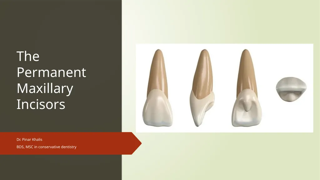
The Permanent Maxillary Incisors Dr. Pinar Khalis






Description: The Permanent Maxillary Incisors Dr. Pinar Khalis BDS, MSC in conservative dentistry Introduction The maxillary incisors are four in number. The maxillary central incisors are centered in the maxilla, one on either side of the median line,
Related Topics
Download Presentation
"The Permanent Maxillary Incisors Dr. Pinar Khalis" is the property of its rightful owner. Permission is granted to download and print the materials on this website for personal, non-commercial use only, and to display it on your personal computer provided you do not modify the materials and that you retain all copyright notices contained in the materials. By downloading content from our website, you accept the terms of this agreement.
Presentation Transcript
slide1. The Permanent Maxillary Incisors Dr. Pinar Khalis
BDS, MSC in conservative dentistry<br>
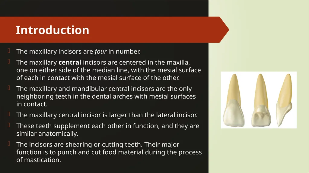
slide2. Introduction The maxillary incisors are four in number.
The maxillary central incisors are centered in the maxilla, one on either side of the median line, with the mesial surface of each in contact with the mesial surface of the other.
The maxillary and mandibular central incisors are the only neighboring teeth in the dental arches with mesial surfaces in contact.
The maxillary central incisor is larger than the lateral incisor.
These teeth supplement each other in function, and they are similar anatomically.
The incisors are shearing or cutting teeth. Their major function is to punch and cut food material during the process of mastication.<br>
slide3. Incisors have incisal ridges or edges Incisal Ridge
The incisal ridge is that portion of the crown which makes up the complete incisal portion.
When an incisor is newly erupted, the incisal portion is rounded and merges with the mesioincisal and distoincisal angles and the labial and lingual surfaces.
This ridge portion of the crown is called the incisal ridge. Incisal Edge
The term edge implies an angle formed by the merging of two flat surfaces.
Therefore, an incisal edge does not exist on an incisor until occlusal wear has created a flattened surface linguoincisally, which surface forms an angle with the labial surface.
The incisal edge is formed by the junction of the linguoincisal surface.<br>
slide4. Incisal edge vs incisal ridge<br>
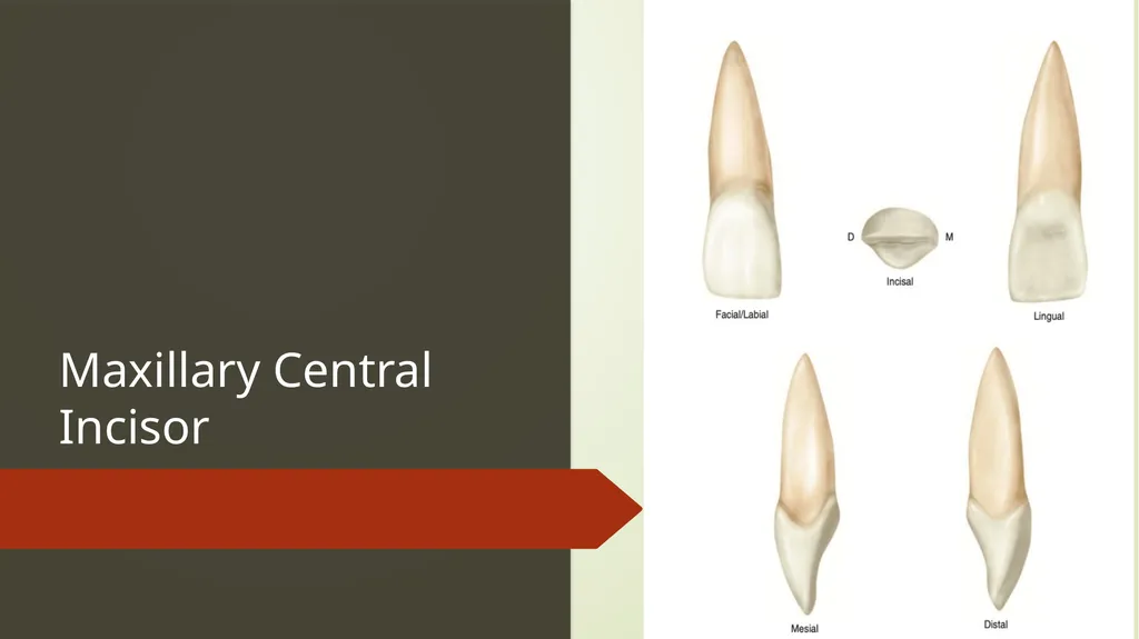
slide5. Maxillary Central Incisor<br>
slide6. Maxillary Central Incisor The maxillary central incisor is the widest mesiodistally of any of the anterior teeth.
Two in number
The labial face is less convex than that of the maxillary lateral incisor or canine, which gives the central incisor a squared or rectangular appearance.<br>
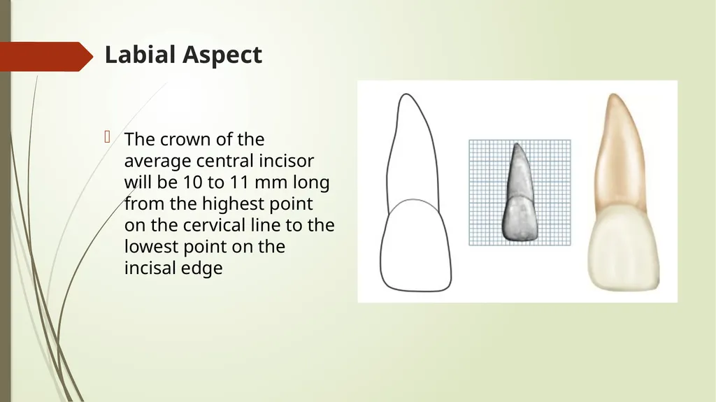
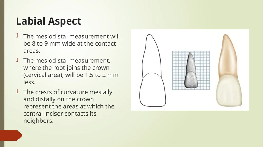
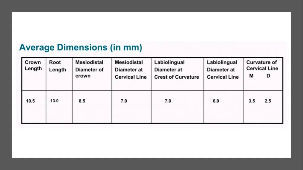
slide7. Labial Aspect The crown of the average central incisor will be 10 to 11 mm long from the highest point on the cervical line to the lowest point on the incisal edge<br>
slide8. Labial Aspect The mesiodistal measurement will be 8 to 9 mm wide at the contact areas.
The mesiodistal measurement, where the root joins the crown (cervical area), will be 1.5 to 2 mm less.
The crests of curvature mesially and distally on the crown represent the areas at which the central incisor contacts its neighbors.<br>
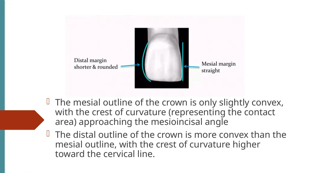
slide9. The mesial outline of the crown is only slightly convex, with the crest of curvature (representing the contact area) approaching the mesioincisal angle
The distal outline of the crown is more convex than the mesial outline, with the crest of curvature higher toward the cervical line.<br>
slide10. The distoincisal angle is not as sharp as the mesioincisal angle,
The incisal outline is usually regular and straight in a mesiodistal direction<br>
slide11. The incisal outline tends to curve downward toward the center of the crown outline, so that the crown length is greater at the center than at the two mesial angles.
The cervical outline of the crown follows a semicircular direction with the curvature rootwise, from the point at which the root outline joins the crown mesially to the point at which the root outline joins the crown distally.<br>
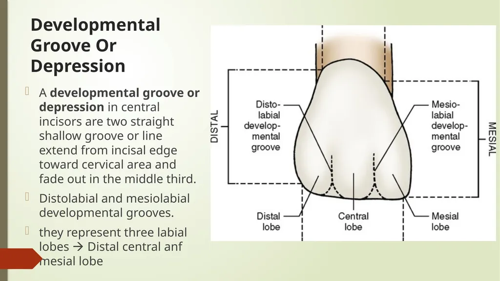
slide13. Developmental Groove Or Depression A developmental groove or depression in central incisors are two straight shallow groove or line extend from incisal edge toward cervical area and fade out in the middle third.
Distolabial and mesiolabial developmental grooves.
they represent three labial lobes Distal central anf mesial lobe<br>
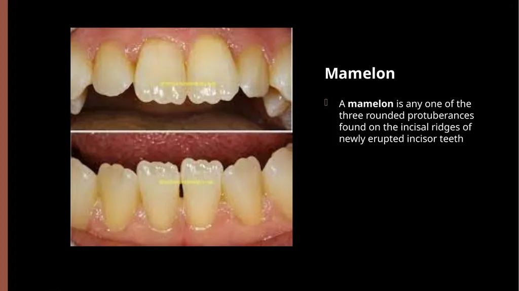
slide14. Mamelon A mamelon is any one of the three rounded protuberances found on the incisal ridges of newly erupted incisor teeth<br>
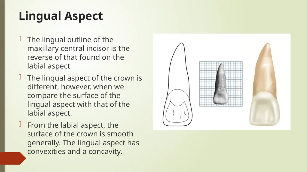
slide15. Lingual Aspect The lingual outline of the maxillary central incisor is the reverse of that found on the labial aspect
The lingual aspect of the crown is different, however, when we compare the surface of the lingual aspect with that of the labial aspect.
From the labial aspect, the surface of the crown is smooth generally. The lingual aspect has convexities and a concavity.<br>
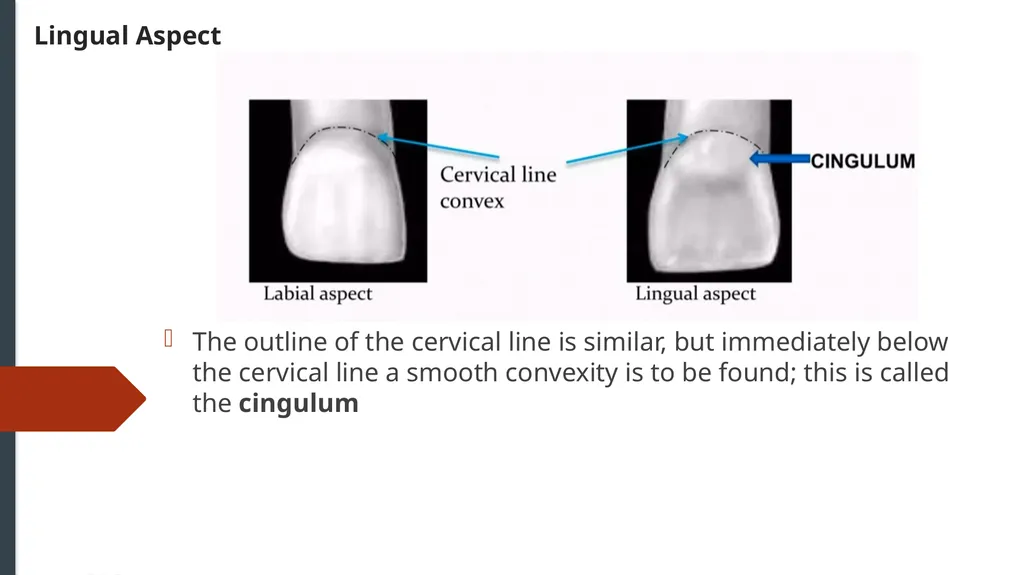
slide16. Lingual Aspect The outline of the cervical line is similar, but immediately below the cervical line a smooth convexity is to be found; this is called the cingulum<br>
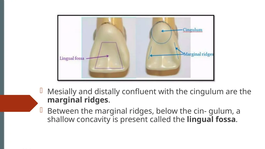
slide17. Mesially and distally confluent with the cingulum are the marginal ridges.
Between the marginal ridges, below the cin- gulum, a shallow concavity is present called the lingual fossa.<br>
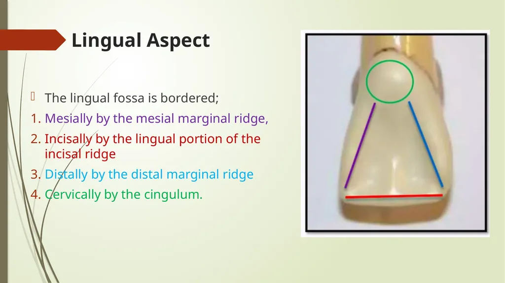
slide18. Lingual Aspect The lingual fossa is bordered;
Mesially by the mesial marginal ridge,
Incisally by the lingual portion of the incisal ridge
Distally by the distal marginal ridge
Cervically by the cingulum.<br>
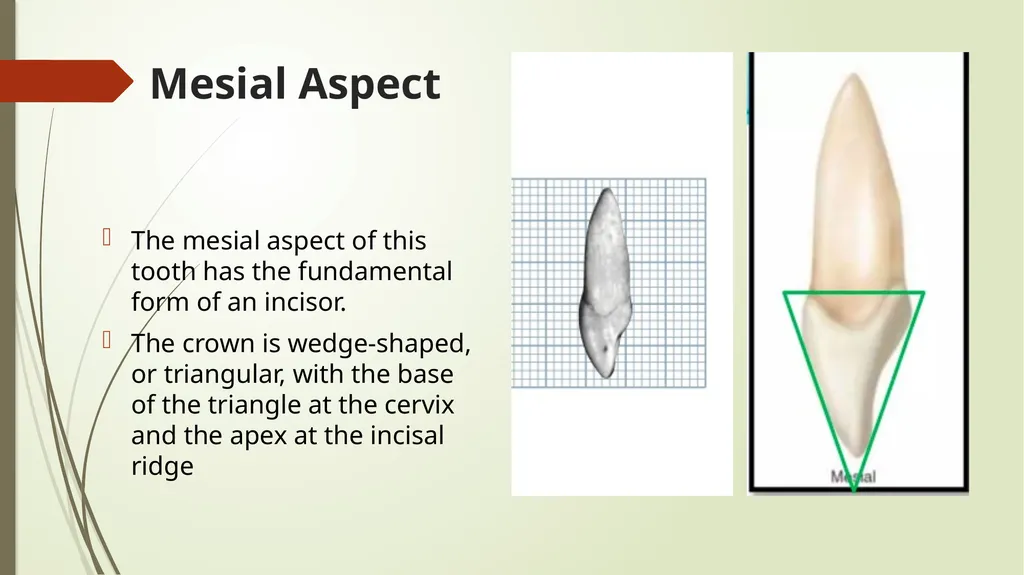
slide19. Mesial Aspect The mesial aspect of this tooth has the fundamental form of an incisor.
The crown is wedge-shaped, or triangular, with the base of the triangle at the cervix and the apex at the incisal ridge<br>
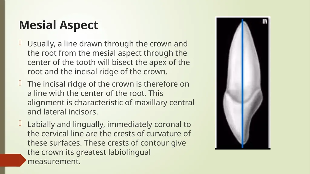
slide20. Mesial Aspect Usually, a line drawn through the crown and the root from the mesial aspect through the center of the tooth will bisect the apex of the root and the incisal ridge of the crown.
The incisal ridge of the crown is therefore on a line with the center of the root. This alignment is characteristic of maxillary central and lateral incisors.
Labially and lingually, immediately coronal to the cervical line are the crests of curvature of these surfaces. These crests of contour give the crown its greatest labiolingual measurement.<br>
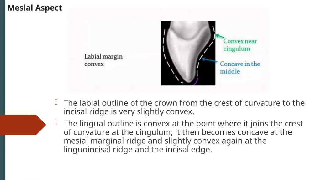
slide21. Mesial Aspect The labial outline of the crown from the crest of curvature to the incisal ridge is very slightly convex.
The lingual outline is convex at the point where it joins the crest of curvature at the cingulum; it then becomes concave at the mesial marginal ridge and slightly convex again at the linguoincisal ridge and the incisal edge.<br>
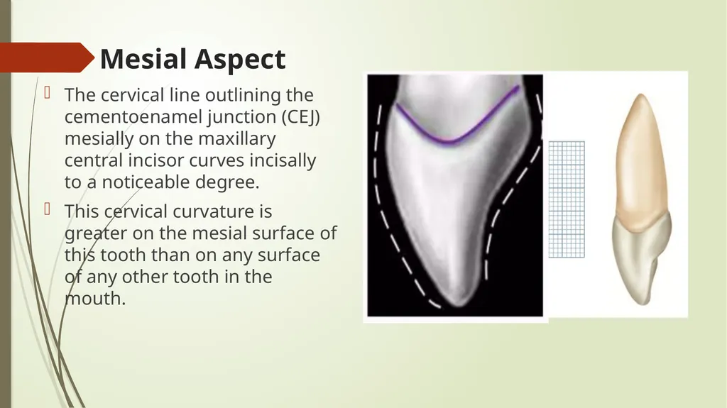
slide22. Mesial Aspect The cervical line outlining the cementoenamel junction (CEJ) mesially on the maxillary central incisor curves incisally to a noticeable degree.
This cervical curvature is greater on the mesial surface of this tooth than on any surface of any other tooth in the mouth.<br>
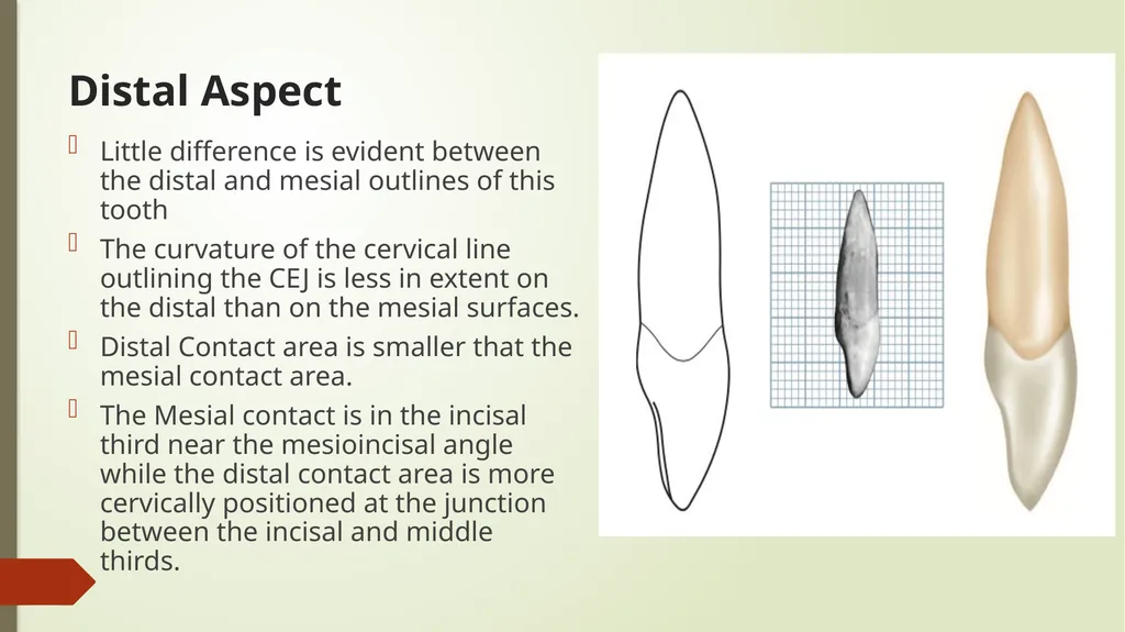
slide23. Distal Aspect Little difference is evident between the distal and mesial outlines of this tooth
The curvature of the cervical line outlining the CEJ is less in extent on the distal than on the mesial surfaces.
Distal Contact area is smaller that the mesial contact area.
The Mesial contact is in the incisal third near the mesioincisal angle while the distal contact area is more cervically positioned at the junction between the incisal and middle thirds.<br>
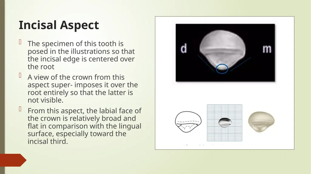
slide24. Incisal Aspect The specimen of this tooth is posed in the illustrations so that the incisal edge is centered over the root
A view of the crown from this aspect super- imposes it over the root entirely so that the latter is not visible.
From this aspect, the labial face of the crown is relatively broad and flat in comparison with the lingual surface, especially toward the incisal third.<br>
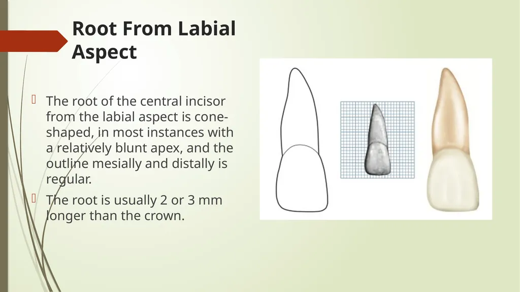
slide25. Root From Labial Aspect The root of the central incisor from the labial aspect is cone-shaped, in most instances with a relatively blunt apex, and the outline mesially and distally is regular.
The root is usually 2 or 3 mm longer than the crown.<br>
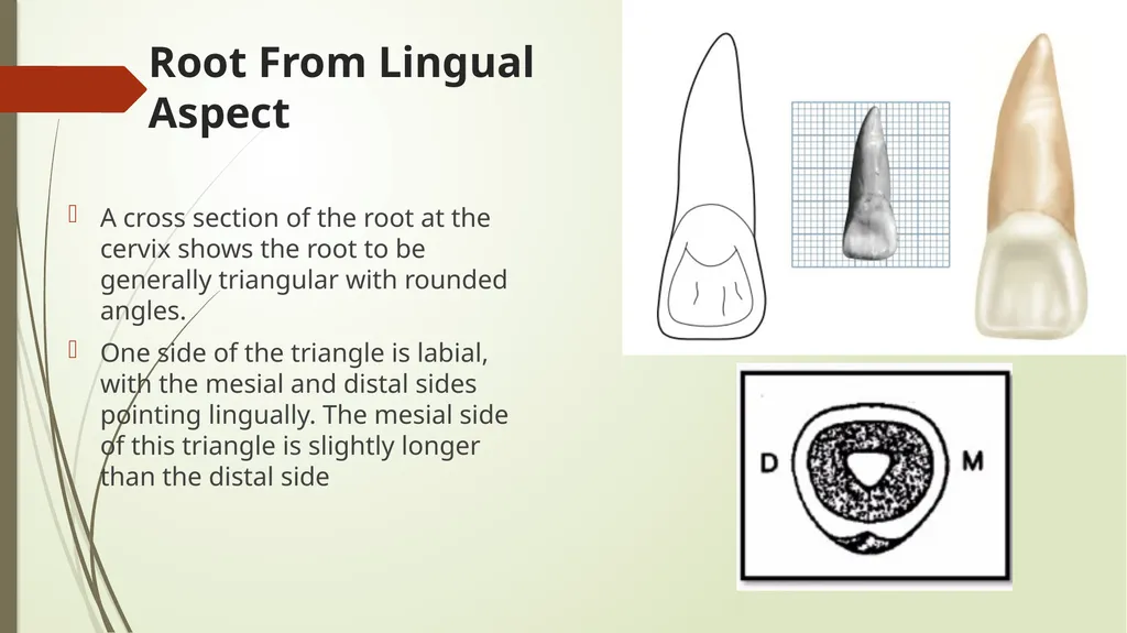
slide26. Root From Lingual Aspect A cross section of the root at the cervix shows the root to be generally triangular with rounded angles.
One side of the triangle is labial, with the mesial and distal sides pointing lingually. The mesial side of this triangle is slightly longer than the distal side<br>
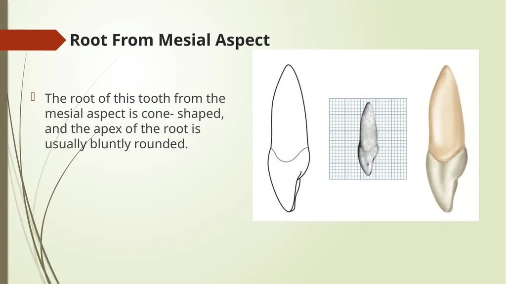
slide27. Root From Mesial Aspect The root of this tooth from the mesial aspect is cone- shaped, and the apex of the root is usually bluntly rounded.<br>
slide28. Thank You For Paying Attention<br>
BDS, MSC in conservative dentistry<br>
slide2. Introduction The maxillary incisors are four in number.
The maxillary central incisors are centered in the maxilla, one on either side of the median line, with the mesial surface of each in contact with the mesial surface of the other.
The maxillary and mandibular central incisors are the only neighboring teeth in the dental arches with mesial surfaces in contact.
The maxillary central incisor is larger than the lateral incisor.
These teeth supplement each other in function, and they are similar anatomically.
The incisors are shearing or cutting teeth. Their major function is to punch and cut food material during the process of mastication.<br>
slide3. Incisors have incisal ridges or edges Incisal Ridge
The incisal ridge is that portion of the crown which makes up the complete incisal portion.
When an incisor is newly erupted, the incisal portion is rounded and merges with the mesioincisal and distoincisal angles and the labial and lingual surfaces.
This ridge portion of the crown is called the incisal ridge. Incisal Edge
The term edge implies an angle formed by the merging of two flat surfaces.
Therefore, an incisal edge does not exist on an incisor until occlusal wear has created a flattened surface linguoincisally, which surface forms an angle with the labial surface.
The incisal edge is formed by the junction of the linguoincisal surface.<br>
slide4. Incisal edge vs incisal ridge<br>
slide5. Maxillary Central Incisor<br>
slide6. Maxillary Central Incisor The maxillary central incisor is the widest mesiodistally of any of the anterior teeth.
Two in number
The labial face is less convex than that of the maxillary lateral incisor or canine, which gives the central incisor a squared or rectangular appearance.<br>
slide7. Labial Aspect The crown of the average central incisor will be 10 to 11 mm long from the highest point on the cervical line to the lowest point on the incisal edge<br>
slide8. Labial Aspect The mesiodistal measurement will be 8 to 9 mm wide at the contact areas.
The mesiodistal measurement, where the root joins the crown (cervical area), will be 1.5 to 2 mm less.
The crests of curvature mesially and distally on the crown represent the areas at which the central incisor contacts its neighbors.<br>
slide9. The mesial outline of the crown is only slightly convex, with the crest of curvature (representing the contact area) approaching the mesioincisal angle
The distal outline of the crown is more convex than the mesial outline, with the crest of curvature higher toward the cervical line.<br>
slide10. The distoincisal angle is not as sharp as the mesioincisal angle,
The incisal outline is usually regular and straight in a mesiodistal direction<br>
slide11. The incisal outline tends to curve downward toward the center of the crown outline, so that the crown length is greater at the center than at the two mesial angles.
The cervical outline of the crown follows a semicircular direction with the curvature rootwise, from the point at which the root outline joins the crown mesially to the point at which the root outline joins the crown distally.<br>
slide13. Developmental Groove Or Depression A developmental groove or depression in central incisors are two straight shallow groove or line extend from incisal edge toward cervical area and fade out in the middle third.
Distolabial and mesiolabial developmental grooves.
they represent three labial lobes Distal central anf mesial lobe<br>
slide14. Mamelon A mamelon is any one of the three rounded protuberances found on the incisal ridges of newly erupted incisor teeth<br>
slide15. Lingual Aspect The lingual outline of the maxillary central incisor is the reverse of that found on the labial aspect
The lingual aspect of the crown is different, however, when we compare the surface of the lingual aspect with that of the labial aspect.
From the labial aspect, the surface of the crown is smooth generally. The lingual aspect has convexities and a concavity.<br>
slide16. Lingual Aspect The outline of the cervical line is similar, but immediately below the cervical line a smooth convexity is to be found; this is called the cingulum<br>
slide17. Mesially and distally confluent with the cingulum are the marginal ridges.
Between the marginal ridges, below the cin- gulum, a shallow concavity is present called the lingual fossa.<br>
slide18. Lingual Aspect The lingual fossa is bordered;
Mesially by the mesial marginal ridge,
Incisally by the lingual portion of the incisal ridge
Distally by the distal marginal ridge
Cervically by the cingulum.<br>
slide19. Mesial Aspect The mesial aspect of this tooth has the fundamental form of an incisor.
The crown is wedge-shaped, or triangular, with the base of the triangle at the cervix and the apex at the incisal ridge<br>
slide20. Mesial Aspect Usually, a line drawn through the crown and the root from the mesial aspect through the center of the tooth will bisect the apex of the root and the incisal ridge of the crown.
The incisal ridge of the crown is therefore on a line with the center of the root. This alignment is characteristic of maxillary central and lateral incisors.
Labially and lingually, immediately coronal to the cervical line are the crests of curvature of these surfaces. These crests of contour give the crown its greatest labiolingual measurement.<br>
slide21. Mesial Aspect The labial outline of the crown from the crest of curvature to the incisal ridge is very slightly convex.
The lingual outline is convex at the point where it joins the crest of curvature at the cingulum; it then becomes concave at the mesial marginal ridge and slightly convex again at the linguoincisal ridge and the incisal edge.<br>
slide22. Mesial Aspect The cervical line outlining the cementoenamel junction (CEJ) mesially on the maxillary central incisor curves incisally to a noticeable degree.
This cervical curvature is greater on the mesial surface of this tooth than on any surface of any other tooth in the mouth.<br>
slide23. Distal Aspect Little difference is evident between the distal and mesial outlines of this tooth
The curvature of the cervical line outlining the CEJ is less in extent on the distal than on the mesial surfaces.
Distal Contact area is smaller that the mesial contact area.
The Mesial contact is in the incisal third near the mesioincisal angle while the distal contact area is more cervically positioned at the junction between the incisal and middle thirds.<br>
slide24. Incisal Aspect The specimen of this tooth is posed in the illustrations so that the incisal edge is centered over the root
A view of the crown from this aspect super- imposes it over the root entirely so that the latter is not visible.
From this aspect, the labial face of the crown is relatively broad and flat in comparison with the lingual surface, especially toward the incisal third.<br>
slide25. Root From Labial Aspect The root of the central incisor from the labial aspect is cone-shaped, in most instances with a relatively blunt apex, and the outline mesially and distally is regular.
The root is usually 2 or 3 mm longer than the crown.<br>
slide26. Root From Lingual Aspect A cross section of the root at the cervix shows the root to be generally triangular with rounded angles.
One side of the triangle is labial, with the mesial and distal sides pointing lingually. The mesial side of this triangle is slightly longer than the distal side<br>
slide27. Root From Mesial Aspect The root of this tooth from the mesial aspect is cone- shaped, and the apex of the root is usually bluntly rounded.<br>
slide28. Thank You For Paying Attention<br>