
VISION INTRODUCTION ACCESSORY STRUCTURES OF THE

Description: VISION INTRODUCTION ACCESSORY STRUCTURES OF THE EYE STRUCTURE OF THE EYE BALL PHYSIOLOGY OF VISION OVERVIEW OF LIGHT AND OPTICS FOCUSING OF LIGHT ON THE RETINA PHOTORECEPTORS AND PHOTOTRANSDUCTION THE VISUAL PATHWAY VISUAL PROCESSING
Related Topics
Download Presentation
"VISION INTRODUCTION ACCESSORY STRUCTURES OF THE" is the property of its rightful owner. Permission is granted to download and print the materials on this website for personal, non-commercial use only, and to display it on your personal computer provided you do not modify the materials and that you retain all copyright notices contained in the materials. By downloading content from our website, you accept the terms of this agreement.
Presentation Transcript
slide1. VISION<br>
slide2. INTRODUCTION
ACCESSORY STRUCTURES OF THE EYE
STRUCTURE OF THE EYE BALL
PHYSIOLOGY OF VISION
OVERVIEW OF LIGHT AND OPTICS
FOCUSING OF LIGHT ON THE RETINA
PHOTORECEPTORS AND PHOTOTRANSDUCTION
THE VISUAL PATHWAY
VISUAL PROCESSING<br>
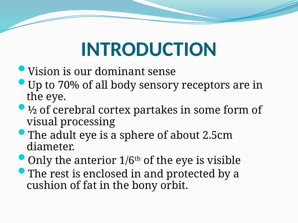
slide3. Introduction Vision is our dominant sense
Up to 70% of all body sensory receptors are in the eye.
½ of cerebral cortex partakes in some form of visual processing
The adult eye is a sphere of about 2.5cm diameter.
Only the anterior 1/6th of the eye is visible
The rest is enclosed in and protected by a cushion of fat in the bony orbit.<br>
slide4. Accessory Structures of the Eye Eye brows
Eye lids
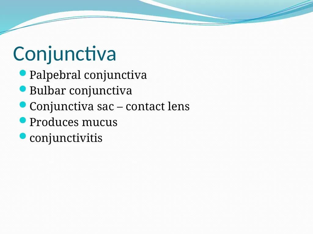
Conjunctiva
Lacrimal apparatus
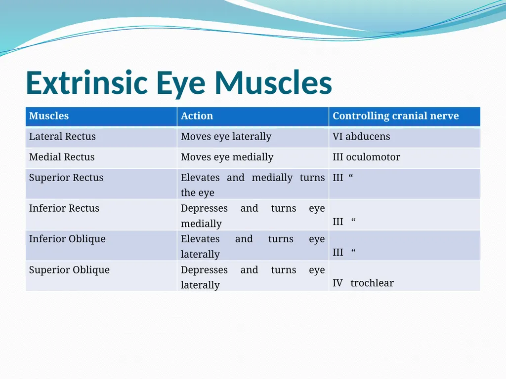
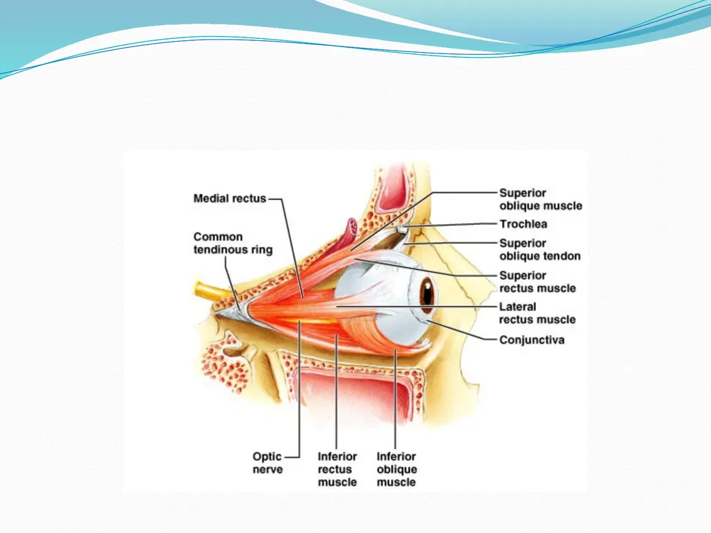
Extrinsic muscles of the eye<br>
slide6. Eyebrows Short coarse hairs over lying supra orbital margin
Sun shade
Keep out perspiration<br>
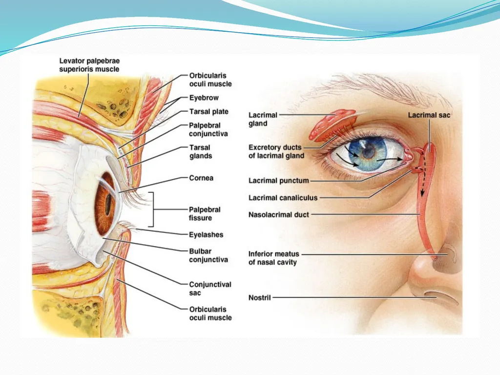
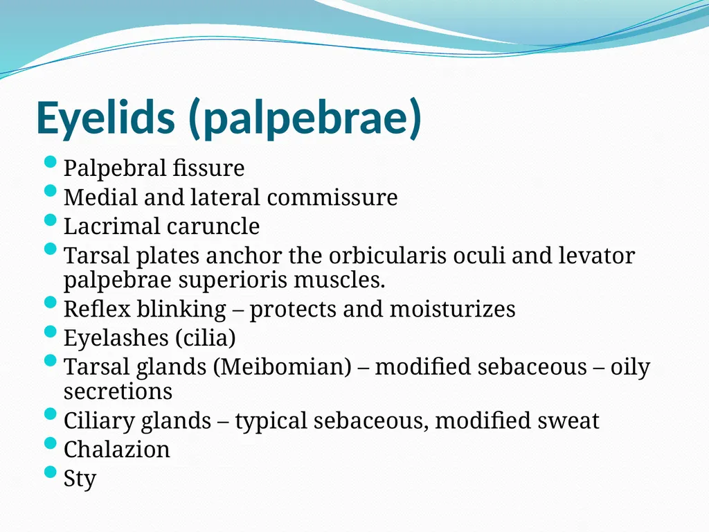
slide7. Eyelids (palpebrae) Palpebral fissure
Medial and lateral commissure
Lacrimal caruncle
Tarsal plates anchor the orbicularis oculi and levator palpebrae superioris muscles.
Reflex blinking – protects and moisturizes
Eyelashes (cilia)
Tarsal glands (Meibomian) – modified sebaceous – oily secretions
Ciliary glands – typical sebaceous, modified sweat
Chalazion
Sty<br>
slide8. Conjunctiva Palpebral conjunctiva
Bulbar conjunctiva
Conjunctiva sac – contact lens
Produces mucus
conjunctivitis<br>
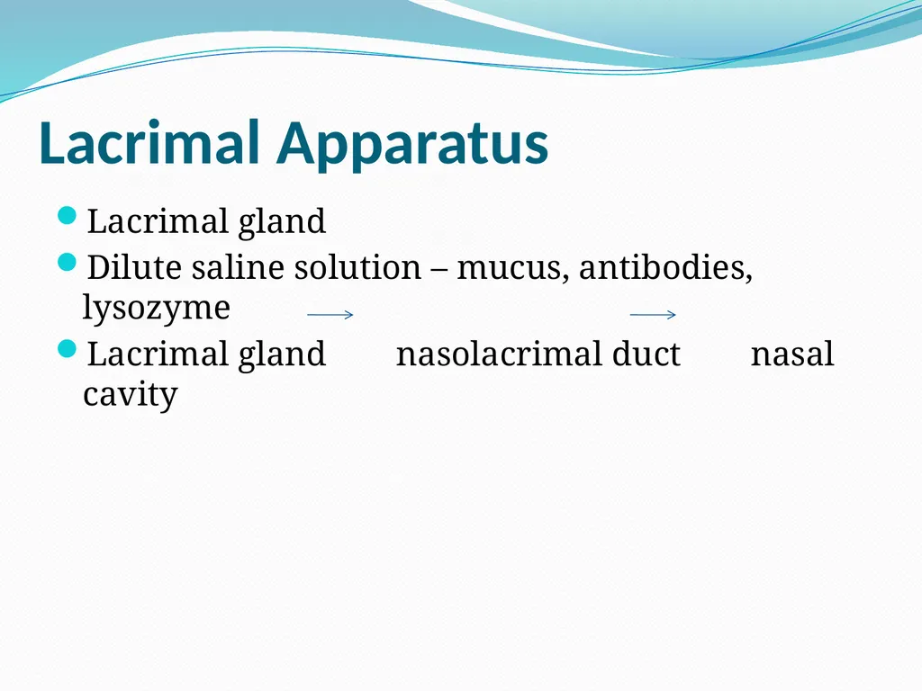
slide9. Lacrimal Apparatus Lacrimal gland
Dilute saline solution – mucus, antibodies, lysozyme
Lacrimal gland nasolacrimal duct nasal cavity<br>
slide10. Extrinsic Eye Muscles<br>
slide12. Structure of The Eye Ball Anterior pole
Posterior pole
Optic axis
Visual axis<br>
slide13. Layers Forming Wall of Eye Ball Fibrous layer
Vascular layer
Inner layer (neural layer)<br>
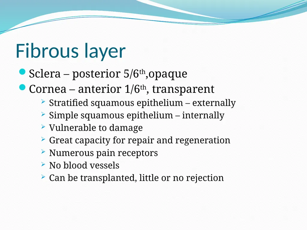
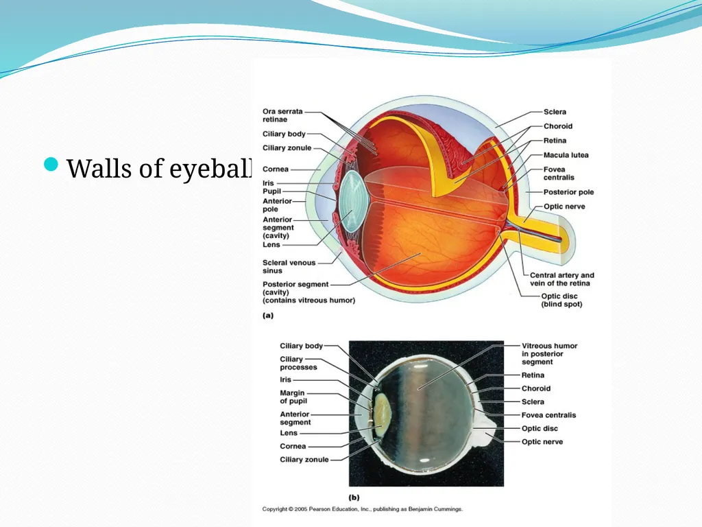
slide14. Fibrous layer Sclera – posterior 5/6th,opaque
Cornea – anterior 1/6th, transparent
Stratified squamous epithelium – externally
Simple squamous epithelium – internally
Vulnerable to damage
Great capacity for repair and regeneration
Numerous pain receptors
No blood vessels
Can be transplanted, little or no rejection<br>
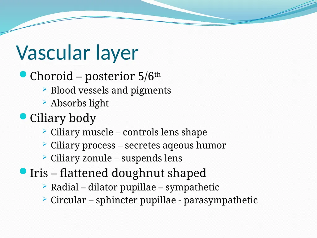
slide15. Vascular layer Choroid – posterior 5/6th
Blood vessels and pigments
Absorbs light
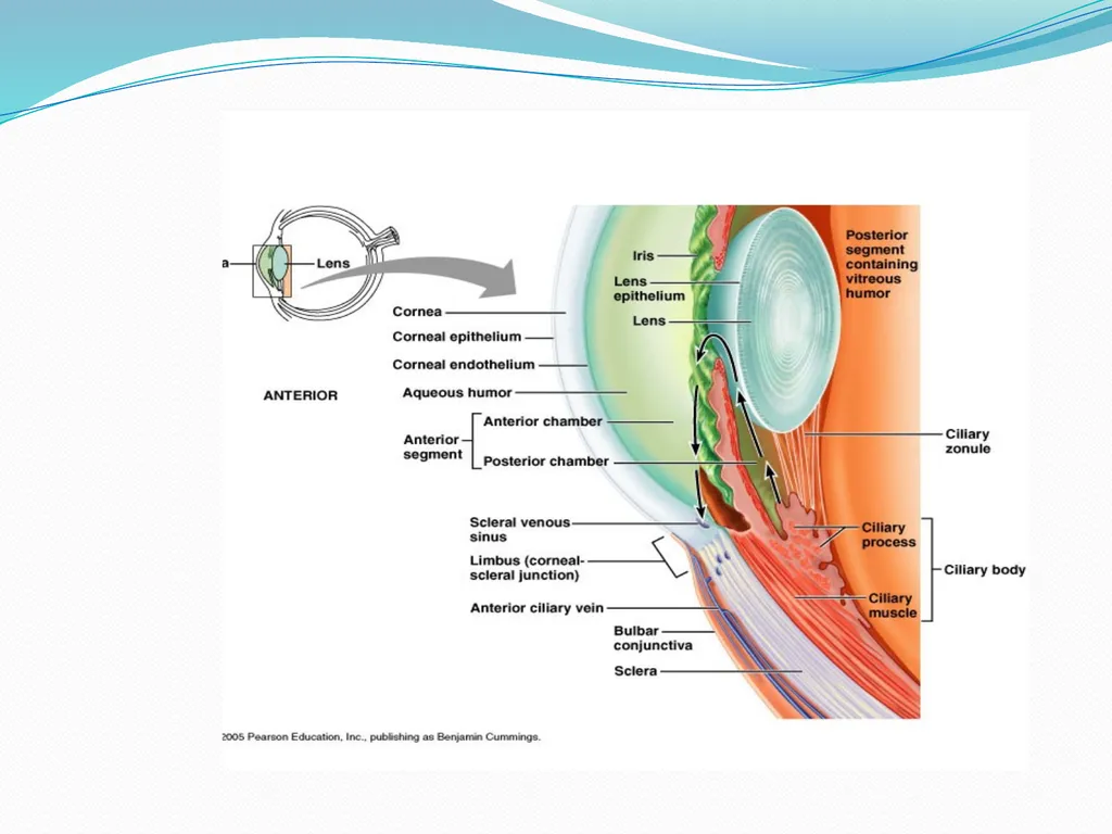
Ciliary body
Ciliary muscle – controls lens shape
Ciliary process – secretes aqeous humor
Ciliary zonule – suspends lens
Iris – flattened doughnut shaped
Radial – dilator pupillae – sympathetic
Circular – sphincter pupillae - parasympathetic<br>
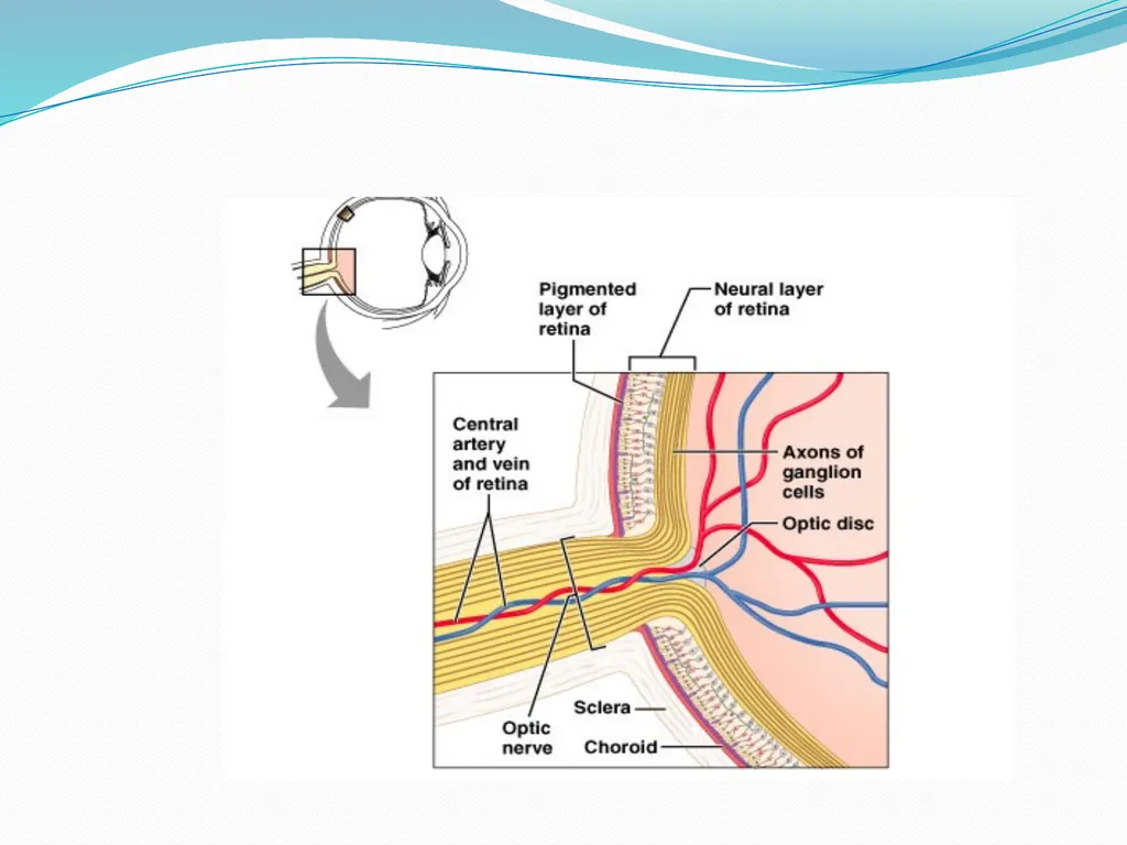
slide16. Walls of eyeball<br>
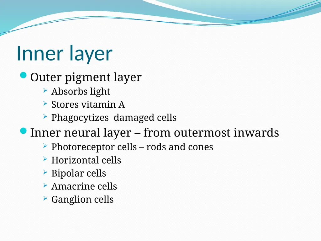
slide17. Inner layer Outer pigment layer
Absorbs light
Stores vitamin A
Phagocytizes damaged cells
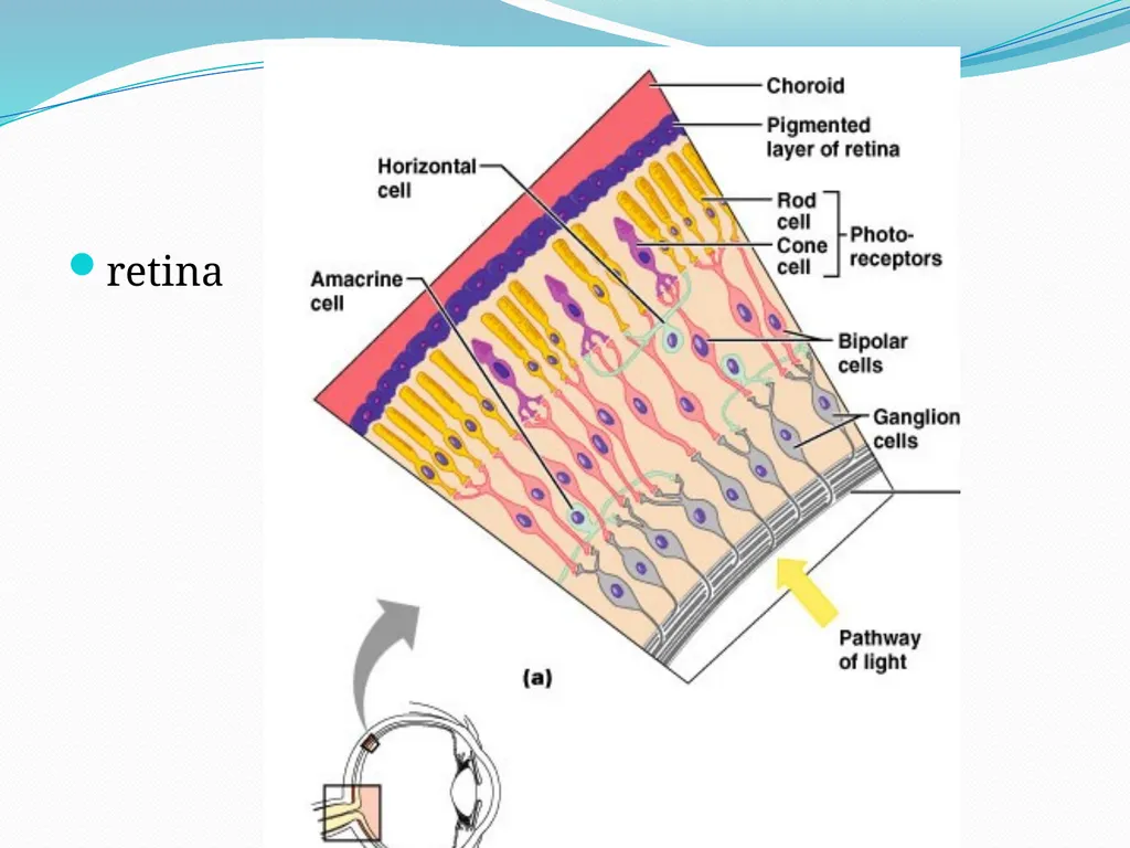
Inner neural layer – from outermost inwards
Photoreceptor cells – rods and cones
Horizontal cells
Bipolar cells
Amacrine cells
Ganglion cells<br>
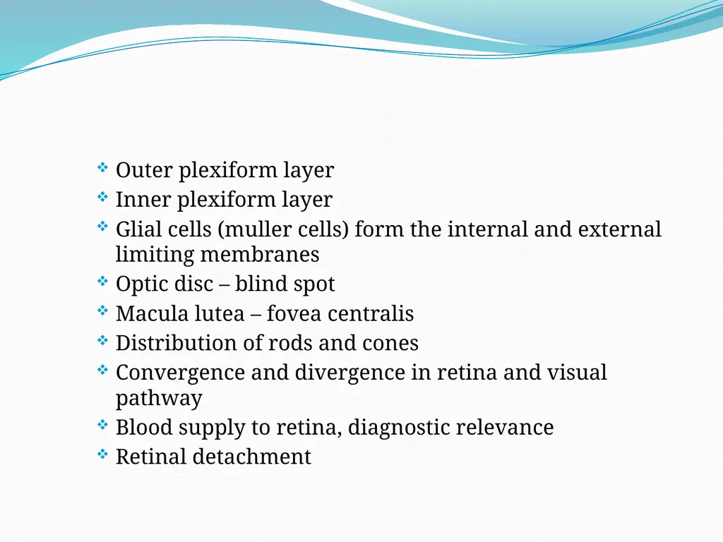
slide18. Outer plexiform layer
Inner plexiform layer
Glial cells (muller cells) form the internal and external limiting membranes
Optic disc – blind spot
Macula lutea – fovea centralis
Distribution of rods and cones
Convergence and divergence in retina and visual pathway
Blood supply to retina, diagnostic relevance
Retinal detachment<br>
slide19. retina<br>
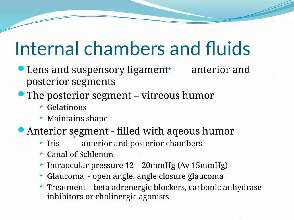
slide21. Internal chambers and fluids Lens and suspensory ligament anterior and posterior segments
The posterior segment – vitreous humor
Gelatinous
Maintains shape
Anterior segment - filled with aqeous humor
Iris anterior and posterior chambers
Canal of Schlemm
Intraocular pressure 12 – 20mmHg (Av 15mmHg)
Glaucoma - open angle, angle closure glaucoma
Treatment – beta adrenergic blockers, carbonic anhydrase inhibitors or cholinergic agonists<br>
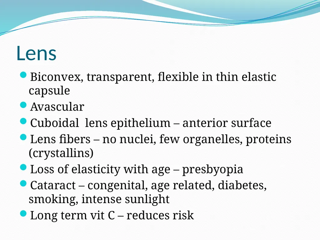
slide23. Lens Biconvex, transparent, flexible in thin elastic capsule
Avascular
Cuboidal lens epithelium – anterior surface
Lens fibers – no nuclei, few organelles, proteins (crystallins)
Loss of elasticity with age – presbyopia
Cataract – congenital, age related, diabetes, smoking, intense sunlight
Long term vit C – reduces risk<br>
slide24. PHYSIOLOGY OF VISION<br>
slide25. Overview of light and optics Electromagnetic spectrum – all energy waves from long radio waves(m) to very short gamma and x – rays(≤1nm)
Visible light – 400 – 700nm
Light energy - photons
Prism – visible spectrum
Red – longest wavelength, lowest energy
Violet – shortest wavelength, greatest energy
Green object – absorbs other wavelengths, reflects green
Black absorbs all, white reflects all<br>
slide26. Speed of light – 300,000km/s increases in less dense medium, decreases denser medium
Refraction of oblique rays at media interface
Convex lens – converges
Concave lens – diverges<br>
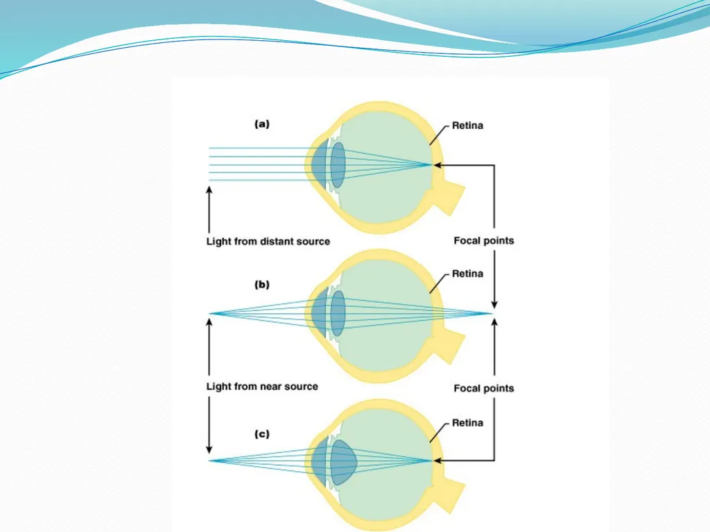
slide27. Focusing of light on the retina Far point of vision(distance beyond which no change in lens shape is required for focusing – 6m(20ft),ciliary muscles relaxed, ciliary zonule tense, lens thin and at lowest refractory power
Close vision – light from close objects diverge, to focus on retina requires:
Accommodation of the lenses
Constriction of the pupil
Convergence of the eyeballs =Near Response
Pupillary light reflexes – direct and consensual
Argyll Robertson pupil<br>
slide29. Near point of vision – the nearest point from the eye at which an object can be brought clearly into focus. Can change from 10cm at 10yrs to 83cm at 60yrs
Pupillary aperture 1.5 – 8mm
Decrease in size – miosis(parasymp)
Increase in size – mydriasis(symp)<br>
slide30. Errors of refraction Mainly due to eyeball shape
Myopia – near sightedness, eyeball too long, near point of vision nearer than normal, correction concave lens
Hyperopia – far sightedness, eyeball too short, far point of vision farther than normal, correction convex lens
Astigmatism – unequal curvatures in cornea or lens, corrected by cylindrical lenses, corneal implants, or laser procedures<br>
slide31. Photoreceptors and phototransduction Functional anatomy of rods and cones
Outer segment – membrane discs(rods) and shelves(cones), visual pigments, Na and Ca ion channels open in dark by cGMP, close in light
Inner segment – cell organelles e.g. nucleus and mitochondria, synthesis of new pigments, provision of energy
Synaptic body or terminal – neurotransmitter - filled vesicles<br>
slide32. Rods Cones More sensitive – one photon sufficient
Sensitive to light from all directions
Best suited for peripheral and night(scotopic) vision
Single type of visual pigment – no color vision
Easily saturated – hyperpolarization is sustained for a longer time
More convergent – 100: 1(ganglion cell) – fuzzy, indistinct vision Less sensitive – 100s of photons required
Light must fall along it’s axis
For vision in bright light(photopic)
3 different types of pigments – t/4 mediates color vision
Hyperpolarize briefly, regenerate pigments faster t/4 mediate variations in light intensity
Less convergent – few: 1 or 1:1 – high resolution, detailed vision<br>
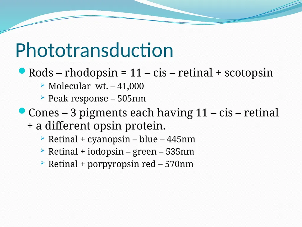
slide33. Phototransduction Rods – rhodopsin = 11 – cis – retinal + scotopsin
Molecular wt. – 41,000
Peak response – 505nm
Cones – 3 pigments each having 11 – cis – retinal + a different opsin protein.
Retinal + cyanopsin – blue – 445nm
Retinal + iodopsin – green – 535nm
Retinal + porpyropsin red – 570nm<br>
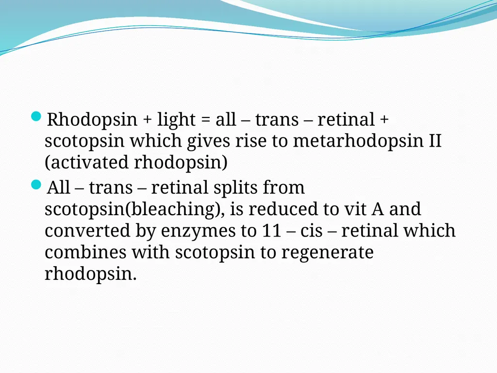
slide34. Rhodopsin + light = all – trans – retinal + scotopsin which gives rise to metarhodopsin II (activated rhodopsin)
All – trans – retinal splits from scotopsin(bleaching), is reduced to vit A and converted by enzymes to 11 – cis – retinal which combines with scotopsin to regenerate rhodopsin.<br>
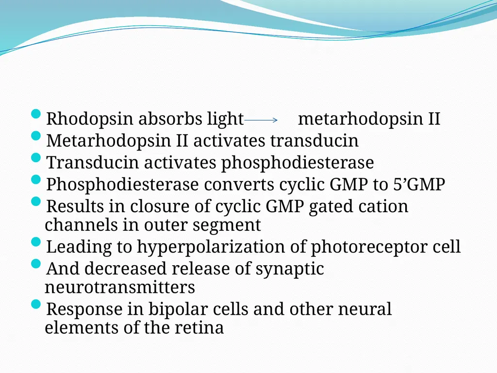
slide35. Rhodopsin absorbs light metarhodopsin II
Metarhodopsin II activates transducin
Transducin activates phosphodiesterase
Phosphodiesterase converts cyclic GMP to 5’GMP
Results in closure of cyclic GMP gated cation channels in outer segment
Leading to hyperpolarization of photoreceptor cell
And decreased release of synaptic neurotransmitters
Response in bipolar cells and other neural elements of the retina<br>
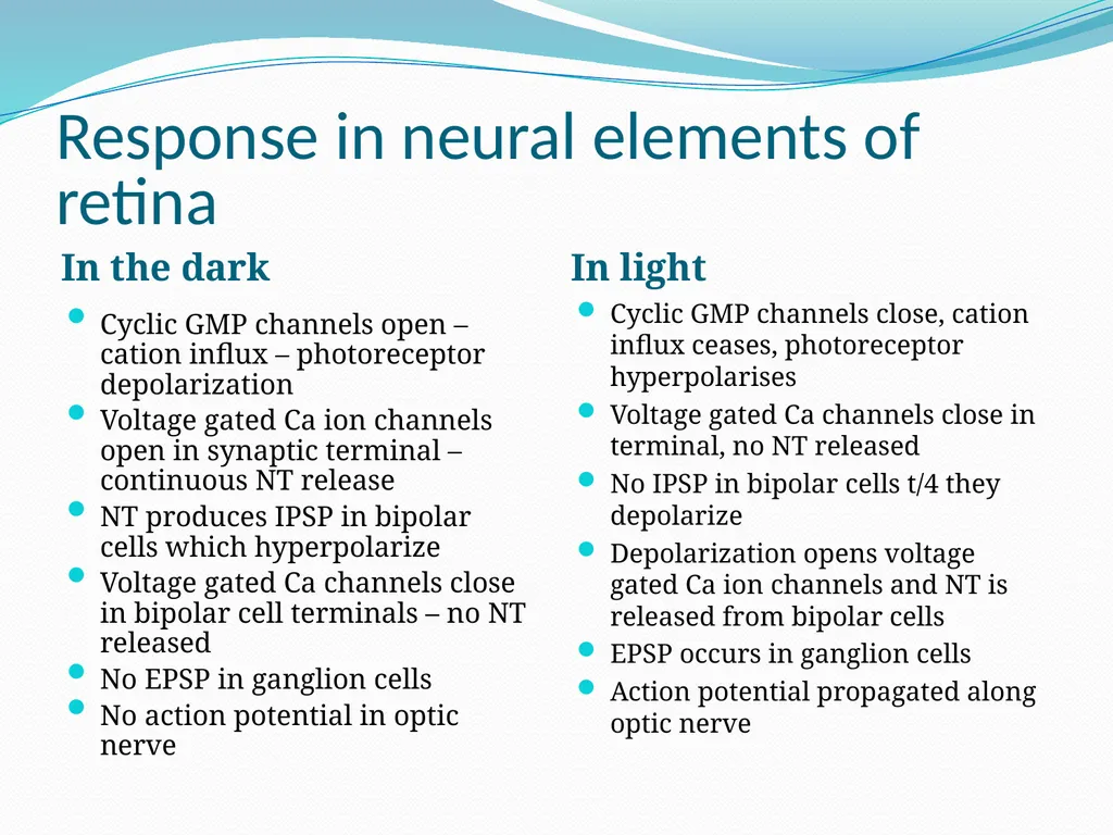
slide36. Response in neural elements of retina In the dark In light Cyclic GMP channels open – cation influx – photoreceptor depolarization
Voltage gated Ca ion channels open in synaptic terminal – continuous NT release
NT produces IPSP in bipolar cells which hyperpolarize
Voltage gated Ca channels close in bipolar cell terminals – no NT released
No EPSP in ganglion cells
No action potential in optic nerve Cyclic GMP channels close, cation influx ceases, photoreceptor hyperpolarises
Voltage gated Ca channels close in terminal, no NT released
No IPSP in bipolar cells t/4 they depolarize
Depolarization opens voltage gated Ca ion channels and NT is released from bipolar cells
EPSP occurs in ganglion cells
Action potential propagated along optic nerve<br>
slide37. Color vision Young – Helmholtz trichromatic theory – the perception of any color depends on the ratio of signals arising from each type of cone
Orange – red(99%), green(42%), blue(0%)
Yellow – red(83%), green(83%), blue(0%)
Color blindness
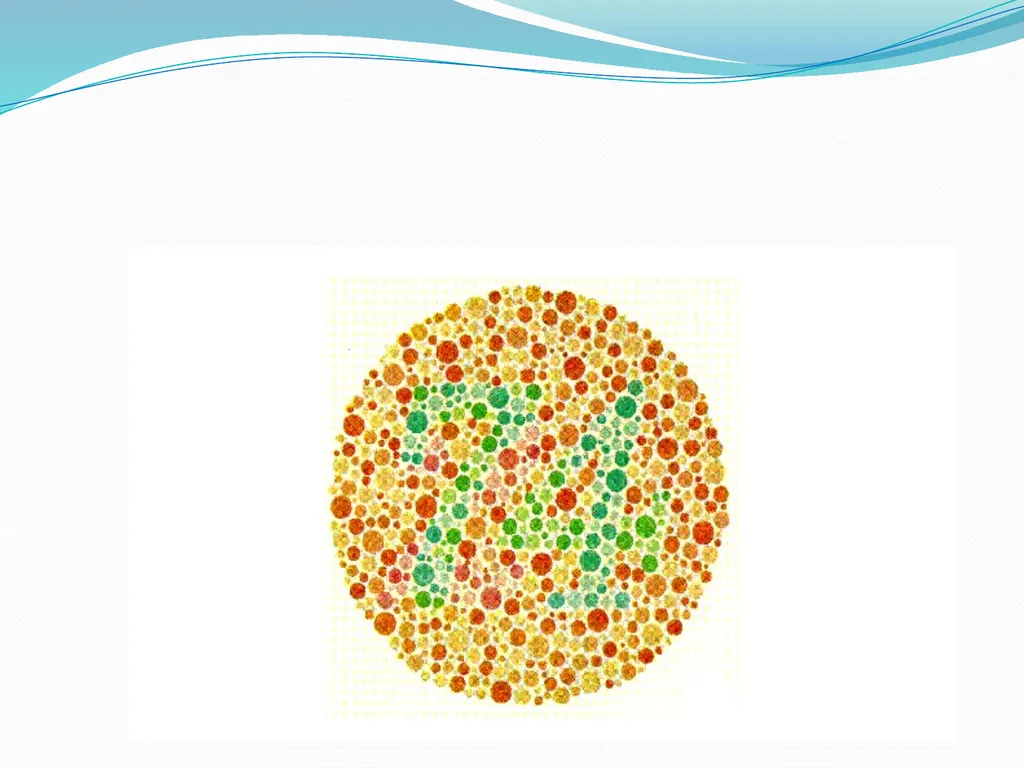
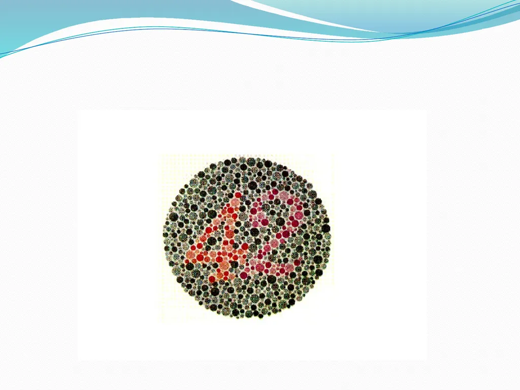
Color vision is tested with Ishihara charts
Inherited as an X – linked recessive trait
More common in males, females mainly carriers
Red/green color blindness is most common<br>
slide40. Melanopsin A small subset of ganglion cells contain the visual pigment melanopsin(circadian pigment)
These ganglion cells respond directly to light
Project directly to the pretectal nuclei which mediates pupillary reflexes
And to the suprachiasmatic nucleus of hypothalamus which mediates circadian rhythms in response to daytime and night<br>
slide41. Dark and light adaptation Dark adaptation
Moving from bright place to dimly – lit place
One is not able to see for some time but begins to see slowly
Max. duration – 20mins
It occurs as a result of : time required for increased rod sensitivity b/c of rhodopsin regeneration and dilatation of the pupils
Light adaptation
moving from dim to brightly lit room produces a dazzling effect and after sometime the eyes adapt and one can see without discomfort
It is due to : decreased sensitivity of rods and constriction of the pupils<br>
slide42. The visual pathway Visual field – the view seen by one eye without movement of the head
Binocular vision – the visual fields of both eyes overlap
Corresponding points on the retina
Diplopia results when light from an object in the binocular field of vision does not fall on corresponding points of the retinae. Possible causes include:
Paralysis of ocular muscles e.g. myaesthenia gravis
Alcoholic intoxication
Lesion in 3rd 4th and 6th cranial nerves, oculomotor nucleus and cerebral peduncles<br>
slide43. The visual field is divided into four parts
Temporal field(lateral)
Nasal field(medial)
Upper field
Lower field
There is a total inversion of the image formed on the retina i.e. top becomes bottom, lateral becomes medial so:
Light rays from temporal field falls on nasal half of retina and vice versa
Light rays from upper right quadrant of field fall on the lower left quadrant of the retina etc.
Light from the center of the visual field fall on the macula(fovea
The shape and extent of the visual field is mapped using the Goldman perimeter, Bjerrum Tangent screen or the confrontation test<br>
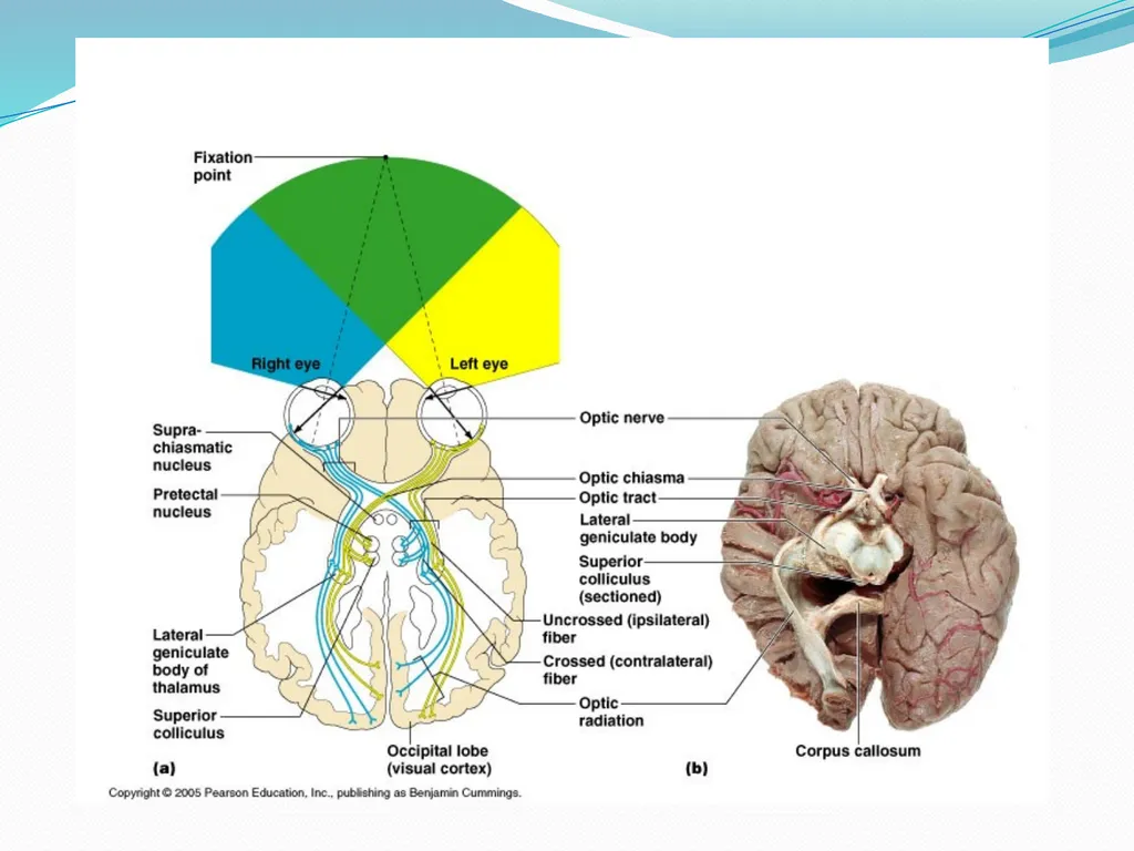
slide45. The visual pathway The visual pathway consists of:
The optic nerve
Optic chiasma
Optic tract
Lateral geniculate body
Optic radiation
Visual cortex<br>
slide46. The optic nerve Leaves retina at optic disc
Fibers from temporal part of retina is in lateral part of nerve and carry impulses from nasal half of visual field
Fibers from nasal part of retina is in the medial part of the nerve and carry impulses from the temporal half of the visual field<br>
slide47. The optic chiasma Medial fibers of each optic nerve cross the midline at the optic chiasma to join the uncrossed lateral fibers of the opposite optic nerve to form the optic tract<br>
slide48. The optic tract Contains fibers from lateral side of ipsilateral retina and fibers from medial side of contralateral retina
Conveys information from the part of the retina on the same(homonymous) side which receives light from the contralateral half of the visual field<br>
slide49. The lateral geniculate body Most fibers of the optic tract end in the LGB of the thalamus where they synapse with neurons of the lat. Geniculate nuclei whose axons form the geniculo – calcarine tract(optic radiation)
Laminated organization of the nuclei of the LGB
Somatotopic representation of the retina to the LGB such that:
Crossed fibers from contralateral retina terminates on laminae 1,4 and 6
Uncrossed fibers terminate in laminae 2,3 and 5<br>
slide50. Some optic tract fibers do not terminate in the LGB but in one of the following areas:
In the superior colliculus – coordinates reflex ocular and head movements in response to visual stimulus
Pretectal area of midbrain – mediates pupillary reflexes
Suprachiasmatic nucleus and tuber cinereum of the hypothalamus – mediates hypothalamic functions related to circadian rhythms<br>
slide51. The visual cortex Primary visual cortex – forms walls and lips of the calcarine fissure in the medial surface of occipital lobes
It consists of primary, secondary and visual association areas
There is somatotopic representation of the retina in the primary visual cortex such that
Upper quadrant of homonymous retinas – above the calcarine fissure calcarine(cuneus gyrus)
Lower quadrant of homonymous retinas – below calcarine fissure(lingular gyrus)
Cortical area for the macula is disproportionately large – occupies posterior 3rd of the of calcarine cortex<br>
slide52. The primary visual – area 17 – perception of visual impulses
Area 18 – interpretation of visual impulses
Area 19 – movement of the eye<br>
slide53. Effect of lesions of the visual pathway Optic nerve – blindness of affected eye
Optic chiasma – bitemporal hemianopia
Right Optic tract – left homonymous hemianopia and vice versa
Optic radiation and visual cortex – contralateral homonymous hemianopia
Damage to lingular gyrus of right cerebral hemisphere – damages fibers from the right lower quadrants of the two retinas<br>
slide54. Macula sparing Lesions that damage visual cortex often spare macula vision b/c fibers from the macula separate from those from peripheral retina and terminate in a large posterior area of the visual cortex.<br>
slide55. Visual processing Retinal processing
Thalamic processing
Cortical processing<br>
slide56. Retinal processing Ganglion cells generate action potential sat a fairly steady rate 20 – 30/sec even in the dark – basal rate
Basal rate does not change when retina is evenly illuminated
Activity of individual ganglion cells changes dramatically when a particular pattern of light falls on it’s receptive field.
The receptive field is that part of the retina that when stimulated influences the activity of the ganglion cell
Different ganglion cells detect different light patterns e.g. lines at a particular angle, or moving in particular direction at a particular speed. The simplest being a spot of light<br>
slide57. Ganglion cells receiving input from rods Have two types (circle within a circle) of receptive fields based on what happens to the ganglion cell when the center of it’s receptive field is illuminated with a spot of light
On - center fields
Off - center fields<br>
slide58. Ganglion cells with on – center
Depolarize(stimulated) when light falls on photoreceptors in the center of their receptive field
Inhibited when light falls on photoreceptors in the periphery
Ganglion cells with off – center
Depolarize(stimulated) when light falls on photoreceptors in the periphery of their receptive field
Inhibited when light falls on photoreceptors in the center of their receptive field
Activation of a neural unit associated with the inhibition of nearby units is an example of lateral or afferent inhibition. It serves to sharpen the edges of a stimulus and improve discrimination<br>
slide59. Thalamic processing The ganglion cells project a detailed spatial representation of the retina on the LGB.
The LGB has 6 layers receiving input from two types of ganglion cells
Small ganglion cells – parvo or P cells relay signals to layers 3 – 6 of the LGB called the parvocellular layers and contains small cells. The P ganglion cells subtract input from one type of cone from input from other types and are concerned with color, texture and shape
Large, magno or M ganglion cells project to layers 1 and 2 of LGB(magnocellular with large cells). These ganglion cells summate responses from different kinds of cones and are concerned with movement and stereopsis(depth perception)
Cells in the interlaminar regions of the LGB also receive input from P ganglion cells and project through a separate component of P pathway to the blobs in the visual cortex<br>
slide60. Cortical processing Like the retina to the LGB, the LGB transmits a similar point for point representation on the primary visual cortex. Many nerve cells in the visual cortex are associated with each incoming fiber and it has 6 layers
Axons from interlaminar regions of LGB end in layers 2 and 3 in cell clusters with high cytochrome oxidase concentration called blobs – concerned with color vision.
Left visual cortex receives input from right visual fields and vice versa<br>
slide61. Two types of areas for processing retinal input are found in the visual cortex
Primary visual cortex(striate cortex, area 17) – responds to dark and bright edges (contrast information) object orientation and provides form, color and motion inputs to visual association areas
Visual association areas(prestriate cortices, areas 18 and 19) continues processing of visual information concerned with form, color and movement<br>
slide62. Complex visual processing extends beyond the occipital lobe to the temporal, parietal and frontal lobes along 2 parallel lines
The ‘what' processing stream extends through the ventral part of the temporal lobe – identification of objects i.e. shape, recognition of forms and faces
The ‘where’ takes a dorsal path through the parietal cortex to the post central gyrus, uses information from primary visual cortex to assess spatial location of objects and motion
Output from these pathways pass to the frontal cortex which uses the information to direct activities
Other parts of the cortex and subcortical structures activated by visual stimuli are the amygdala, pulvinar, caudate nuclei, putamen and claustrum<br>
slide2. INTRODUCTION
ACCESSORY STRUCTURES OF THE EYE
STRUCTURE OF THE EYE BALL
PHYSIOLOGY OF VISION
OVERVIEW OF LIGHT AND OPTICS
FOCUSING OF LIGHT ON THE RETINA
PHOTORECEPTORS AND PHOTOTRANSDUCTION
THE VISUAL PATHWAY
VISUAL PROCESSING<br>
slide3. Introduction Vision is our dominant sense
Up to 70% of all body sensory receptors are in the eye.
½ of cerebral cortex partakes in some form of visual processing
The adult eye is a sphere of about 2.5cm diameter.
Only the anterior 1/6th of the eye is visible
The rest is enclosed in and protected by a cushion of fat in the bony orbit.<br>
slide4. Accessory Structures of the Eye Eye brows
Eye lids
Conjunctiva
Lacrimal apparatus
Extrinsic muscles of the eye<br>
slide6. Eyebrows Short coarse hairs over lying supra orbital margin
Sun shade
Keep out perspiration<br>
slide7. Eyelids (palpebrae) Palpebral fissure
Medial and lateral commissure
Lacrimal caruncle
Tarsal plates anchor the orbicularis oculi and levator palpebrae superioris muscles.
Reflex blinking – protects and moisturizes
Eyelashes (cilia)
Tarsal glands (Meibomian) – modified sebaceous – oily secretions
Ciliary glands – typical sebaceous, modified sweat
Chalazion
Sty<br>
slide8. Conjunctiva Palpebral conjunctiva
Bulbar conjunctiva
Conjunctiva sac – contact lens
Produces mucus
conjunctivitis<br>
slide9. Lacrimal Apparatus Lacrimal gland
Dilute saline solution – mucus, antibodies, lysozyme
Lacrimal gland nasolacrimal duct nasal cavity<br>
slide10. Extrinsic Eye Muscles<br>
slide12. Structure of The Eye Ball Anterior pole
Posterior pole
Optic axis
Visual axis<br>
slide13. Layers Forming Wall of Eye Ball Fibrous layer
Vascular layer
Inner layer (neural layer)<br>
slide14. Fibrous layer Sclera – posterior 5/6th,opaque
Cornea – anterior 1/6th, transparent
Stratified squamous epithelium – externally
Simple squamous epithelium – internally
Vulnerable to damage
Great capacity for repair and regeneration
Numerous pain receptors
No blood vessels
Can be transplanted, little or no rejection<br>
slide15. Vascular layer Choroid – posterior 5/6th
Blood vessels and pigments
Absorbs light
Ciliary body
Ciliary muscle – controls lens shape
Ciliary process – secretes aqeous humor
Ciliary zonule – suspends lens
Iris – flattened doughnut shaped
Radial – dilator pupillae – sympathetic
Circular – sphincter pupillae - parasympathetic<br>
slide16. Walls of eyeball<br>
slide17. Inner layer Outer pigment layer
Absorbs light
Stores vitamin A
Phagocytizes damaged cells
Inner neural layer – from outermost inwards
Photoreceptor cells – rods and cones
Horizontal cells
Bipolar cells
Amacrine cells
Ganglion cells<br>
slide18. Outer plexiform layer
Inner plexiform layer
Glial cells (muller cells) form the internal and external limiting membranes
Optic disc – blind spot
Macula lutea – fovea centralis
Distribution of rods and cones
Convergence and divergence in retina and visual pathway
Blood supply to retina, diagnostic relevance
Retinal detachment<br>
slide19. retina<br>
slide21. Internal chambers and fluids Lens and suspensory ligament anterior and posterior segments
The posterior segment – vitreous humor
Gelatinous
Maintains shape
Anterior segment - filled with aqeous humor
Iris anterior and posterior chambers
Canal of Schlemm
Intraocular pressure 12 – 20mmHg (Av 15mmHg)
Glaucoma - open angle, angle closure glaucoma
Treatment – beta adrenergic blockers, carbonic anhydrase inhibitors or cholinergic agonists<br>
slide23. Lens Biconvex, transparent, flexible in thin elastic capsule
Avascular
Cuboidal lens epithelium – anterior surface
Lens fibers – no nuclei, few organelles, proteins (crystallins)
Loss of elasticity with age – presbyopia
Cataract – congenital, age related, diabetes, smoking, intense sunlight
Long term vit C – reduces risk<br>
slide24. PHYSIOLOGY OF VISION<br>
slide25. Overview of light and optics Electromagnetic spectrum – all energy waves from long radio waves(m) to very short gamma and x – rays(≤1nm)
Visible light – 400 – 700nm
Light energy - photons
Prism – visible spectrum
Red – longest wavelength, lowest energy
Violet – shortest wavelength, greatest energy
Green object – absorbs other wavelengths, reflects green
Black absorbs all, white reflects all<br>
slide26. Speed of light – 300,000km/s increases in less dense medium, decreases denser medium
Refraction of oblique rays at media interface
Convex lens – converges
Concave lens – diverges<br>
slide27. Focusing of light on the retina Far point of vision(distance beyond which no change in lens shape is required for focusing – 6m(20ft),ciliary muscles relaxed, ciliary zonule tense, lens thin and at lowest refractory power
Close vision – light from close objects diverge, to focus on retina requires:
Accommodation of the lenses
Constriction of the pupil
Convergence of the eyeballs =Near Response
Pupillary light reflexes – direct and consensual
Argyll Robertson pupil<br>
slide29. Near point of vision – the nearest point from the eye at which an object can be brought clearly into focus. Can change from 10cm at 10yrs to 83cm at 60yrs
Pupillary aperture 1.5 – 8mm
Decrease in size – miosis(parasymp)
Increase in size – mydriasis(symp)<br>
slide30. Errors of refraction Mainly due to eyeball shape
Myopia – near sightedness, eyeball too long, near point of vision nearer than normal, correction concave lens
Hyperopia – far sightedness, eyeball too short, far point of vision farther than normal, correction convex lens
Astigmatism – unequal curvatures in cornea or lens, corrected by cylindrical lenses, corneal implants, or laser procedures<br>
slide31. Photoreceptors and phototransduction Functional anatomy of rods and cones
Outer segment – membrane discs(rods) and shelves(cones), visual pigments, Na and Ca ion channels open in dark by cGMP, close in light
Inner segment – cell organelles e.g. nucleus and mitochondria, synthesis of new pigments, provision of energy
Synaptic body or terminal – neurotransmitter - filled vesicles<br>
slide32. Rods Cones More sensitive – one photon sufficient
Sensitive to light from all directions
Best suited for peripheral and night(scotopic) vision
Single type of visual pigment – no color vision
Easily saturated – hyperpolarization is sustained for a longer time
More convergent – 100: 1(ganglion cell) – fuzzy, indistinct vision Less sensitive – 100s of photons required
Light must fall along it’s axis
For vision in bright light(photopic)
3 different types of pigments – t/4 mediates color vision
Hyperpolarize briefly, regenerate pigments faster t/4 mediate variations in light intensity
Less convergent – few: 1 or 1:1 – high resolution, detailed vision<br>
slide33. Phototransduction Rods – rhodopsin = 11 – cis – retinal + scotopsin
Molecular wt. – 41,000
Peak response – 505nm
Cones – 3 pigments each having 11 – cis – retinal + a different opsin protein.
Retinal + cyanopsin – blue – 445nm
Retinal + iodopsin – green – 535nm
Retinal + porpyropsin red – 570nm<br>
slide34. Rhodopsin + light = all – trans – retinal + scotopsin which gives rise to metarhodopsin II (activated rhodopsin)
All – trans – retinal splits from scotopsin(bleaching), is reduced to vit A and converted by enzymes to 11 – cis – retinal which combines with scotopsin to regenerate rhodopsin.<br>
slide35. Rhodopsin absorbs light metarhodopsin II
Metarhodopsin II activates transducin
Transducin activates phosphodiesterase
Phosphodiesterase converts cyclic GMP to 5’GMP
Results in closure of cyclic GMP gated cation channels in outer segment
Leading to hyperpolarization of photoreceptor cell
And decreased release of synaptic neurotransmitters
Response in bipolar cells and other neural elements of the retina<br>
slide36. Response in neural elements of retina In the dark In light Cyclic GMP channels open – cation influx – photoreceptor depolarization
Voltage gated Ca ion channels open in synaptic terminal – continuous NT release
NT produces IPSP in bipolar cells which hyperpolarize
Voltage gated Ca channels close in bipolar cell terminals – no NT released
No EPSP in ganglion cells
No action potential in optic nerve Cyclic GMP channels close, cation influx ceases, photoreceptor hyperpolarises
Voltage gated Ca channels close in terminal, no NT released
No IPSP in bipolar cells t/4 they depolarize
Depolarization opens voltage gated Ca ion channels and NT is released from bipolar cells
EPSP occurs in ganglion cells
Action potential propagated along optic nerve<br>
slide37. Color vision Young – Helmholtz trichromatic theory – the perception of any color depends on the ratio of signals arising from each type of cone
Orange – red(99%), green(42%), blue(0%)
Yellow – red(83%), green(83%), blue(0%)
Color blindness
Color vision is tested with Ishihara charts
Inherited as an X – linked recessive trait
More common in males, females mainly carriers
Red/green color blindness is most common<br>
slide40. Melanopsin A small subset of ganglion cells contain the visual pigment melanopsin(circadian pigment)
These ganglion cells respond directly to light
Project directly to the pretectal nuclei which mediates pupillary reflexes
And to the suprachiasmatic nucleus of hypothalamus which mediates circadian rhythms in response to daytime and night<br>
slide41. Dark and light adaptation Dark adaptation
Moving from bright place to dimly – lit place
One is not able to see for some time but begins to see slowly
Max. duration – 20mins
It occurs as a result of : time required for increased rod sensitivity b/c of rhodopsin regeneration and dilatation of the pupils
Light adaptation
moving from dim to brightly lit room produces a dazzling effect and after sometime the eyes adapt and one can see without discomfort
It is due to : decreased sensitivity of rods and constriction of the pupils<br>
slide42. The visual pathway Visual field – the view seen by one eye without movement of the head
Binocular vision – the visual fields of both eyes overlap
Corresponding points on the retina
Diplopia results when light from an object in the binocular field of vision does not fall on corresponding points of the retinae. Possible causes include:
Paralysis of ocular muscles e.g. myaesthenia gravis
Alcoholic intoxication
Lesion in 3rd 4th and 6th cranial nerves, oculomotor nucleus and cerebral peduncles<br>
slide43. The visual field is divided into four parts
Temporal field(lateral)
Nasal field(medial)
Upper field
Lower field
There is a total inversion of the image formed on the retina i.e. top becomes bottom, lateral becomes medial so:
Light rays from temporal field falls on nasal half of retina and vice versa
Light rays from upper right quadrant of field fall on the lower left quadrant of the retina etc.
Light from the center of the visual field fall on the macula(fovea
The shape and extent of the visual field is mapped using the Goldman perimeter, Bjerrum Tangent screen or the confrontation test<br>
slide45. The visual pathway The visual pathway consists of:
The optic nerve
Optic chiasma
Optic tract
Lateral geniculate body
Optic radiation
Visual cortex<br>
slide46. The optic nerve Leaves retina at optic disc
Fibers from temporal part of retina is in lateral part of nerve and carry impulses from nasal half of visual field
Fibers from nasal part of retina is in the medial part of the nerve and carry impulses from the temporal half of the visual field<br>
slide47. The optic chiasma Medial fibers of each optic nerve cross the midline at the optic chiasma to join the uncrossed lateral fibers of the opposite optic nerve to form the optic tract<br>
slide48. The optic tract Contains fibers from lateral side of ipsilateral retina and fibers from medial side of contralateral retina
Conveys information from the part of the retina on the same(homonymous) side which receives light from the contralateral half of the visual field<br>
slide49. The lateral geniculate body Most fibers of the optic tract end in the LGB of the thalamus where they synapse with neurons of the lat. Geniculate nuclei whose axons form the geniculo – calcarine tract(optic radiation)
Laminated organization of the nuclei of the LGB
Somatotopic representation of the retina to the LGB such that:
Crossed fibers from contralateral retina terminates on laminae 1,4 and 6
Uncrossed fibers terminate in laminae 2,3 and 5<br>
slide50. Some optic tract fibers do not terminate in the LGB but in one of the following areas:
In the superior colliculus – coordinates reflex ocular and head movements in response to visual stimulus
Pretectal area of midbrain – mediates pupillary reflexes
Suprachiasmatic nucleus and tuber cinereum of the hypothalamus – mediates hypothalamic functions related to circadian rhythms<br>
slide51. The visual cortex Primary visual cortex – forms walls and lips of the calcarine fissure in the medial surface of occipital lobes
It consists of primary, secondary and visual association areas
There is somatotopic representation of the retina in the primary visual cortex such that
Upper quadrant of homonymous retinas – above the calcarine fissure calcarine(cuneus gyrus)
Lower quadrant of homonymous retinas – below calcarine fissure(lingular gyrus)
Cortical area for the macula is disproportionately large – occupies posterior 3rd of the of calcarine cortex<br>
slide52. The primary visual – area 17 – perception of visual impulses
Area 18 – interpretation of visual impulses
Area 19 – movement of the eye<br>
slide53. Effect of lesions of the visual pathway Optic nerve – blindness of affected eye
Optic chiasma – bitemporal hemianopia
Right Optic tract – left homonymous hemianopia and vice versa
Optic radiation and visual cortex – contralateral homonymous hemianopia
Damage to lingular gyrus of right cerebral hemisphere – damages fibers from the right lower quadrants of the two retinas<br>
slide54. Macula sparing Lesions that damage visual cortex often spare macula vision b/c fibers from the macula separate from those from peripheral retina and terminate in a large posterior area of the visual cortex.<br>
slide55. Visual processing Retinal processing
Thalamic processing
Cortical processing<br>
slide56. Retinal processing Ganglion cells generate action potential sat a fairly steady rate 20 – 30/sec even in the dark – basal rate
Basal rate does not change when retina is evenly illuminated
Activity of individual ganglion cells changes dramatically when a particular pattern of light falls on it’s receptive field.
The receptive field is that part of the retina that when stimulated influences the activity of the ganglion cell
Different ganglion cells detect different light patterns e.g. lines at a particular angle, or moving in particular direction at a particular speed. The simplest being a spot of light<br>
slide57. Ganglion cells receiving input from rods Have two types (circle within a circle) of receptive fields based on what happens to the ganglion cell when the center of it’s receptive field is illuminated with a spot of light
On - center fields
Off - center fields<br>
slide58. Ganglion cells with on – center
Depolarize(stimulated) when light falls on photoreceptors in the center of their receptive field
Inhibited when light falls on photoreceptors in the periphery
Ganglion cells with off – center
Depolarize(stimulated) when light falls on photoreceptors in the periphery of their receptive field
Inhibited when light falls on photoreceptors in the center of their receptive field
Activation of a neural unit associated with the inhibition of nearby units is an example of lateral or afferent inhibition. It serves to sharpen the edges of a stimulus and improve discrimination<br>
slide59. Thalamic processing The ganglion cells project a detailed spatial representation of the retina on the LGB.
The LGB has 6 layers receiving input from two types of ganglion cells
Small ganglion cells – parvo or P cells relay signals to layers 3 – 6 of the LGB called the parvocellular layers and contains small cells. The P ganglion cells subtract input from one type of cone from input from other types and are concerned with color, texture and shape
Large, magno or M ganglion cells project to layers 1 and 2 of LGB(magnocellular with large cells). These ganglion cells summate responses from different kinds of cones and are concerned with movement and stereopsis(depth perception)
Cells in the interlaminar regions of the LGB also receive input from P ganglion cells and project through a separate component of P pathway to the blobs in the visual cortex<br>
slide60. Cortical processing Like the retina to the LGB, the LGB transmits a similar point for point representation on the primary visual cortex. Many nerve cells in the visual cortex are associated with each incoming fiber and it has 6 layers
Axons from interlaminar regions of LGB end in layers 2 and 3 in cell clusters with high cytochrome oxidase concentration called blobs – concerned with color vision.
Left visual cortex receives input from right visual fields and vice versa<br>
slide61. Two types of areas for processing retinal input are found in the visual cortex
Primary visual cortex(striate cortex, area 17) – responds to dark and bright edges (contrast information) object orientation and provides form, color and motion inputs to visual association areas
Visual association areas(prestriate cortices, areas 18 and 19) continues processing of visual information concerned with form, color and movement<br>
slide62. Complex visual processing extends beyond the occipital lobe to the temporal, parietal and frontal lobes along 2 parallel lines
The ‘what' processing stream extends through the ventral part of the temporal lobe – identification of objects i.e. shape, recognition of forms and faces
The ‘where’ takes a dorsal path through the parietal cortex to the post central gyrus, uses information from primary visual cortex to assess spatial location of objects and motion
Output from these pathways pass to the frontal cortex which uses the information to direct activities
Other parts of the cortex and subcortical structures activated by visual stimuli are the amygdala, pulvinar, caudate nuclei, putamen and claustrum<br>