/
Antenatal assessment  of fetal wellbeing Antenatal assessment  of fetal wellbeing

Antenatal assessment of fetal wellbeing - PowerPoint Presentation

zoe
zoe . @zoe
Follow
349 views
Uploaded On 2022-06-15

Antenatal assessment of fetal wellbeing - PPT Presentation

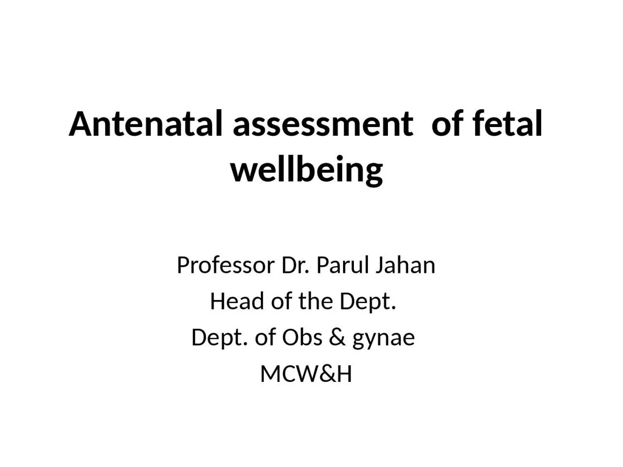
Professor Dr Parul Jahan Head of the Dept Dept of Obs amp gynae MCWampH Definition These are some tools for assessment of fetal condition in ante natal period Antenatal Fetal Monitoring ID: 919553

test fetal fluid pregnancy fetal test pregnancy fluid biophysical movement amniotic nst risk movements msafp stress normal variability weeks

Share:

Link:

Embed:

Download Presentation from below link

Download Presentation The PPT/PDF document "Antenatal assessment of fetal wellbeing" is the property of its rightful owner. Permission is granted to download and print the materials on this web site for personal, non-commercial use only, and to display it on your personal computer provided you do not modify the materials and that you retain all copyright notices contained in the materials. By downloading content from our website, you accept the terms of this agreement.


Presentation Transcript

Slide1

Antenatal assessment of fetal wellbeing

Professor Dr.

Parul

Jahan

Head of the Dept.

Dept. of

Obs

& gynae

MCW&H

Slide2

Definition

These are some tools for assessment of fetal condition in ante natal period.

Slide3

Antenatal Fetal Monitoring

Aims :

• Ensure growth & well being of fetus

• Screen high risk factors that may affect fetal growth

Primary objective :

Avoid fetal death

Slide4

Indications

1)Pregnancy with obstetric complications:

IUGR

Multiple pregnancy

Polyhydramnios

Oligohydramnios

Rhesus

alloimmunization

Slide5

Indications

1)Pregnancy with obstetric complications:

IUGR

Multiple pregnancy

Polyhydramnios

Oligohydramnios

Rhesus

alloimmunization

Slide6

Contd…

2)

Pregnancy with medical complications:

Diabetes mellitus

Hypertension

Epilepsy

Renal or Cardiac disease

Infection (Tuberculosis)

SLE

Slide7

Contd…

3)Others:

Advanced maternal age (> 35 years)

Previous still birth or recurrent abortion Structural (anencephaly,

spina

bifida) Chromosomal abnormalities

4)Routine

antenatal testing

Slide8

Rationality

Tests must provide superior information than clinical evaluation

Should be helpful in management

Benefits of tests must outweigh potential risks and costs.

Slide9

Clinical Assessment

Maternal weight

measurement in each F/U :

2

nd

half of pregnancy - 1 kg a fortnight.

Blood pressure

:

Prior to 12 weeks helps to differentiate a

pre-existing chronic HTN from PIH.

Slide10

Contd…

Symphysio-fundal

height

:

- After 24 weeks corresponds to period of gestation.

- Variation of 1–2 cm acceptable.

Clinical assessment of

liquor

Girth

of abdomen

Slide11

Special investigations

Biochemical

Biophysical

Cytogenetic analysis

Slide12

Biochemical Tests

Maternal serum alpha fetoprotein (MSAFP)

Triple test:

MSAFP, hCG and UE3.

Quadruple test (MSAFP, UE3, Total

hCG

,

Inhibin

A)

Acetylcholine esterase - ↑in open neural tube defects.

Inhibin

A - ↑ in Down’s syndrome

Slide13

First trimester screening:

↑ hCG, ↓ MSAFP, ↓ PAPP

Second trimester screening (15–18 weeks):

Triple test (↓ MSAFP, ↓ UE3, ↑Total hCG) Quadruple test (↓ MSAFP, ↓ UE3,↑Total hCG, ↑

Inhibin

A)

Slide14

Cytogenetic Analysis :

Amniocentesis

Chorion

villus sampling (CVS)

Cordocentesis

Fluorescence In Situ Hybridization

Slide15

Biophysical Tests in late pregnancy

(I) Fetal movement count

(II)

Cardiotocography

(CTG)

(III) Non-stress test (NST)

(IV) USG

(V) Fetal biophysical profile (BPP)

(VI) Doppler ultrasound

(VII)

Vibro

acoustic stimulation test

(VIII) Contraction stress test (CST)

Slide16

Fetal movement count

Any of the two methods can be applied:

Cardif

‘count 10’ formula:

The patient counts fetal movements starting at 9 am.

The counting comes to an end as soon as 10 movements are perceived.

She is instructed to report the physician if—

(

i

) less than 10 movements occur during 12 hours on 2 successive days or

(ii) no movement is perceived even after 12 hours in a single day.

Slide17

• Daily fetal movement count (DFMC): 3 counts each of one hour duration (morning, noon and evening) are recommended.

Total counts multiplied by four gives daily (12 hour) fetal movement count (DFMC).

If there is diminution of the number of ‘kicks’ to less than 10 in 12 hours (or less than 3 in each hour), it indicates fetal compromise.

Slide18

Role of USG

Late Pregnancy :

Measurement of BPD, AC, HC, FL and AFI.

IUGR can be diagnosed accurately with serial measurement of BPD, AC, HC, FL and amniotic fluid volume.

AC is the single measurement which best reflects fetal nutrition

.

When the HC/AC ratio is elevated (> 1.0) after 34 weeks, IUGR is suspected.

Slide19

Non-stress test (NST)

In non-stress test, a continuous electronic monitoring of the fetal heart rate along with recording of fetal movement is undertaken.

Slide20

Fetal Cardiotocography (CTG):

Defition

: Is the graphical presentation of the

foetal

heart activity and the uterine contraction to detect the

foetal

hypoxia.

A normal CTG tracing after 32 weeks,

Base line heart rate of 110–150 b/m

Base line variability 5–25

bpm

.

No deceleration or early deceleration of very short duration.

2 or more accelerations during a 20 minute period

Slide21

Reactive trace with acceleration

Slide22

Cont..

Interpretation

Reactive (Reassuring)—

When 2 or more accelerations of > 15 b/m above baseline and >15 sec in duration present in a 20 minute observation.

Non-reactive (Non-reassuring)

—Absence of any fetal reactivity.

Slide23

Slide24

Persistent late deceleration with loss of variability

Slide25

Interpretation of a CTG

I

. Accelerations and normal baseline variability denote a healthy fetus.

II. Absence of accelerations is the first feature to denote hypoxia.

III. Absence of accelerations, reduced base line variability of < 5

bpm

for > 90 minutes denote a hypoxic fetus.

IV. Decreased baseline variability may be due to fetal sleep,

infection,hypoxia

, anomalies or due to maternal medications.

V. Repeated late decelerations increase the risk of low

Apgar

score and cerebral palsy (CP).

VI.Reduced

baseline variability, with late or variable deceleration increases the risk of CP.

Slide26

Biophysical profile

Definition

: Screening test for

utero

-placental insufficiency.

Pathophysiology

:

Fetal biophysical activities are initiated, modulated and regulated through fetal nervous system.

Fetal CNS is very much sensitive to diminished oxygenation.

Hypoxia → metabolic acidosis → CNS depression → changes in fetal biophysical activity.

Slide27

Fetal Biophysical Profile (BPP)

BPP using real time ultrasonography has a high predictive value for fetal assessment.

Indication

— Non-reactive NST

High risk pregnancy

Test frequency

weekly - normal NST

twice weekly - an abnormal test

Slide28

Total

5

components : Each component carries 2 points. They are ------

NST

Fetal breathing movement

Gross body movement

Fetal muscle tone

Amniotic fluid volume

Slide29

Biophysical profile

Parameters

Minimal normal criteria

BPP Score

Non-stress Test (NST)

Reactive pattern

2

Fetal breathing

movements

≥ 1 episode lasting

> 30 second

2

Gross body

movements

≥ 3 discrete body/limb

movements

2

Fetal muscle

tone

≥ 1 episode of extension (limb or

trunk) with return of flexion

2

Amniotic fluid

≥ 1 pocket measuring 2 cm in

two perpendicular planes

2

Slide30

Modified Biophysical Profile

Consists of

NST

Amniotic fluid index

Slide31

Amniotic fluid volume (AFV)

Amniotic fluid volume is primarily dependent upon the fetal urine, pulmonary fluid production and fetal swallowing.

Decreasing AFV may be the result of fetal hypoxia and

placental insufficiency.

A vertical pocket of amniotic fluid > 2 cm is considered normal.

Slide32

Amniotic fluid index (AFI)

: is the sum of vertical

pockets from 4 quadrants of uterine cavity.

Norma value : 5 - 20

AFI < 5 is associated with increased risk

of

perinatal

mortality and morbidity.

Slide33

Contraction stress test (CST)

Based on the response of the fetus at risk for

utero

placental insufficiency in relation to uterine contractions

Slide34

Doppler Ultrasound Velocimetry

Doppler flow velocity wave forms are obtained from arterial and venous beds in the fetus.

Arterial Doppler :

waveforms are helpful to assess the downstream vascular resistance.

It is used to measure the peak systolic(s), peak diastolic (D) and mean (M) volumes.

From these values

S/D ratio

,

pulsatility

index (PI),

Resistance Index (RI)

are calculated.

Slide35

In a normal pregnancy the S/D ratio

,

PI and RI decreases as the gestational age advances

. Higher values greater than 2 SDs above the gestational age mean indicates reduced diastolic velocities and increased placental vascular resistance. These features are at increased risk for adverse pregnancy outcome.

Slide36

Venous Doppler

provide information about cardiac forward function (cardiac compliance, contractility and after load).

Fetuses with abnormal cardiac function show

pulsatile

flow in the umbilical vein (UV) instead of

monophasic

flow.

Slide37

Slide38

Thank

YoU