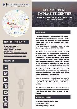
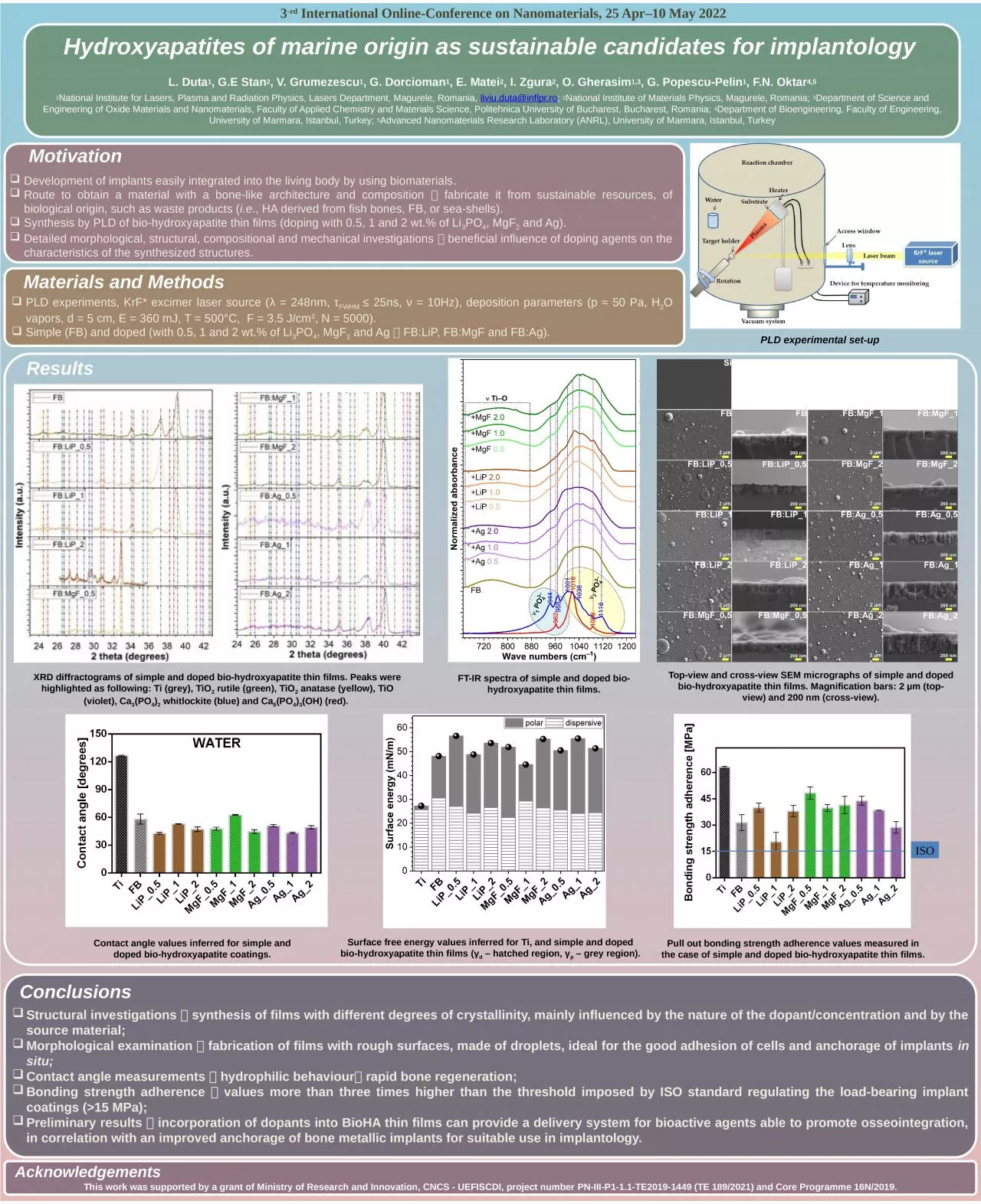
/
Results Motivation Development of implants easily integrated into the living body by using Results Motivation Development of implants easily integrated into the living body by using

Results Motivation Development of implants easily integrated into the living body by using - PowerPoint Presentation

SereneBeauty
SereneBeauty . @SereneBeauty
Follow
342 views
Uploaded On 2022-08-03

Results Motivation Development of implants easily integrated into the living body by using - PPT Presentation

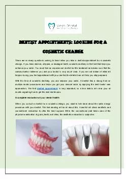
Route to obtain a material with a bonelike architecture and composition fabricate it from sustainable resources of biological origin such as waste products ie HA derived from fish bones FB or seashells ID: 934312

thin films bio simple films thin simple bio hydroxyapatite doped values materials view implants nanomaterials bone pld romania tio

Share:

Link:

Embed:

Download Presentation from below link

Download Presentation The PPT/PDF document "Results Motivation Development of implan..." is the property of its rightful owner. Permission is granted to download and print the materials on this web site for personal, non-commercial use only, and to display it on your personal computer provided you do not modify the materials and that you retain all copyright notices contained in the materials. By downloading content from our website, you accept the terms of this agreement.


Presentation Transcript

Slide1

Results

Motivation

Development of implants easily integrated into the living body by using biomaterials

.

Route to obtain a material with a bone-like architecture and composition

fabricate it from sustainable resources, of biological origin, such as waste products (

i.e

., HA derived from fish bones, FB, or sea-shells). Synthesis by PLD of bio-hydroxyapatite thin films (doping with 0.5, 1 and 2 wt.% of Li3PO4, MgF2 and Ag).Detailed morphological, structural, compositional and mechanical investigations  beneficial influence of doping agents on the characteristics of the synthesized structures.

Materials and Methods

PLD experiments, KrF* excimer laser source (λ = 248nm, τFWHM ≤ 25ns, ν = 10Hz), deposition parameters (p ≈ 50 Pa, H2O vapors, d = 5 cm, E = 360 mJ, T = 500°C, F = 3.5 J/cm2, N = 5000).Simple (FB) and doped (with 0.5, 1 and 2 wt.% of Li3PO4, MgF2 and Ag  FB:LiP, FB:MgF and FB:Ag).

Conclusions

Structural investigations  synthesis of films with different degrees of crystallinity, mainly influenced by the nature of the dopant/concentration and by the source material;Morphological examination  fabrication of films with rough surfaces, made of droplets, ideal for the good adhesion of cells and anchorage of implants in situ;Contact angle measurements  hydrophilic behaviour rapid bone regeneration;Bonding strength adherence  values more than three times higher than the threshold imposed by ISO standard regulating the load-bearing implant coatings (>15 MPa);Preliminary results  incorporation of dopants into BioHA thin films can provide a delivery system for bioactive agents able to promote osseointegration, in correlation with an improved anchorage of bone metallic implants for suitable use in implantology.

Acknowledgements

This work was supported by a grant of Ministry of Research and Innovation, CNCS - UEFISCDI, project number PN-III-P1-1.1-TE2019-1449 (TE 189/2021) and Core Programme 16N/2019.

Hydroxyapatites of marine origin as sustainable candidates for implantology

L. Duta1, G.E Stan2, V. Grumezescu1, G. Dorcioman1, E. Matei2, I. Zgura2, O. Gherasim1,3, G. Popescu-Pelin1, F.N. Oktar4,51National Institute for Lasers, Plasma and Radiation Physics, Lasers Department, Magurele, Romania, liviu.duta@inflpr.ro; 2National Institute of Materials Physics, Magurele, Romania; 3Department of Science and Engineering of Oxide Materials and Nanomaterials, Faculty of Applied Chemistry and Materials Science, Politehnica University of Bucharest, Bucharest, Romania; 4Department of Bioengineering, Faculty of Engineering, University of Marmara, Istanbul, Turkey; 5Advanced Nanomaterials Research Laboratory (ANRL), University of Marmara, Istanbul, Turkey

3

-rd International Online-Conference on Nanomaterials, 25 Apr–10 May 2022

PLD experimental set-up

Top-view and cross-view SEM micrographs of simple and doped bio-hydroxyapatite thin films. Magnification bars: 2 µm (top-view) and 200 nm (cross-view).

FT-IR spectra of simple and doped bio-hydroxyapatite thin films.

XRD diffractograms of simple and doped bio-hydroxyapatite thin films. Peaks were highlighted as following: Ti (grey), TiO

2

rutile (green), TiO

2

anatase (yellow), TiO (violet), Ca3(PO4)2 whitlockite (blue) and Ca5(PO4)3(OH) (red).

Contact angle values inferred for simple and doped bio-hydroxyapatite coatings.

Surface free energy values inferred for Ti, and simple and doped bio-hydroxyapatite thin films (γd – hatched region, γp – grey region).

Pull out bonding strength adherence values measured in the case of simple and doped bio-hydroxyapatite thin films.

ISO