/
Blood cells – Platelets (Thrombocytes) Blood cells – Platelets (Thrombocytes)

Blood cells – Platelets (Thrombocytes) - PowerPoint Presentation

alyssa
alyssa . @alyssa
Follow
29 views
Uploaded On 2024-02-09

Blood cells – Platelets (Thrombocytes) - PPT Presentation

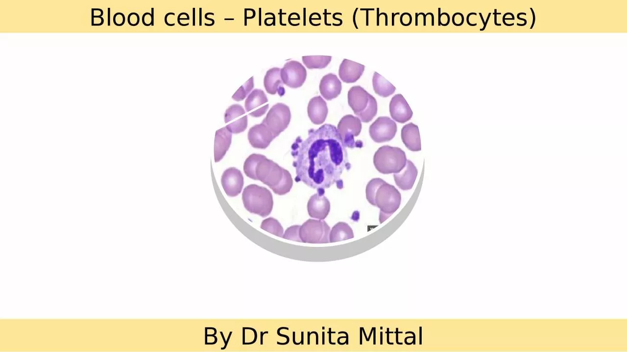
By Dr Sunita Mittal Learning Objectives Platelets Morphology Hemostasis Primary hemostasis Platelet plug formation Disorders of p rimary hemostasis Secondary hemostasis Disorders of secondary hemostasis ID: 1045087

platelet blood bleeding platelets blood platelet platelets bleeding clotting time hemostasis amp coagulation factors test formation count purpura factor

Share:

Link:

Embed:

Download Presentation from below link

Download Presentation The PPT/PDF document "Blood cells – Platelets (Thrombocytes)" is the property of its rightful owner. Permission is granted to download and print the materials on this web site for personal, non-commercial use only, and to display it on your personal computer provided you do not modify the materials and that you retain all copyright notices contained in the materials. By downloading content from our website, you accept the terms of this agreement.


Presentation Transcript

1. Blood cells – Platelets (Thrombocytes)By Dr Sunita Mittal

2. Learning ObjectivesPlatelets MorphologyHemostasisPrimary hemostasisPlatelet plug formationDisorders of primary hemostasisSecondary hemostasisDisorders of secondary hemostasisTests to assess hemostasisAnticoagulants

3. Platelets-Characteristics▪Small 2-3 µm disc-shaped, anuclear, reddish-purple cells.▪ Platelet life span 7-10 days▪ Platelets count: 1.5‐4 lakh/ mm3 of blood (Platelets are released from bone marrow to enter 2/3- peripheral blood and 1/3 seized in spleen.)

4. Platelets are formed within the cytoplasm ofmegakaryocytes (1 megakaryocyte produces about 2000 platelets)within bone marrow and released into the circulation.& IL3Platelets-Production

5. Platelets structurePeripheral zone:Responsible for platelet adhesion and aggregationGlycocalyx: Fluffy surface coat contains glycoprotein receptors: GPIa & IIa binds to collagen,GPIb binds von Willebrand’s factor ; GPIIb/IIIa binds fibrinogen Membrane ZoneLayer called PF3 (platelet factor) surface for interactionof plasma coagulation factors, Initiation of formation of thromboxane A2 2 systems :Surface-connected open canalicular system (OCS)Dense tubular system(DTS). CytoskeletonResponsible for platelet retraction and platelet shapeMicrotubules/MicrofilamentsBinding proteins: Actin & MyosinOrganelle zoneResponsible for storage and platelet release functionsGranules:Dense bodies, alpha granules, lysosomal granules and microperoxisomes. Mitochondria and Glycogen.

6. HemostasisHemostasis is the natural process that stops blood loss when an injury occurs. It involves three steps that occur in a rapid sequence: Primary Hemostasis: (1) vascular spasm, or vasoconstriction, a brief and intense contraction of blood vessels; (2) formation of a platelet plug; and Secondary Hemostasis: blood clotting or coagulation, which reinforces the platelet plug with fibrin mesh that acts as a glue to hold the clotfibrinolytic proteins system - Excessive blood clotting is prevented.

7. VasoconstrictionThe vasoconstriction that occurs during hemostasis is a brief reflexive contraction that causes a decrease in blood flow to the area and restrict blood loss.The vasoconstriction response is triggered by factors such as secretion of serotonin from activated platelets, a direct injury to vascular smooth muscle and nervous system reflexes initiated by local pain receptors. Primary Hemostasis - Vasoconstriction

8. Platelets Function- Pri. Hemostasis - Platelets plug formationPlatelets plug formation1) Adhesion – exposed collagen (GpIa & GpIIa and vWF-GpIb) 2) Activation –Initiation of formation of thromboxane A2 and secretion of granule contents specially ADP & Ca++3) Aggregation - Expression of GpIb-IIIa receptors on platelets to glue together

9. Formation of thromboxane A2 and Antiplatelet activity of Aspirin (ASPIRIN)Thromboxane synthetase

10. Platelets Function- Pri. Hemostasis - Platelets plug formation

11. Abnormalities in primary hemostasis-PurpuraPurpura is purple-colored spots and patches that occur on the skin, and in mucus membranes, including the lining of the mouth. Purpura occurs when small blood vessels leak blood under the skin.Purpura measure between 4 and 10 mm (millimeters) in diameter. When purpura spots are less than 4 mm in diameter, they are called petechiae. Purpura spots larger than 1 cm (centimeter) are called ecchymoses.

12. Defects in the blood vessel wall- lead to hemorrhage from mucosal surfaces (epistaxis, gingival bleeding, melaena, haematuria), and into skin and mucosae (petechial or ecchymotic haemorrhages - Purpura). Decreased platelets number (Thrombocytopenia) or function (Thrombosthenia)Abnormal vWF- Bernard Soulier SyndromeAbnormalities in primary hemostasisPrimary haemostasis inhibitors - natural inhibitors of platelet function - such as prostacyclin, bradykinin released by endothelial cells. Induced inhibition of platelet function - more commonly, platelet function is inhibited intentionally by the administration of agents such as aspirin for the prevention of thrombosis.

13. Bone Marrow Dysfunction –Many conditions and factors can damage stem cellsCancer- leukemia or lymphoma. Cancer treatments, such as radiation and chemotherapy.Toxic Chemicals- pesticides, arsenic, and benzeneMedicines-diuretics and chloramphenicol (aspirin or ibuprofen, also can affect platelets functioning)Alcohol-especially if they're eating foods that are low in iron, vitamin B12, or folate.Viruses-Chickenpox, mumps, rubella, Epstein-Barr virus, HIV and dengue fever virus etc often develop thrombocytopenia.Genetic Conditions-Wiskott-Aldrich syndromes.The Body Destroys Platelets at a higher rateITP (Idiopathic thrombocytopenic purpura)Autoimmune Diseases-immune thrombocytopenia . An autoimmune response is thought to cause most cases of ITP eg SLE and RAMargination in congestive splenomegalyMedicines-quinine; antibiotics that contain sulfa; dilantin, vancomycin, and rifampin. Infection-a widespread bacterial infection septicemia, mononucleosis or cytomegalovirusSurgery-Prosthetic heart valves, blood vessel grafts, or machines and tubing used - blood transfusions or bypass surgery.Pregnancy-About 5 percent of pregnant women develop mild thrombocytopenia when they're close to delivery. Causes of Thrombocytopenia

14. Process of secondary hemostasis-Blood Coagulation-Clotting Factors

15. Process of secondary hemostasis-Blood Coagulation

16. Disorders of Blood Coagulation- Coagulopathy Coagulopathy is a condition in that the blood’s ability to coagulate is impaired. This condition can cause a tendency toward prolonged or excessive bleeding (bleeding diathesis), which may occur spontaneously or following an injury

17. Disorders of Blood Coagulation- Hypocoagulopathy -CAUSES Major causes of coagulation disorders resulting in bleeding include:Hemophilia-Factor VIII deficiency (X chromosome linked disorder)Von Willebrand diseaseOther clotting factor deficiencies (Christmas disease) Disseminated intravascular coagulationLiver Disease Vit K deficiency

18. Conditions increasing the risk of a blood clot: Inherited (genetic) abnormalities that cause an increased tendency to clotA surgical procedureSitting or lying down for long periods (more than 4 hours)- for example, after surgery or on long plane or car rides. This lack of movement reduces blood flow in the legs by 50%.Major injuryIncreasing ageCancerHeart failurePregnancyUse of hormone therapy such as birth control pills or hormone replacement therapyHaving a history of DVTTobaccoDisorders of Blood Coagulation- Hyper-coagulopathy - CAUSES

19. Fibrinolytic System

20. Clotting factor deficiencies may cause the development of a significant bleeding tendency.Formation of clots inside blood vessels is called thrombosis due to hyper coagulability of blood Thromboses are a major medical problem. They frequently occlude the arterial supply to the organs in which they form, and bits of thrombus (emboli) sometimes break off and travel in the bloodstream to distant sites, damaging other organs. An example is obstruction of the pulmonary artery or its branches by thrombi from the leg veins (pulmonary embolism). Process of Thrombosis & Bleeding (fibrinolysis) should be balanced

21. Tests of Hemostasis-Laboratory InvestigationsBleeding Time • It is the time taken from the puncture of the blood vessel to the stoppage of bleeding. N : 1-6 minutes • The bleeding time test is a useful tool to test for platelet plug formation and capillary integrity. • BT is more imp. than CT. • CT concerns the blood only i.e. how firm the clot is formed, whereas BT involves the interaction of blood with injured tissues.• People with von Willebrand disease usually experience increased bleeding time. • Von Willebrand factor is a platelet agglutination protein, but this is not considered an effective diagnostic test for this condition. Capillary Fragility Test • The test is defined by the WHO as one of the necessary requisites for diagnosis of Dengue fever. • A blood pressure cuff is applied and inflated to a point between the systolic and diastolic blood pressures for five minutes. • The test is positive if there are 10 or more petechiae per square inch. • In DHF the test usually gives a definite positive result with 20 petechiae or more. Indicative of hemorrhagic tendency of a personPlatelet Count • Platelet Count can be determined by improved Neubauer’s counting chamber with RBC pipette & 1% ammonium oxalate. • They can be seen as tiny diameter, well seprated, highly refractile rounded bodies with silvery appearance. • N : 1.5 – 4 lacs / cumm. • Leishman stain : in clumps, blue cytoplasm, reddish purple granules, no nucleus. • Surgical bleeding usually does not occur until the platelet count is less than 50,000/mm3. • Spontaneous bleeding does not occur until the platelet count is less than 10,000-20,000/mm3. ↓Platelet Count • THOMBOCYTOPENIA : • Bone marrow depression – Aplastic anemia • Hypersplenism • Viral infections • Drugs : Aspirin, Heparin, Chemotherapy • ITP • TTP • DIC

22. Tests of Hemostasis-Laboratory InvestigationsProthombin Time • The prothrombin time (PT) and its derived measures of prothrombin ratio (PR) and international normalised ratio (INR) are measures of the extrinsic pathway of coagulation. • They are used to determine the clotting tendency of blood, in the measure of warfarin dosage, liver damage and vitamin K status. • PT measures factors I, II, V, VII and X. • The reference range for prothrombin time is usually around 11-16 seconds; the normal range for the INR is 0.8–1.2. APTT • The partial thromboplastin time (PTT) or activated partial thromboplastin time (aPTT or APTT) is a performance indicator measuring the efficacy of both the intrinsic and the common coagulation pathways. • Apart from detecting abnormalities in blood clotting, it is also used to monitor the treatment effects with heparin, a major anticoagulant. • Normal PTT times require the presence of the following coagulation factors: I, II, V, VIII, IX, X, XI & XII. • Deficiencies in factors VII or XIII will not be detected with the PTT test. Clotting Time It is the time taken from the puncture of the blood vessel to the formation of a fibrin thread. • A. Capillary Glass Tube Method : Here the blood is collected in capillary tube & total time is noted to form FIBRIN THREADS on breaking tube every 30 seconds. N : 3-8 minutes • Mechanism Involved is INTRINSIC Pathway. • CT depends on presence of all clotting factors. • It gets prolonged in : - 1. Deficiency of clotting factors – Hemophilia. - 2. Vitamin K Deficiency – Factor II, VII, IX & X. - 3. Anticoagulant overdose. • BT & CT is measured before surgery & liver or bone marrow biopsy. • PURPURA : BT increased, CT normal. • HEMOPHILIA : BT normal, CT increased.

23. ●Heparin is a naturally occurring anticoagulantthat facilitates the action of anti-thrombin III. Low-molecular weight fragments with an average molecular weight of 5000 have been produced from unfractionated heparin, and these low-molecular-weight heparins are seeing increased clinical use because they have a longer half-life and produce a more predictable anticoagulant response than unfractionated heparin. ● In vivo, a plasma Ca2+ level low enough to interfere with blood clotting is incompatible with life, but clotting can be prevented in vitro if Ca2+ is removed from the blood by the addition of substances such as oxalates, which form insoluble salts with Ca2+, or chelating agents, which bind Ca2+. Used in vitro only.● Coumarin derivatives such as dicumarol and warfarin are also effective anticoagulants. They inhibit the action of vitamin K, which is a necessary cofactor for the enzyme that catalyzes the conversion of glutamic acid residues to γ-carboxyglutamic acid residues. Six of the proteins involved in clotting require conversion of a number of glutamic acid residues to γ-carboxyglutamic acid residues before being released into the circulation, and hence all six are vitamin K-dependent. These proteins are factors II (prothrombin),VII, IX, and X, protein C, and protein S.ANTICOAGULANTS

24. Self Assessment▪Megakaryocyte produces about ……………………..platelets.▪ Platelet life span ………………….. Days.▪ ………………………….receptors causes gluing of platelets together.• Spontaneous bleeding does not occur until the platelet count is less than ……………………../mm3.• Surgical bleeding does not occur until the platelet count is less than ……………………………../mm3.• Secondary Hemostasis includes ………………………………processes.• Liver Disease results in hyp0coagulability of blood because………………………

25. Thank you