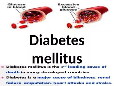
/
All forms of diabetes have very serious effects on health  In addition All forms of diabetes have very serious effects on health  In addition

All forms of diabetes have very serious effects on health In addition - PDF document

clara
clara . @clara
Follow
343 views
Uploaded On 2021-09-28

All forms of diabetes have very serious effects on health In addition - PPT Presentation

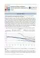
Figure 1 T1D Incidence Rates Worldwide 0510152025303540FINSARSWENORUSWIUSPAITAISRJAPCHIare being diagnosed with the T1D around the world each year Although the peak age at onset is at puberty T1D ID: 888644

t2d diabetes t1d risk diabetes t2d risk t1d type 2004 2003 glucose insulin genes age class factors 2000 gene

Share:

Link:

Embed:

Download Presentation from below link

Download Pdf The PPT/PDF document "All forms of diabetes have very serious ..." is the property of its rightful owner. Permission is granted to download and print the materials on this web site for personal, non-commercial use only, and to display it on your personal computer provided you do not modify the materials and that you retain all copyright notices contained in the materials. By downloading content from our website, you accept the terms of this agreement.


Presentation Transcript

1 All forms of diabetes have very serious
All forms of diabetes have very serious effects on health. In addition to the consequences of abnormal metabolism of glucose (e.g., hyperlipidemia, glycosylation of proteins, etc.), there are a number of long-term complications associated with the disease. These include cardiovascular, peripheral vascular, ocular, neurologic and renal abnormalities, which are responsible for morbidity, disability and premature death in young adults. Furthermore, the disease is associated with reproductive complications causing problem Figure 1. T1D Incidence Rates Worldwide 0510152025303540 FINSARSWENORUS-WIUS-PAITAISRJAPCHI are being diagnosed with the T1D around the world each year. Although the peak age at onset is at puberty, T1D can also develop in adults. Epidemiologic studies have revealed no significant gender differences in incidence among individuals diagnosed before age 15 (Kyvik et al., 2004). However, after age 25, the male to female incidence ratio is approximately 1.5. There is also a notable seasonal variation in the incidence of T1D in many countries, with lower rates in the warm summer months, and higher rates during the cold winter (Dorman et al., 2003). Environmental Risk Factors . The epidemiological patterns described above suggest that environmental factors contribute to the etiology of the T1D. In particular, the recent temporal increase in T1D incidence points to a changing global environment rather than variation in the gene pool, which require the passage of multiple generations. Twin studies also p

2 rovide evidence for the importance of en
rovide evidence for the importance of environmental risk factors for T1D. T1D concordance rates for monozygous twins are higher than those for dizygous twins (approximately 30% vs. 10%, respectively) (Hirschhorn, 2003). However, most monozygous twin pairs remain discordant. Thus, T1D cannot be completely genetically determined. Environmental risk factors are thought to act as either ‘initiators’ or ‘accelerators’ of beta cell autoimmunity, or ‘precipitators’ of overt symptoms in individuals who already have evidence of beta cell destruction. They also may function by mechanisms that are directly harmful to the pancreas, or by indirect methods that produce an abnormal immune response to proteins normally present in cells. The T1D environmental risk factors that have received most attention are viruses and infant nutrition. Enteroviruses, especially Coxsackie virus B (CVB), have been the focus of numerous ecologic and case-control studies (Dahlquist et al., 1998). CVB infections are frequent during childhood and are known to have systemic effects on the pancreas. Recent prospective studies are helping to elucidate the role of viruses to the etiology of T1D. For example, enteroviral infections occurring as early as in utero appear to increase a child’s subsequent risk of developing the disease (Dahlquist et al., 1995, Hyoty et al., 1995). Other viruses, including mumps (Hyoty et al., 1993), cytomegalovirus (Pak et al., 1988), rotavirus (Honeyman et al., 2000) and rubella, (McIntos

3 h and Menser, 1992) have also been assoc
h and Menser, 1992) have also been associated with the disease. Another hypothesis that has been the subject of considerable interest relates to early exposure to cow’s milk protein and the subsequent development of T1D. The first epidemiologic observation of such a relationship was by Borch-Johnsen et al., who found that T1D children were breast-fed for shorter periods of time than their non-diabetic siblings or children from the general population (Borsh-Johnsen et al., 1984). The authors postulated that the lack of immunologic protection from insufficient breast-feeding may increase risk for T1D later during childhood. It was also postulated that shorter duration of breast feeding may indirectly reflect early exposure to dietary proteins that stimulate an abnormal immune response in newborns. Most recently it has been hypothesized that the protective effect of breast-feeding may be due, in part, to its role in gut maturation (Kolb and Pozzilli, 1999; Harrison and Honeyman, 1999; Vaarala, 1999). Breast milk contains growth factors, cytokines, and other substances necessary for the maturation of the intestinal mucosa. Breast-feeding also protects against enteric infections during infancy, and promotes proper colonization of the gut. Interestingly, enteroviral infections can also interfere with gut immunoregulation, which may explain the epidemiologic associations between viral infections and T1D. The role of hygiene in the etiology of T1D is also currently being explored (McKinney et al., 1997; Marshall et a

4 l., 2004). It has been hypothesized tha
l., 2004). It has been hypothesized that delayed exposure to microorganisms due to improvements in standard of living hinders the development of the immune system, such that it is more 2 likely to respond inappropriately when introduced to such agents at older (compared to younger) ages. This explanation is consistent with recent reports indicating that factors such as day care attendance (McKinney et al. 2000), sharing a bedroom with a sibling, and contact with pets are protective against T1D (Marshall et al., 2004). Further studies are needed to determine if improved hygiene can explain the temporal increase in the incidence of T1D worldwide. Type 2 Diabetes Epidemiology . T2D is the most common form of the disease, accounting for approximately 90% of all affected individuals. A diagnosis of T2D is made if a fasting plasma glucose concentration is � 7.0 mmol/L ( � 126 mg/dl) or plasma glucose 2 hours after a standard glucose challenge is � 11.1 mmol/L ( � 200 mg/dl) (WHO, 1999) T2D is caused by relative impaired insulin secretion and peripheral insulin resistance. Typically, T2D is managed with diet, exercise, oral hypoglycemic agents and sometimes exogenous insulin. However, it is associated with the same long-term complications as T1D. The highest rates of T2D are found among Native Americans, particularly the Pima Indians who reside in Arizona in the US, and in natives of the South Pacific islands, such as Nauru (Wild et al., 2004). T2D is also known to be more predom

5 inant in Hispanic and African American p
inant in Hispanic and African American populations than in Caucasians. In 2000, it is estimated that 171 million people (2.8% of the worlds population) had diabetes and that by 2030 this number will be 366 million (4.4% of the world's population). The vast majority of this increase will occur in men and women aged 45 to 64 years living in developing countries. According to Wild et al.(2004), the ‘top’ three countries in terms of the number of T2D individuals with diabetes are India (31.7 million in 2000; 79.4 million in 2030), China (20.8 million in 2000; 42.3 million in 2030) and the US (17.7 million in 2000; 30.3 million in 2030). Clearly, T2D has become an epidemic in the 21 st century. In addition to the burden of T2D there is an even larger number of people with raised levels of blood glucose but below the level for diabetes. The World Health Organization defines impaired fasting glucose as a fasting plasma glucose level of � 6.1 mmoll -1 and less than 7 mmoll -1 , and impaired glucose tolerance as 2 hour plasma glucose, post glucose challenge, of 7.8 to less than 11.1 mmoll -1 (WHO, 1999). The prevalence of T2D increases with age of population (Wild et al., 2004). In developing countries, the largest number of people with diabetes are in the age group 45 to 64 years, while in developed the largest number is found in those aged 65 years and over. These differences largely reflected differences in population age structure between developed and developing countries. Worldwide rates are simila

6 r in men and women, although they are sl
r in men and women, although they are slightly higher in men e&#x 60 ;&#xyear;&#xs of;&#x age;&#x and;&#x in ;&#xwom0;n age 65 years. Of great concern is the recent increase in T2D in children (Bloomgarden, 2004). A report based on the Pima Indians in Arizona noted that between 1967-76 and 1987-96, the prevalence of T2D increased 6-fold in adolescents (Fagot-Campagna et al., 2000). In the US, the incidence of T2D increased from 0.3-1.2/100,000/yr before 1992 to 2.4/100,000/yr in 1994 (Weill et al., 2004). Most T2D children diagnosed during this period were females from minority populations, with a mean age of onset at around puberty. They were also likely to have a positive family history of the disease, particularly maternal diabetes. 3 Environmental Risk Factors . As early as 1962, Neel hypothesized that T2D represented a ‘thrifty genotype’, which had a selective advantage (Neel, 1962). He postulated that in primitive times, individuals who were ‘metabolically thrifty’ and able to store a high proportion of energy as fat when food was plentiful were more likely to survive times of famine. However, in recent years, most populations experience a continuous supply of calorie-dense processed foods, as well as a decrease in physical activity. This likely explains the rise in T2D prevalence worldwide. The major environmental risk factors for T2D are obesity ( � 120% ideal body weight or a body mass index � 30 k/m 2 ) and a sedentary lifestyle (van Dam, 2003; Shaw and Chish

7 olm, 2003). Thus, the tremendous increa
olm, 2003). Thus, the tremendous increase in the rates of T2D in recent years has been attributed, primarily, to the dramatic rise in obesity worldwide (Zimmet et al., 2001). It has been estimated that approximately 80% of all new T2D cases are due to obesity (Lean, 2000). This is true for adults and children. In the Pima Indians, 85% of the T2D children were either overweight or obese (Fagot-Campagna et al., 2000). Another study in the US reported that IGT was detected in 25% of obese children age 4-10 years, and in 21% of obese adolescents (Sinha et al., 2002). Undiagnosed T2D was detected in 4% of the adolescents. In addition to general obesity, the distribution of body fat, estimated by the ratio of waist-to-hip circumference (WHR), also has an impact on T2D risk. WHR is a reflection of abdominal (central) obesity, which is more strongly associated with T2D than the standard measures of obesity, such as those based on body mass index. The other major T2D risk factor is physical inactivity. In addition to controlling weight, exercise improves glucose and lipid metabolism, which decreases T2D risk. Physical activity, such as daily walking or cycling for more than 30 minutes, has been shown to significantly reduce the risk of T2D (Hu et al., 2003). Physical activity has also been inversely related to body mass index and IGT. Recently, intervention studies in China (Pan et al., 1997), Finland (Tuomilehto J et al., 2001) and the US (Diabetes Prevention Program Study Group, 2002) have shown that lifestyle interv

8 entions targeting diet and exercise decr
entions targeting diet and exercise decreased the risk of progression from IGT to T2D by approximately 60% . In contrast, oral hypoglycemic medication only reduced the risk of progression by about 30%. There is also considerable evidence suggesting that the intrauterine environment is an important predictor of T2D risk (Hales and Barker, 2001; Sobngwi et al., 2003), Numerous studies have shown that low birth weight, which is an indicator of fetal malnutrition, is associated with IGT and T2D later in life. However, it is unclear whether low birth weight is causal or related to potential confounding factors that contribute to both poor fetal growth and T2D (Frayling and Hattersley, 2001). Role of Genetics in the Development of Diabetes Type 1 Diabetes First degree relatives have a higher risk of developing T1D than unrelated individuals from the general population (approximately 6% vs. ) (Dorman and Bunker, 2000). These data suggest that genetic factors are involved with the development of the disease. At present, there is evidence that more than 20 regions of the genome may be involved in genetic susceptibility to T1D. However, none of the candidates identified have a greater influence on T1D risk than that conferred by genes in the HLA region of chromosome 6. This region contains several hundred genes known to be involved in 4 immune response. Those most strongly associated with the disease are the HLA class II genes (i.e., HLA-DR, DQ, DP). IDDM1. The HLA class II genes, also referred to as I

9 DDM1, contribute approximately 40-50% of
DDM1, contribute approximately 40-50% of the heritable risk for T1D (Hirschhorn et al., 2003). When evaluated as haplotypes, DQA1*0501-DQB1*0201 and DQA1*0301-DQB1*0302 are most strongly associated T1D in Caucasian populations. They are in linkage disequilibrium with DRB1*03 and DRB1*04, respectively. Specific DRB1*04 alleles also modify the risk associated with the DQA1*0301-DQB1*0302 haplotype. Other reported high risk haplotypes for T1D include DRB1*07-DQA1*0301-DQB1*0201 among African Americans, DRB1*09-DQA1*0301-DQB1*0303 among Japanese, and DRB1*04-DQA1*0401-DQB1*0302 among Chinese. DRB1*15-DQA1*0602-DQB1*0102 is protective and associated with a reduced risk of T1D in most populations. Recent reports suggest that other genes in the central, class I and extended class I regions may also increase T1D risk independent of HLA class II genes (Nejentsev et al., 1997; Lie et al., 1999). Individuals with two high risk DRB1-DQA1-DQB1 haplotypes have a significantly higher T1D risk than individuals with no high risk haplotype. The T1D risk among those with only one susceptibility haplotype is also increased, but effect is more modest. Relative risk estimates range from 10 – 45 and 3-7, respectively, for these groups, depending on race (Dorman and Bunker, 2000). In terms of absolute risk, Caucasian individuals with two susceptibility haplotypes have an approximately 6% chance of developing T1D through age 35 years. However, this figure is substantially lower in populations where T1D is rare (i.e., ong Asians). In

10 addition to IDDM1, two other genes are
addition to IDDM1, two other genes are now known to influence T1D risk (Anjos and Polychronakos, 2004). These include INS and CTLA-4. Table 1. Several T1D Susceptibility Genes Gene Locus Variant Estimated RR † HLA-DQB1 6p21.3 *0201 & *0302 3 – 45 INS 11p15. 5 Class I 1 – 2 CTLA4 2q31-35 Thr17Ala 1 – 2 † RR = relative risk INS (insulin). The INS gene, located on chromosome 11p15.5, has been designated as IDDM2. Positive associations have been observed with a non-transcribed variable number of tandem repeat (VNTR) in the 5’ flanking region (Bennett et al., 1997; Pugliese et al., 1997) . There are two common variants. The shorter class I variant predisposes to T1D (relative increase: 1 – 2), whereas the longer class III variant appears to be dominantly protective. The biological plausibility of these associations may relate to the expression of insulin mRNA in the thymus. Class III variants appear to generate higher levels of insulin mRNA than class I variants. Such differences could contribute to a better immune tolerance for class III positive individuals by increasing the likelihood of negative selection for autoreactive T-cell clones. The effect of INS appears to vary by ethnicity, with lesser effects in non-Caucasian populations (Undlien et al. 1994). CTLA-4 (cytotoxic T lymphocyte-associated 4) . The CTLA-4 gene is located on chromosome 2q31-35 (Anjos and Polychronakos, 2004), where multiple T1D genes may be located. CTLA-4 variants have been

11 associated with T1D, as well as other a
associated with T1D, as well as other autoimmune disease. CTLA-4 negatively regulates T-cell 5 channel’s activity and insulin secretion, ultimately leading to the development of T2D. Interestingly, are only 4.5 kb apart, and not far from the gene. Variant forms of KCNJ11Because of the close proximity of these genes, current studies are evaluating whether they work in concert with each other, or rather have an independent effect on T2D susceptibility. and inely in the treatment of T2D, there are pharmacogenetic implications for maintaining good glycemic control. Response to determine who is at high risk for developing T2D, but may also be useful in guiding treatment regimens for T2D. encodes an intracellular calcium-depeintronic A to G mutation at position amino acid polymorphisms (Thr504Ala and Phe200Thr) have also been associated with T2D risk. ng and noncoding polymorphisms do not independently is. Physiological studies suggest that variations in calpain 10 activity effects insulin secretion, and therefore, susceptibility to T2D. may be much larger in Mexican-American than Caucasian populations. Maturity-Onset Diabetes of the Young An uncommon form of T2D (accounting for % of all T2D cases) that generally occurs before age 25 onset of symptoms, the absence of obesity, no l autoimmunity. It is most often managed without the need for exogenous insulin. MODY displays an autosomal domthree generations (Stride and Hattersley, 2002). Because of advances in molecular genetics, it is now known that there are at leas

12 t six forms of MODY, each of which cause
t six forms of MODY, each of which caused by a mutation in a different with beta cell function (Winter, 2003). Table 3 lists the MODY genes ry mutations in one of (Demenais et al., 2003; Frayling et only MODY gene that does not in glucose metabolism and 2 patients differs from the prognosis assoMODY2 patients have a mild fasting hyperglycemia that is present from birth, and generally stable throughout life. There may be a mild deterioration of normoglycemiaType Gene Locus # Mutations % 20q12-q13.1 12 ~5% 7p15-p13 ~200 ~15% �12q24.2 100 ~65% 13q12.1 Few 17cen-q21.3 Few NEUROD1 2q32 Few 41. Lehto, M., Wipemo, C., Ivarsson, S.A., et al. High frequency of mutations in MODY and mitochondrial genes in Scandinavian patients with familial early-onset diabetes. Diabetologia, 1999. 42: 1131-1137. 42. Lie, B.A., Todd, J.A., Pociot, F., et al. The predisposition to type 1 diabetes linked to the human leukocyte antigen complex includes at least one non-class II gene. Am J Hum Genet, 1999. 64: 793-800. 43. Marshall, A.L., Chetwynd, A., Morris, J., et al. Type 1 diabetes mellitus in childhood: a matched case control study in Lancashire and Cumbria, UK. Diabet Med, 2004. 21: 1035-1040. 44. McIntosh, E.D.G., Menser, M. A fifty-year follow-up of congenital rubella. Lancet, 1992. 340: 414-415. 45. McKinney, P.A., Okasha, M., Parslow, R., et al. Ante-natal risk factors for childhood diabetes mellitus, a case-control study of medical record data in Yorkshire, UK. Diabetologia, 1997. 40: 933-939. 46. Moller, A.M., Dalgaard,

13 L.T., Pociot, F., et al. Mutations in t
L.T., Pociot, F., et al. Mutations in the hepatocyte nuclear factor-1a gene in Caucasian families originally classified as having type 1 diabetes. Diabetologia, 1998. 41: 1528-1531. 47. Neel, J. Diabetes mellitus: a thrifty genotype rendered detrimental by "progress"? Am J Hum Genet, 1962. 14: 353-362. 48. Nejentsev, S., Reijonen, H., Adojaan, B., et al. The effect of HLA-B allele on the IDDM risk defined by DRB1*04 subtypes and DQB1*0302. Diabetes, 1997. 46: 1888-1892. 49. (NIDDK), N.I.o.D.D.K.D. Oral Insulin Does Not Prevent Type 1 Diabetes. News Briefs (June 15, 2003): Available at: www.niddk.nih.gov/welcome/releases/6-15-03.htm March 8, 2004. 50. Onkamo, P., Vaananen, S., Karvonen, M., et al. Worldwide increase in incidence of type 1 diabetes--the analysis of the data on published incidence trends. Diabetologia, 1999. 42: 1395-1403. 51. Pak, C.Y., McArthur, R.G., Eun, H.M. Association of cytomegalovirus infection with autoimmune type 1 diabetes. Lancet, 1988. 2: 1-4. 52. Pan, X.R., Li, G.W., Hu, Y.H., et al. Effects of diet and exercise in preventing NIDDM in people with impaired glucose tolerance. The Da Qing IGT and Diabetes Study. Diabetes Care, 1997. 20: 537-544. 53. Paronen, J., Knip, M., Savilahti, E., et al. Effect of cow's milk exposure and maternal type 1 diabetes on cellular and humoral immunization to dietary insulin in infants at genetic risk for type 1 diabetes. Diabetes, 2000. 49: 1657-1665. 54. Pearson, E.R., Badman, M.K., Lockwood, C., et al. Contrasting diabetes phenotypes associated with hepato

14 cyte nuclear factor-1a and -1B mutations
cyte nuclear factor-1a and -1B mutations. Diabetes Care, 2004. 27: 1102-1107. 55. Pugliese, A., Zeller, M., Ferndandez, J.A. The insulin gene is transcribed in human thymus and transcription levels correlated with allelic variation at the INS VNTR-IDDM 2 susceptibility locus for type 1 diabetes. Nat Genet, 1997. 15: 293-297. 56. Shaw, J., Chisholm, D. Epidemiology and prevention of type 2 diabetes and the metabolic syndrome. MJA, 2003. 179: 379-383. 57. Shepherd, M. 'I'm amazed I've been able to come off injections': Patients' perceptions of genetic testing in diabetes. Pract Diab Int, 2003. 20: 338-342. 58. Shepherd, M. Genetic testing in maturity onset diabetes of the young (MODY)-practical guidelines for professionals. Pract Diab Int, 2003. 20: 108-110. 59. Shepherd, M., Hattersley, A.T. 'I don't feel like a diabetic any more': the impact of stopping insulin in patients with maturity onset diabetes of the young following genetic testing. Clin Med, 2004. 4: 144-147. 60. Silander, K., Mohlke, K.L., Scott, L.J., et al. Genetic variation near the hepatocyte nuclear factor-4a gene predicts susceptibility to type 2 diabetes. Diabetes, 2004. 53: 1141-1149. 14 61. Sinha, R., Fisch, G., Teague, B., et al. Prevalence of impaired glucose tolerance among children and adolescents with marked obesity. N Engl J Med, 2002. 346: 802-810. 62. Sobngwi, E., Boudou, P., Mauvais-Jarvis, F., et al. Effect of a diabetic environment in utero on predisposition to type 2 diabetes. Lancet, 2003. 361: 1861-1865. 63. Stride, A., Hattersley, A

15 .T. Different genes, different diabetes:
.T. Different genes, different diabetes: lessons from maturity-onset diabetes of the young. Ann Med, 2002. 34: 207-216. 64. Stumvoll, M. Control of glycaemia: from molecules to men. Minkowski Lecture 2003. Diabetologia, 2004. 47: 770-781. 65. Tuomilehto, J., Lindstrom, J., Eriksson, J.G., et al. Prevention of type 2 diabetes mellitus by changes in lifestyle among subjects with impaired glucose tolerance. N Engl J Med, 2001. 344: 1343-1350. 66. Undlien, D.E., Hamaguchi, K., Kimura, A. Type 1 diabetes susceptibility associated with polymorphism in the insulin gene region: a study of blacks, Caucasians, and orientals. Diabetologia, 1994. 37: 745-749. 67. Vaarala, O. Gut and the induction of immune tolerance in type 1 diabetes. Diabetes Metab Res Rev, 1999. 15: 353-361. 68. van Dam, R.M. The epidemiology of lifestyle and risk for type 2 diabetes. Eur J Epidemiol, 2003. 18: 1115-1125. 69. Weill, J., Vanderbecken, S., Froguel, P. Understanding the rising incidence of type 2 diabetes in adolescence. Arch Dis Child, 2004. 89: 502-504. 70. WHO, 1999. Definition, Diagnosis and Classification of Diabetes Mellitus and its Complications. WHO/NCD/NCS/99.2. 71. Wild, S., Roglic, G., Green, A., et al. Global prevalence of diabetes. Diabetes Care, 2004. 27: 1047-1053. 72. Winter, W.E. Newly defined genetic diabetes syndromes: maturity onset diabetes of the young. Rev Endocr Metab Disord, 2003. 4: 43-51. 73. Zimmett, P., Alberti, K.G.M.M., Shaw, J. Global and societal implication of the diabetes epidemic. Nature, 2001. 414: 782-787.