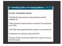
/
 Chapter 12 Section 1  DNA: The Genetic Material  Chapter 12 Section 1  DNA: The Genetic Material

Chapter 12 Section 1 DNA: The Genetic Material - PowerPoint Presentation

danika-pritchard
danika-pritchard . @danika-pritchard
Follow
349 views
Uploaded On 2020-04-02

Chapter 12 Section 1 DNA: The Genetic Material - PPT Presentation

Ms Diana and Ms Suad Discovery of the Genetic Material Since Mendels work was rediscovered scientists have tried to figure out which of the macromolecules nucleic acids DNA or proteins was the actual source of genetic information ID: 774595

dna cells bacteria strain dna cells bacteria strain structure experiment mouse live genetic scientists amount nucleotides sugar watson killed

Share:

Link:

Embed:

Download Presentation from below link

Download Presentation The PPT/PDF document " Chapter 12 Section 1 DNA: The Genetic ..." is the property of its rightful owner. Permission is granted to download and print the materials on this web site for personal, non-commercial use only, and to display it on your personal computer provided you do not modify the materials and that you retain all copyright notices contained in the materials. By downloading content from our website, you accept the terms of this agreement.


Presentation Transcript

Slide1

Chapter 12Section 1 DNA: The Genetic Material

Ms. Diana and Ms. Su’ad

Slide2

Discovery of the Genetic Material

Since Mendel’s work was rediscovered, scientists have tried to figure out which of the macromolecules- nucleic acids (DNA) or proteins was the actual source of genetic information. Scientists knew that genetic information was found on chromosomes and the main components of chromosomes are DNA and protein.

Slide3

Griffith’s Experiment

The first major experiment that led to the discovery of DNA was performed by Frederick Griffith in 1928. He studied two strains of the bacteria Streptococcus pneumoniae, which causes pneumonia. He found that one strain could be transformed (changed) into the other form. Smooth strain (S) – has a sugar coat and causes pneumonia. Rough strain (R) – does not have a sugar coat and does not cause pneumonia.

Slide4

Griffith’s Experiment

Griffith’s Experiment:

Step 1 – When the mouse was injected with the live S strain, the mouse died.

Step 2 – When the mouse was injected with the R strain, the mouse lived.

Step 3 – When the mouse was injected with the killed S strain (by using heat), the mouse lived.

Step 4 – When a mixture of live R cells and killed S cells were injected into the mouse, the mouse died.

It became obvious that there was a disease-causing factor that was passed from the killed S bacteria to the live R bacteria.

Griffith concluded then, that there had been a transformation from live R bacteria to live S bacteria.

Slide5

Griffith’s Experiment

Slide6

Avery Experiment

In 1944, Oswald Avery and his colleagues identified the molecule that transformed the R strain of bacteria into the S strain.

Avery separated different macromolecules, such as DNA, proteins and lipids, from killed S cells. Then he exposed live R cells to each of the macromolecules separately.

When the live R cells were exposed to the S strain DNA, they were transformed into S cells. Avery concluded that when the S cells in Griffith’s experiments were killed, DNA was released.

Some of the R bacteria kept this DNA in their cells, and this changed the bacteria into S cells.

Slide7

A Closer Look

Slide8

Hershey and Chase

In 1952, Alfred Hershey and Martha Chase published results of experiments that provided definite evidence that DNA was the actual transforming factor.

Their experiments involved a

bacteriophage

, a type of virus that attacks bacteria.

Two components made the experiment perfect for confirming that DNA is the genetic material.

1. The

bacteriophage

is used in the experiment was made of DNA and protein.

2. Viruses cannot replicate themselves. They must inject their genetic material into a living cell to reproduce.

Slide9

A Closer Look

Slide10

DNA Structure

Scientists were convinced that DNA was the genetic material. The questions that remained were how nucleotides came together to form DNA and how DNA could communicate information remained. Nucleotides are the subunits of nucleic acids and consist of a five-carbon sugar, a phosphate group, and a nitrogenous base.

Slide11

DNA Structure

The two nucleic acids found in living cells are DNA and RNA. DNA nucleotides contain the sugar deoxyribose, a phosphate, and one of four nitrogen bases: AdenineGuanineCytosineThymine

Slide12

RNA Structure

RNA nucleotides contain the sugar ribose, a phosphate, and one of four nitrogen bases: AdenineGuanineCytosineUracil

Slide13

Chargaff

Erwin Chargaff analyzed the amount of adenine, guanine, thymine, and cytosine in the DNA of various species. He found that the amount of guanine nearly equals the amount of cytosine. He also found that the amount of adenine nearly equals the amount of thymine within a species. This finding is known as Chargaff’s rule: C=G and T=A.

Slide14

The Structure Question

Four scientists joined together to search for the structure of DNA. Soon, Chargaff’s data became important and clear.The four scientists are: Rosalind Franklin, a British chemistMaurice Wilkins, a British physicistFrancis Crick, a British physicistJames Watson, an American biologist

Slide15

X-ray Diffraction

Wilkins was working at King’s College in London with a technique called X-ray diffraction (aiming X rays at a DNA molecule). In 1951, Franklin joined the staff and used the same technique and the famous Photo 51. It shows that DNA was a double helix, or twisted laddershape, formed by two strandsof nucleotides twisted aroundeach other.

Slide16

Watson and Crick

Watson and Crick were working at Cambridge University when they saw Franklin’s picture. Using Chagraff’s data, they measured the width of the helix and the spaces between the bases. Together, they built a model of the double helix that looked like this:

Slide17

DNA Structure

DNA is often compared to a twisted ladder. The rails represent the alternating deoxyribose and phosphate. The steps represent the pairs of bases (cytosine-guanine or thymine-adenine).

Slide18

DNA ORIENTATION

Another unique feature of the DNA molecule is the direction (orientation) of the two strands.

On the top rail, the sugars run from 5’ to 3’ (“

five prime to three prime”

).

On the bottom rail, the sugars run from 3’ to 5’ (“

three to five prime”

).

Since they run in opposite directions, they are said to be

antiparallel

.

Slide19

DNA Orientation

Slide20

The Announcement

In 1953, Watson and Crick surprised the scientific community by publishing a one-page article in the journal Nature that showed their DNA structure. In the same journal, Wilkins and Franklin presented evidence that supported Watson and Crick’s structure.

Slide21

Chromosome Structure

In prokaryotes, the DNA molecule is contained in the cytoplasm and consists mainly of a ring of DNA and some proteins.

Eurkaryotic

DNA is organized into individual chromosomes.

The length of a human chromosome ranges from 51 million to 245 million base pairs.

In order for the DNA to fit into the nucleus, the DNA tightly coils around a group of proteins that look like beads called

histones

.

Slide22

A Closer Look

Slide23

Questions