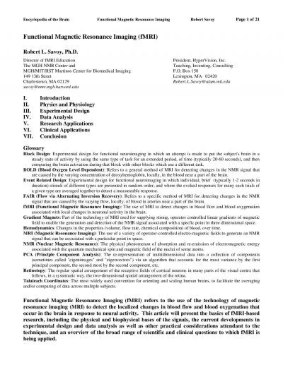
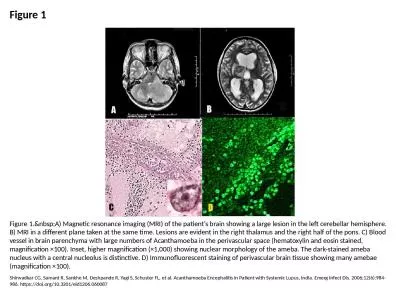
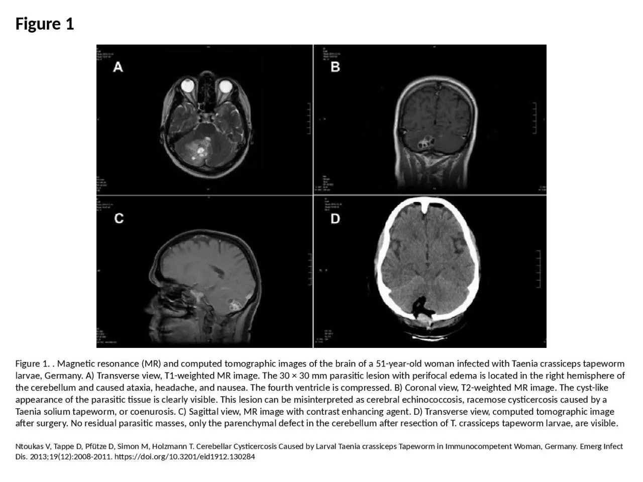
PPT-Figure 1 Figure 1. . Magnetic resonance (MR) and computed tomographic images of the brain
Author : faith | Published Date : 2023-05-20
Ntoukas V Tappe D Pfütze D Simon M Holzmann T Cerebellar Cysticercosis Caused by Larval Taenia crassiceps Tapeworm in Immunocompetent Woman Germany Emerg Infect
Presentation Embed Code
Download Presentation
Download Presentation The PPT/PDF document "Figure 1 Figure 1. . Magnetic resonance ..." is the property of its rightful owner. Permission is granted to download and print the materials on this website for personal, non-commercial use only, and to display it on your personal computer provided you do not modify the materials and that you retain all copyright notices contained in the materials. By downloading content from our website, you accept the terms of this agreement.
Figure 1 Figure 1. . Magnetic resonance (MR) and computed tomographic images of the brain: Transcript
Ntoukas V Tappe D Pfütze D Simon M Holzmann T Cerebellar Cysticercosis Caused by Larval Taenia crassiceps Tapeworm in Immunocompetent Woman Germany Emerg Infect Dis 2013191220082011 httpsdoiorg103201eid1912130284. PRESENTING AUTHOR. Amrita Ajani. CO-AUTHORS. Rohit. . Shetty. , . Abhijit. . Sinha Roy, . Purnima. . Srivatsa. , . Natasha . Pahuja. NO . FINANCIAL DISCLOSURE. PURPOSE OF THE STUDY. To evaluate correlation between tomographic gradation of keratoconic . Department of Chemistry. Dylan W. Benningfield. Electron Spin Resonance (ESR). Electron spin resonance (ESR. ). Electron . paramagnetic resonance (EPR) . Study of paramagnetic materials. Radicals, bi-radicals, triplet states, unfilled conduction bands, transition metal ions, impurities in semi-conductors, etc.. Images. Gillian R. Foulger, . Giuliano. F. . Panza. , Irina M. . Artemieva. , Ian D. . Bastow. , Fabio . Cammarano. , John R. Evans, Warren B. Hamilton, Bruce R. Julian, Michele . Lustrino. , Hans . Part 1. Magnetic Resonance Imaging. A great multidisciplinary application of physics and other sciences. And example of a “pure science” discovery given a practical application. Based on NMR – Nuclear Magnetic Resonance – discovered in the 1940s. Nuclear Magnetic Resonance Spectroscopy. Over the past fifty years nuclear magnetic resonance spectroscopy, commonly referred to as nmr, has become the preeminent technique for determining the structure of organic compounds. . Magnetic Resonance Imaging. Gerald R. Aben, MD FACR. Department of Radiology. College of Osteopathic Medicine. 2/13/2012. 1. Department of Radiology. Magnetic Resonance Imaging. No ionizing radiation. An MRI Machine. I. Computerized Axial Tomography (CAT/CT). CT scanning adds . many X-ray . images with the aid of a computer to generate cross-sectional views of a patient's anatomy.. CT scans are performed to analyze the internal structures of various parts of the . neuro. deficits, and a . negative CT. High clinical suspicion and severe . c. ervical . s. pondylosis. -> . MRI. Otherwise . use clinical judgment, but likely no benefit from MRI and increased risk of . Page 1of 10UnitedHealthcare Commercial Medical PolicyEffective 01/01/2021Proprietary Information of UnitedHealthcare Copyright 2021United HealthCare Services IncUnitedHealthcareCommercial MedicalPolic Nuclear magnetic resonance. The use of NMR in chemical research was pioneered by . Herbert S. . Gutowski. of Department of Chemistry, University of Illinois, who established the relationship between chemical shifts and molecular structures. He also discovered spin-spin coupling.. Printe 09/01/2000 LAST UPDATED 05/17/2007 A Page 1of 2 9/16/2011 p Published:, August 2005 , June 2006 , July 2007 References: A merican College of Cardiology/American Heart Association Expert Consen Encyclopedia of the Brain Functional Magnetic Resonance Imaging Robert Savoy Page 2 of 21I. Introduction It has long been known that there is some degree of localization of function in the human brain Shirwadkar CG, Samant R, Sankhe M, Deshpande R, Yagi S, Schuster FL, et al. Acanthamoeba Encephalitis in Patient with Systemic Lupus, India. Emerg Infect Dis. 2006;12(6):984-986. https://doi.org/10.3201/eid1206.060087. ot. the sample. What is the Hamiltonian for the system? The magnetic fields are as below.. The Hamiltonian is given by,. . where,. The spin matrices are:. Magnetic Resonance. The Hamiltonian is:.
Download Document
Here is the link to download the presentation.
"Figure 1 Figure 1. . Magnetic resonance (MR) and computed tomographic images of the brain"The content belongs to its owner. You may download and print it for personal use, without modification, and keep all copyright notices. By downloading, you agree to these terms.
Related Documents