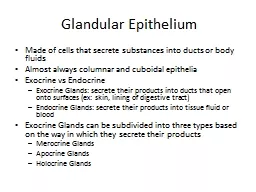
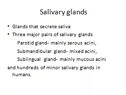
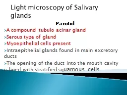
Three major pairs of salivary glands Parotid gland mainly serous acini Submandibular gland mixed acini Sublingual gland mainly mucous acini and hundreds of minor salivary glands in humans ID: 919986
Download Presentation The PPT/PDF document "Salivary glands Glands that secrete sali..." is the property of its rightful owner. Permission is granted to download and print the materials on this web site for personal, non-commercial use only, and to display it on your personal computer provided you do not modify the materials and that you retain all copyright notices contained in the materials. By downloading content from our website, you accept the terms of this agreement.
Slide1
Salivary glands
Glands that secrete saliva
Three major pairs of salivary glands
Parotid gland- mainly serous acini,
Submandibular
gland- mixed acini,
Sublingual gland- mainly mucous acini
and hundreds of minor salivary glands in humans.
Slide2parotid glands
Located near ear in parotid
fossa
Shape – inverted pyramid
Weight- 25-30 gm
Capsule- investing layer of deep cervical fascia
Superficial layer
Deep layer-
stylomandibular ligament
Slide3Structure
Base or superior surface
Apex
Lateral surface or superficial surface
anteromedial
surface
posteromedial
surface
Anterior border
Posterior border
medial border
Slide4Strucrures
within the gland
Facial nerve
External carotid artery
Retromandibular
vein
Duct of gland
stensons
duct
Thick walled
5 cm long
Opens into vestibule of mouth
opposite crown
of 2
nd
upper molar tooth
Slide5Arterial supply
Small branches from
External carotid artery
Superficial temporal artery
Maxillary artery
venous drainage
Retromandibular
vein
External jugular vein
Slide6Lymphatic drainage
Deep cervical nodes through
Superficial parotid nodes
Deep parotid nodes
i
Slide7Nerve supply
Parasympathetic-
secretomotor
-produces thin watery saliva
Preganglionic
fibres
Inferior
salivatory
nucleus-
glossopharyngeal
nerve-tympanic branch of
glossopharyngeal
nerve-tympanic plexus-
lessor
petrosal
nerve-
otic
ganglion
Slide8Postganglionic
fibres
Auriculotemporal
nerve- its parotid branches –parotid gland
sympathetic- vasoconstrictor and reduce secretion
Cervical
ganglia-external carotid nerve plexus-parotid gland
Sensory
– great auricular and
auriculotemporal
nerves
Slide9Applied aspect
Parotid swellings- mumps
Parotid abscess
Parotiditis
-very painful, referred to
Auricle
External acoustic
meatus
Tumours
Benign
malignant
Slide10Facial palsy
Sialography
-
sialogram
Slide11Submandibular gland
Walnut shaped
Weight- 10-20 g
location- partly below and partly deep to body of mandible
Parts- superficial and deep parts
seperated
by
myelohoid
muscle
Slide12Submandibular
duct- Wharton's duct
7 cm long
From superficial part-sublingual papillae
Arterial supply- facial and lingual arteries
venous drainage- internal jugular vein
lymphatic drainage-
submandibular
lymph nodes to deep cervical lymph nodes (
jugulo-omohyoid
nodes)
Slide13Nerve supply
Parasympathetic-for watery secretion
Preganglionic
fibres
. Superior
salivatory
nucleus-facial nerve-
chorda
tympanic branch of facial nerve-lingual nerve-
submandibular
ganglion
Post
ganglionic
fibres
- cells of
ganglionic-submandibular
gland
Slide14sympathetic-for mucous secretion
Post
ganglionic
fibres
from superior cervical ganglion-sympathetic plexus around facial artery-
submandibular
ganglion-
submandibular
gland
Applied aspect
Calculus in duct
sialography
Slide15Sublingual gland
Smallest
Almond shaped
3-4 gm in weight
Location- anterior part of oral cavity
Arterial supply – sublingual and
submental
arteries
venous drainage- internal jugular vein
Slide16lymphatic drainage-
submental
and
submandibular
lymph nodes to deep cervical lymph nodes (
jugulo-omohyoid
nodes)
Nerve supply- same as that of
submandibular
gland