/
CHAPTER 12 THE CELL CYCLE CHAPTER 12 THE CELL CYCLE

CHAPTER 12 THE CELL CYCLE - PowerPoint Presentation

lindy-dunigan
lindy-dunigan . @lindy-dunigan
Follow
355 views
Uploaded On 2018-11-08

CHAPTER 12 THE CELL CYCLE - PPT Presentation

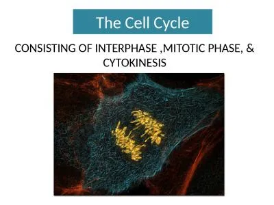
Unicellular organism cell division reproduces an entire organism Multicellular organisms cell division can produce growth or progeny Cell division functions in reproduction growth and repair ID: 723097

cells cell chromosomes cycle cell cells cycle chromosomes spindle division dna microtubules proteins mitosis phase growth cancer chromosome cyclin control divide interphase

Share:

Link:

Embed:

Download Presentation from below link

Download Presentation The PPT/PDF document "CHAPTER 12 THE CELL CYCLE" is the property of its rightful owner. Permission is granted to download and print the materials on this web site for personal, non-commercial use only, and to display it on your personal computer provided you do not modify the materials and that you retain all copyright notices contained in the materials. By downloading content from our website, you accept the terms of this agreement.


Presentation Transcript

Slide1

CHAPTER 12THE CELL CYCLESlide2

Unicellular organism- cell division reproduces an entire organism

Multicellular

organisms- cell division can produce growth or progeny

Cell division functions in reproduction, growth, and repair

AmoebaSlide3

Multicellular organism Begin as a fertilized egg or zygote.Repair and renew cells that die


Sand dollar embryo- dividing fertilized egg

Bone marrow cells dividingSlide4

A dividing cell duplicates its DNA, allocates the two identical copies

to opposite ends of the cell, and then splits into two daughter cells.Slide5

Genome- a cell’s genetic information, packaged as

DNA.

In prokaryotes, the genome is often a single long DNA molecule.In eukaryotes, the genome consists of several DNA molecules.A human cell must duplicate about 3 m of DNA and separate the two copies such that each daughter cell ends up with a complete genome.

Cell division distributes identical sets of chromosomes to daughter cellsSlide6

DNA molecules are packaged into chromosomes.

Every eukaryotic species has a characteristic number of chromosomes in the nucleus.

Human somatic cells (body cells) have 46 chromosomes.

Human gametes (sperm or eggs) have 23 chromosomes,

half the number in a somatic cell.

Chromosomes in the nucleus of a kangaroo rat epithelial cell. The cell is about to divide.Slide7

A eukaryotic chromosome is a long, linear DNA molecule.

has

hundreds or thousands of genes, the units that specify an organism’s inherited traits.Chromatin- DNA-protein

complex, a long thin fiber, helps maintain structure and control gene activity.After the DNA duplication, chromatin condenses,

coils and folds DNA to

make a smaller package. Slide8

Each duplicated chromosome consists of two sister chromatids

which contain identical copies of the chromosome’s DNA.

Centromere- the region where the strands

connect, a small narrow area

Later, the sister chromatids are pulled

apart and repackaged into two new nuclei at

opposite ends of

the parent cell. Slide9

Mitosis- the process of the formation of the two daughter

nuclei

Cytokinesis- division of the cytoplasmThese processes take one cell and produce two cells that are the genetic equivalent of the parent. Slide10

Human developmentMitosis

Humans inherit 23

chromosomes from each parent: one set in an egg and one set in sperm.The fertilized egg (zygote) undergoes trillions of cycles of mitosis and cytokinesis to produce a fully developed

multicellular human.These processes continue every day to replace dead and damaged cell.Essentially, these processes produce clones - cells with the same genetic information.Slide11

Human development- Meiosis

Gametes

(eggs or sperm) are produced only in gonads (ovaries or testes).In the gonads, cells undergo meiosis, which yields four daughter cells, each with half the chromosomes of the parent.

Meiosis reduces the number of chromosomes from 46 to 23.Fertilization fuses two gametes together and doubles the number of chromosomes to 46 again.Slide12

The mitotic (M

)

phase of the cell cycle alternates with the much longer interphase.The M phase includes mitosis

and cytokinesis.Interphase accounts for 90% of the cell

cycle.

The Mitotic Cell Cycle

-

The

mitotic phase alternates with

interphase

in the cell cycle:

an overviewSlide13

Interphase-

the cell grows by producing proteins and

cytoplasmic organelles, copies its chromosomes, and prepares for cell division.Interphase has three subphases:

G1 phase (“first gap”) centered on growth,S

phase (“synthesis”) when the chromosomes are copied, G

2 phase (“second gap”) where the cell completes preparations for cell division,

and divides (M

).Slide14

Mitosisfive subphases

:

prophaseprometaphasemetaphase

anaphase telophase.Slide15

G2 of Interphase

By

late interphase, the chromosomes have been duplicated but are loosely packed.The centrosomes have been duplicated and begin to organize microtubules into an aster (“star”).Slide16

ProphaseThe chromosomes are tightly coiled, with sister

chromatids

joined together.The nucleoli disappear.The mitotic spindle begins to form and appears to push

the centrosomes away from each other toward opposite ends (poles)

of the cell.Slide17

PrometaphaseThe

nuclear envelope fragments and microtubules from the spindle interact with the chromosomes.

Microtubules from one pole attach to one of two kinetochores, special

regions of the centromere, while microtubules from the other pole attach to

the other kinetochore.Slide18

MetaphaseThe spindle fibers push the sister

chromatids

until they are all arranged at the metaphase plate, an imaginary plane equidistant between the poles, defining metaphase.Slide19

AnaphaseThe centromeres

divide, separating the sister

chromatids.Each is now pulled toward the pole to which it is attached by spindle fibers.By the end, the two poles have equivalent

collections of chromosomes.Slide20

TelophaseThe

cell continues to elongate as free spindle fibers from each

centrosome push off each other.Two nuclei begin for form, surrounded by the fragments of the parent’s nuclear envelope.Chromatin becomes

less tightly coiled.Cytokinesis, division of the cytoplasm, begins.Slide21
Slide22
Slide23

Mitotic spindle, fibers composed of

microtubules

and associated proteins, is a major driving force in mitosis.As the spindle assembles during prophase, the elements come from partial disassembly of the cytoskeleton.The spindle fibers elongate by incorporating more subunits of the protein tubulin.

The mitotic spindle distributes chromosomes to daughter cells:

a closer look Slide24

Assembly of the spindle microtubules starts in the centrosome.

The

centrosome (microtubule-organizing center) of animals has a pair of centrioles at the center, but the function of the

centrioles is somewhat undefined.Slide25

As mitosis starts, the two centrosomes are located near the nucleus.

As the spindle fibers grow from them, the

centrioles are pushed apart.By the end of prometaphase they develop as the spindle poles

at opposite ends of the cell.Slide26

Each sister chromatid has a

kinetochore

of proteins and chromosomal DNA at the centromere.During prometaphase,

some spindle microtubules attach to thekinetochores.Slide27

When a chromosome’s kinetochore is

captured

by microtubules, the chromosome moves toward the pole from which those microtubules come.When microtubules attach to the other pole, this movement stops and a tug-of-war ensues.Eventually, the chromosome settles midway between the two poles of the cell, the metaphase plate.

Other microtubules from opposite poles interact as well, elongating the cell.Slide28

One hypothesis for the movement of chromosomes in anaphase is that motor proteins at the kinetochore “walk” the attached chromosome along the microtubule toward the opposite pole.

The excess microtubule sections

depolymerize.Slide29

Experiments support the hypothesis that spindle fibers shorten during anaphase from the end attached to the chromosome, not the centrosome.Slide30

Nonkinetichore

microtubules are responsible for lengthening the cell along the axis defined by the poles.

These microtubules interdigitate across the metaphase plate.During anaphase motor proteins push microtubules from opposite sides away from each other.

At the same time, the addition of new tubulin monomers extends their length. Slide31

Cytokinesis, division of the cytoplasm, typically follows mitosis.In animals, the first sign of

cytokinesis

(cleavage) is the appearance of a cleavage furrow in the cell surface near the old metaphase plate.

Cytokinesis

divides the cytoplasm: a closer look Slide32

On the cytoplasmic side of the cleavage furrow a contractile ring of

actin

microfilaments and the motor protein myosin form.Contraction of the ring pinches the cell in two.Slide33

Cytokinesis in plants, which have cell walls, involves a completely different mechanism.

During

telophase, vesicles from the Golgi coalesce at the metaphase plate, forming a

cell plate.The plate enlarges until its membranes fuse with the plasma membrane at the

perimeter, with the contents of the vesicles forming new

wall material in between.Slide34
Slide35

Prokaryotes reproduce by binary fission, not mitosis.

Most bacterial genes are located on a single bacterial chromosome which consists of a circular DNA molecule and associated proteins.

Their circular chromosome is highly folded and coiled in the cell.

Mitosis in eukaryotes may have evolved from binary fission in bacteria Slide36

In binary fission, chromosome replication begins at one point in the circular chromosome, the origin of replication site.

These copied regions begin to move to opposite ends of the cell.Slide37

The mechanism behind the movement of the bacterial chromosome is still an open question.

As

the bacterial chromosome is replicating and the copied regions are moving to opposite ends of the cell, the bacterium continues to grow until it reaches twice its original size.Slide38

Cell division involves inward growth of the plasma membrane, dividing the parent cell into two daughter cells, each with a complete genome. Slide39

Possible intermediate evolutionary steps between binary fission and mitosis:

Unicellular algae-

In dinoflagellates, replicated chromosomes are attached to the nuclear envelope. In

diatoms, the spindle develops within the nucleus.Slide40
Slide41

The timing and rates of cell division in different parts of an animal or plant are crucial for normal growth, development, and maintenance.The frequency of cell division varies with cell type

.

Regulation of the Cell Cycle- IntroductionSlide42

The cell cycle appears to be driven by specific chemical signals in the cytoplasm.Fusion of an S phase and a G

1

phase cell, induces the G1 nucleus to start S phase.Fusion of a cell in mitosis with one in interphase induces the second cell to enter mitosis.

A molecular control system drives the cell cycleSlide43

Cell cycle control system- directs events of the cell

cycle

The control cycle has a built-in clock, but it is also regulated by

external adjustments and internal controls.Slide44

A checkpoint in the cell cycle is a critical control point where stop and go signals regulate the cycle.

Many signals registered at checkpoints come from cellular surveillance mechanisms

indicating whether key cellular processes have been completed correctly.Checkpoint also register signals from outside the cell.Three major checkpoints are found in the G

1, G2, and M phases.Slide45

For many cells, the G1 checkpoint, the restriction point in mammalian cells, is the most important.

If the cells receives a go-ahead signal, it usually completes the cell cycle and divides.

If it does not receive a go-ahead signal, the cell exits the cycle and switches to a nondividing state, the G0

phase.Slide46

Rhythmic fluctuations in the abundance and activity of control molecules pace the cell cycle.Some molecules are

protein

kinases that activate or deactivate other proteins by phosphorylating them.The levels of these kinases

are present in constant amounts, but these kinases require a second protein, a cyclin, to become activated.Level of

cyclin proteins fluctuate cyclically.The complex of

kinases and cyclin forms

cyclin

-dependent

kinases

(

Cdks

).Slide47

Cyclin levels rise sharply throughout interphase

, then fall abruptly during mitosis.

Peaks in the activity of one cyclin-Cdk complex, MPF, correspond to peaks in cyclin concentration.Slide48

MPF (“maturation-promoting factor” or “M-phase-promoting-factor”) triggers the cell’s passage past the G2 checkpoint to the M phase.

MPF promotes mitosis by

phosphorylating a variety of other protein kinases.MPF stimulates

fragmentation of the nuclear envelope.It also triggers the breakdown of

cyclin, dropping

cyclin and MPF levels during

mitosis and

inactivating MPF.Slide49

The key G1 checkpoint is regulated by at least three

Cdk

proteins and several cyclins.Slide50

While research scientists know that active Cdks function by

phosphorylating

proteins, the identity of all these proteins is still under investigation.Scientists do not yet know what Cdks actually do in most cases.

Internal and external cues help regulate the cell cycleSlide51

The M phase checkpoint ensures that all the chromosomes are properly attached to the spindle at the metaphase plate before anaphase.A

signal to delay anaphase originates at

kinetochores that have not yet attached to spindle microtubules.This keeps the anaphase-promoting complex (APC) in an inactive state.

When all kinetochores are attached, the APC activates, triggering breakdown of cyclin and inactivation of proteins uniting sister chromatids together.Slide52

Particularly important for mammalian cells are growth factors

, proteins released by one group of cells that stimulate other cells to divide.

For example, platelet-derived growth factors (PDGF), produced by platelet blood cells, bind to tyrosine-kinase receptors of fibroblasts, a type of connective tissue cell.

This triggers a signal-transduction pathway that leads to cell division.Each cell type probably responds specifically to a certain growth factor or combination of factors.Slide53

Fibroblasts in culture will only divide in the presence of medium that also contains PDGF.Slide54

In a living organism, platelets release PDGF in the vicinity of an injury.

The resulting proliferation of fibroblasts help heal the wound.

Slide55

Growth factors appear to be a key in density-dependent inhibition of cell division.

Cultured cells normally

divide until they form a single layer on the inner surface of the culture

container.If a gap is created, the cells will grow to fill the gap.

At high densities, the amount of growth factors

and nutrients is insuffi-

cient

to allow continued

cell growth. Slide56

Most animal cells also exhibit anchorage dependence for cell division.

To divide they must be anchored to a substratum, typically the extracellular matrix of a tissue.

Control appears to be mediated by connections between the extracellular matrix and plasma membrane proteins and cytoskeletal elements.Cancer cells are free of both density-dependent inhibition and anchorage dependence.Slide57

Cancer cells divide excessively and invade other tissues because they are free of the body’s control mechanisms. Cancer cells do not stop dividing when growth factors are depleted either because they manufacture their own, have an abnormality in the signaling pathway, or have a problem in the cell cycle control system.

If and when cancer cells stop dividing, they do so at random points, not at the normal checkpoints in the cell cycle.

Cancer

cells have escaped from cell cycle controlsSlide58

Cancer cell may divide indefinitely if they have a continual supply of nutrients.In contrast, nearly all mammalian cells divide 20 to 50 times under culture conditions before they stop, age, and die.

Cancer cells may be “immortal”.

Cells (HeLa) from a tumor removed from a woman (Henrietta Lacks) in 1951 are still reproducing in culture.Slide59
Slide60
Slide61
Slide62

The abnormal behavior of cancer cells begins when a single cell in a tissue undergoes a transformation that converts it from a normal cell to a cancer cell.

Normally, the immune system recognizes and destroys transformed cells.

However, cells that evade destruction proliferate to form a tumor, a mass of abnormal cells.If the abnormal cells remain at the originating site, the lump is called a

benign tumor. Most do not cause serious problems and can be removed by surgery.Slide63

In a malignant tumor, the cells leave the original site to impair the functions of one or more organs.

This typically fits the colloquial definition of cancer.

In addition to chromosomal and metabolic abnormalities, cancer cells often lose attachment to nearby cells, are carried by the blood and lymph system to other tissues, and start more tumors in a event called metastasis.Slide64
Slide65

Treatments for metastasizing cancers include high-energy radiation and chemotherapy with toxic drugs.These treatments target actively dividing cells.

Researchers are beginning to understand how a normal cell is transformed into a cancer cell.

The causes are diverse.However, cellular transformation always involves the alteration of genes that influence the cell cycle control system.Slide66

p5353,000 dalton molecular weight“guardian angel of the genome”DNA damage leads to p53’s expression

Turns on p21 which stops cell division by binding to CDKs

Turns on genes for DNA repairIf damage is too severe, p53 turns on suicide genes (apoptosis- cell programed death)People without p53 develop tumors in early adulthood. 50% of all tumors show a missing or malfunctioning p53Slide67

UbiquitinRegulatory protein that binds to proteins and labels them for destruction. It exists in all eukaryotic cells. Human and yeast ubiquitin is 96% genetically similar.

It attaches to

cyclin during G1 phase and assists in regulating the cell cycle. Also involved in DNA repair, transcription regulation, and apoptosis.