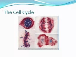
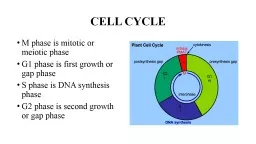
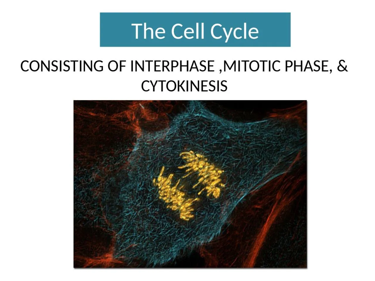
/
The Cell Cycle CONSISTING OF INTERPHASE ,MITOTIC PHASE, & CYTOKINESIS The Cell Cycle CONSISTING OF INTERPHASE ,MITOTIC PHASE, & CYTOKINESIS

The Cell Cycle CONSISTING OF INTERPHASE ,MITOTIC PHASE, & CYTOKINESIS - PowerPoint Presentation

leah
leah . @leah
Follow
342 views
Uploaded On 2022-06-07

The Cell Cycle CONSISTING OF INTERPHASE ,MITOTIC PHASE, & CYTOKINESIS - PPT Presentation

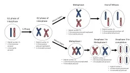
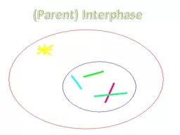
The Mitosis Puzzle Lay blank sheet lengthwise Write Interphase Prophase Metaphase Anaphase amp Telophase across the top of the sheet Cut out the cell diagrams and tapeglue them across the top under the appropriate phase label ID: 913808

cell cells cycle amp cells cell amp cycle cyclin cancer stem growth divide phase mitosis kinases embryonic factors dna

Share:

Link:

Embed:

Download Presentation from below link

Download Presentation The PPT/PDF document "The Cell Cycle CONSISTING OF INTERPHASE ..." is the property of its rightful owner. Permission is granted to download and print the materials on this web site for personal, non-commercial use only, and to display it on your personal computer provided you do not modify the materials and that you retain all copyright notices contained in the materials. By downloading content from our website, you accept the terms of this agreement.


Presentation Transcript

Slide1

The Cell Cycle

CONSISTING OF INTERPHASE ,MITOTIC PHASE, & CYTOKINESIS

Slide2

The Mitosis Puzzle

Lay blank sheet lengthwise .Write Interphase, Prophase, Metaphase, Anaphase, &

Telophase

across the top of the sheet.

Cut out the cell diagrams and tape/glue them across the top under the appropriate phase label.

Arrange each description beneath the appropriate picture to describe the cellular changes of that phase.

Slide3

Why do cells divide?

Bacteria cells & unicellular eukaryotic organisms divide & produce an entire organism.Multi-cellular organisms: Development -Growth -Repair

Slide4

What do you get at the end of the cell cycle?

2 genetically identical daughter cells

Slide5

Cellular Organization

PROKARYOTIC

EUKARYOTIC

Approximately 2 m of DNA to copy & be separated

Slide6

Replication of so much DNA is manageable because of how DNA is packaged.

chromosomes

chromatin

Slide7

Does the number of chromosomes in an organism determine how complex the organism is?

Bat

Herring gull

Human

Crayfish

Fern

Reptiles

Dog

Organize the following organisms in order from complex to simple

Slide8

Parts of a chromosome

THIS CHROMOSOME IS A DUPLICATED CHROMOSOME WITH 4 CHROMOSOMAL ARMS

Slide9

Slide10

The Cell Cycle

Interphase Accounts for 90% of the cycleDivided up into 3 subunits

Slide11

The Cell Cycle

Mitotic phaseIncludes both mitosis and cytokinesisUsually the shortest part of the cell cycle.

and Cytokinesis

Slide12

Mitotic Spindle

Begins to form during prophase & is complete during metaphase

Starts here

Form from the breakdown of microtubules of the cytoskeleton

Not present in plant cells

Slide13

Slide14

What is the significance of the fact that chromosomes condense before they are moved?

Slide15

WHICH OF THESE WOULD YOU RATHER ORGANIZE WITHOUT BREAKING?

Slide16

Slide17

CYTOKINESIS

ANIMAL CELLS

PLANT CELLS

Slide18

BINARY FISSION

PROKARYOTES (BACTERIA & ARCHEA)

Slide19

Evolution of Mitosis

A protein is thought to anchor the DNA to specific spot on membrane

Slide20

Timing is everything!

The frequency of cell division varies with cell type.EX: human skin cells

vs

liver cells

Some cells do not divide at all in a mature human.

EX: nerve cells and muscle cells

Slide21

What was concluded?

Molecules present in the cytoplasm of cells in the S or M phase control the progression of phases.

What Drives The Cell Cycle?

Hypothesis:

-

Each event in the cycle

triggers the next.

Slide22

CELL CYCLE CONTROL SYSTEM

Slide23

THE G

1

CHECKPOINT

Slide24

The Cell Cycle Clock

Maturation/Mitosis Promoting Factor (MPF) are regulatory molecules (mainly proteins)Protein kinases and cyclins

plus a phosphate group

Slide25

Kinases are enzymes

Activate or inactivate other proteins by phosphorylating them.Give the go ahead signals at the G1

and G

2

checkpoints.

Present in a constant concentration in a growing

cell, but are mostly inactive.

To be active they must attach to a

cyclin

(a

protein)

=

cyclin-dependent kinases, or Cdks

Cyclin

and Kinases

Slide26

Slide27

Cyclin

D

triggers cells to move from G

0

to G

1

into S phase

Cyclin

E

prepares the cell for DNA replication in S phase

Cyclin

A

activates DNA replication inside the nucleus in S phase

Cyclin

B

promotes the assembly of the mitotic spindle & other

tasks in the cytoplasm to prepare for mitosis

Slide28

Cyclins and cyclin-dependent

kinases control the cell cycle.

After the MPF does its job the

cyclin

degrades. Why?

The cell would continue to divide even when not ready

Slide29

In summaryInternally:

The fluctuation of cyclin & cyclin-dependent kinases seems to control the cell cycle internally using checkpoints to tell the cell to proceed or not

The MPF complexes (

cyclin

+

Cdk

) initiates mitosis & can then go on to produce a cascade of other cell responses including phosphorylation of other proteins which:

Promotes fragmentation of nuclear envelope

Chromosome condensation and spindle formation

Slide30

What about external factors?

Slide31

Example of a growth factor is PDGF

(platelet-derived growth factor)

PDGF is required for the division of fibroblasts

(a type of connective tissue)

Triggers a transduction pathway allowing the cells to pass the G

1

checkpoint & divide.

An injury can instigate this growth factor to help heal a wound.

Slide32

MORE EXTERNAL FACTORS ON CELL DIVISION:

DENSITY-DEPENDENT INHIBITION (in culture)

Slide33

In summaryExternally:

Growth factors secreted from the endocrine glands or blood cells are presentA substrate to attach to is neededDensity of neighboring cells are a factorReceptors (on outside of cell that’s dividing) are needed to receive each of the cell signals mentioned above.

Slide34

When do cells fail to divide?

If essential nutrients are missing.If growth factors

( protein released by certain cells to stimulate other cells to divide)

are missing.

Note: there are more than 50 growth factors

Let’s look at a cell gone wrong in the video Non

disjunction or non segregation in Mitosis

How does a cell divide “wrong”?

Slide35

What is the definition of cancer?The unregulated cell division of

an organism’s cell

Slide36

LOSS OF CELL CYCLE CONTROLS

Cancer cells do not follow the normal signals that regulate the cell cycle.

They don’t stop dividing even when there are no growth factors present.

Can continue dividing indefinitely in culture with ample nutrients.

EX:

HeLa

cells of 1951

Slide37

MALIGNANT VS BENIGN TUMOR

What does it mean if the cancer has metastasized?

Have too few genetic & cellular changes to survive elsewhere.

Have genetic & cellular changes that enable the cells to spread to new tissues & impair functions of organs = cancer

Slide38

Cancer most often results from mutations in genesProto-oncogenes:

they often code for proteins that stimulate cell division, prevent cell differentiation or regulate programmed cell death (apoptosis).

Tumor suppressor genes-

produce proteins that signal cells when they are getting too crowded.

Slide39

How does someone “get cancer”?

It can be triggered by:Carcinogens- Substances and environmental exposures that can lead to cancer

Teratogens-Any

agent that can disturb the development of an embryo or fetus. Teratogens may cause a birth defect in the child. Or a teratogen may halt the pregnancy outright. The classes of teratogens include radiation, maternal infections, chemicals, and drugs.

Viruses: ex: HPV

(human papilloma virus)

causes cervical cancer

& EBV

(Epstein Barr virus)

is associated with Hodgkin’s lymphoma, and gastric cancer.

Aging: ex: breast cancer increases the older you get

Slide40

How can we kill cancer cells?Radiation

ChemotherapyCDKs are considered a potential target for anti-cancer medication. If it is possible to selectively interrupt the cell cycle regulation in cancer cells by interfering with CDK action, the cell will die

.

Targets fast dividing cells

Slide41

Slide42

Differentiation of Human Cells

A zygote starts development by dividing over and over until you get a few dozen identical cells. These cells are embryonic stem cells.

Slide43

What are stem cells?

Slide44

Embryonic Stem Cells

Cells that start to take different development paths to become specialized cells, such as blood stem cells, which means they can no longer produce any other type of cell.

Can

give rise to any and all tissues in the

body

they can differentiate into some, but not all, cell types

.

Slide45

Totipotent cells can form all the cell types in a body, plus the extra-embryonic, or

placental cells. Embryonic cells within the first couple of cell divisions

after fertilization are the only cells that are totipotent.

Pluripotent

cells can give rise to all of the cell types that make up the body; embryonic stem cells are considered pluripotent.

Multipotent

cells can develop into more than one cell type, but are more limited than pluripotent cells; adult stem cells and cord blood stem cells are considered

multipotent

TOTIPOTENT

VS

PLURIPOTENT

VS

MULTIPOTENT

Stem Cells

Video

Slide46

To

generate cultures of specific types of differentiated cells—heart muscle cells, blood cells, or nerve cells, for example—scientists try to control the differentiation of embryonic stem cells. They change the chemical composition of the culture medium, alter the surface of the culture dish, or modify the cells by inserting specific genes.

Slide47

What side of stem cell research do you fall?

Slide48

Slide49