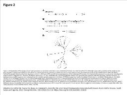
/
Figure 2 Figure 2. A) Organization of the viral genome of novel paramyxovirus related Figure 2 Figure 2. A) Organization of the viral genome of novel paramyxovirus related

Figure 2 Figure 2. A) Organization of the viral genome of novel paramyxovirus related - PowerPoint Presentation

reese
reese . @reese
Follow
27 views
Uploaded On 2024-02-02

Figure 2 Figure 2. A) Organization of the viral genome of novel paramyxovirus related - PPT Presentation

Albariño CG Foltzer M Towner JS Rowe LA Campbell S Jaramillo CM et al Novel Paramyxovirus Associated with Severe Acute Febrile Disease South Sudan and Uganda 2012 Emerg Infect Dis 2014202211216 httpsdoiorg103201eid2002131620 ID: 1044159

sequences virus viral protein virus sequences protein viral paramyxovirus tuhoko parainfluenza algorithm terminal vrna vcrna achimota clc version disease

Share:

Link:

Embed:

Download Presentation from below link

Download Presentation The PPT/PDF document "Figure 2 Figure 2. A) Organization of th..." is the property of its rightful owner. Permission is granted to download and print the materials on this web site for personal, non-commercial use only, and to display it on your personal computer provided you do not modify the materials and that you retain all copyright notices contained in the materials. By downloading content from our website, you accept the terms of this agreement.


Presentation Transcript

1. Figure 2Figure 2. A) Organization of the viral genome of novel paramyxovirus related to rubula-like viruses isolated from fruit bats was determined from the full-length sequenceB) Localization of the predicted viral genes and open reading frames (ORFs)The V/P edition site is predicted from the similarity to Tuhoko virus 3C) Terminal sequences were determined by standard rapid amplification of cDNA ends (RACE) methodsThe complementarity of terminal sequences is shown in vRNA and vcRNA senseD) Amino acid sequences of the nucleocapsid (N) protein of 22 representative paramyxovirus sequences were aligned by using the MUSCLE algorithm (CLC Genomics Workbench version 6.0.1; CLC bio, Cambridge, MA, USA)The phylogenetic analysis was conducted with a Bayesian algorithm (Mr.Bayes, Geneious version 6.1.5, www.geneious.com/)NP sequences were extracted from the complete genomic sequences in GenBank: KF774436 (Sosuga virus [SosV]), GU128082 (Tuhoko virus 3 ), GU128081 (Tuhoko virus 2), GU128080 (Tuhoko virus 1), AF298895 (Tioman virus), NC_007620 (Menangle virus), JX051319 (Achimota virus 1), JX051320 (Achimota virus 2), NC_003443 (human parainfluenza virus type 2), AF052755 (simian parainfluenza virus 5), HQ660095 (bat paramyxovirus Epo_spe/AR1/DRC/2009), NC_002200 (mumps virus), NC_009489 (Mapuera virus), NC_009640 (porcine rubulavirus), NC_001498 (measles virus), NC_006296 (rinderpest virus), NC_001921 (canine distemper virus), NC_001552 (Sendai virus), NC_003461 (human parainfluenza virus type 1), NC_002728 (Nipah virus), NC_001906 (Henra virus), NC_002617 (Newcastle disease virus)vcRNA, viral complementary RNA; N, nucleocapsid protein; V/P, V protein; M, matrix protein; F, fusion protein; HN, hemagglutinin-neuraminidase; L, molecular weight DNA ladder; CDS, coding sequence; nt posnucleotide position; vRNA, viral RNA.Albariño CG, Foltzer M, Towner JS, Rowe LA, Campbell S, Jaramillo CM, et al. Novel Paramyxovirus Associated with Severe Acute Febrile Disease, South Sudan and Uganda, 2012. Emerg Infect Dis. 2014;20(2):211-216. https://doi.org/10.3201/eid2002.131620