Explore
Featured
Recent
Articles
Topics
Login
Upload
Featured
Recent
Articles
Topics
Login
Upload
Search Results for 'mri figure'
mri figure published presentations and documents on DocSlides.
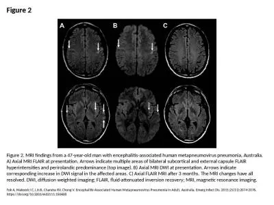
Figure 2 Figure 2. MRI findings from a 47-year-old man with encephalitis-associated human metapneum
by arya
Fok A, Mateevici C, Lin B, Chandra RV, Chong V. En...
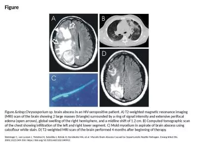
Figure Figure. Chrysosporium sp. brain abscess in an HIV-seropositive patient. A) T2-weigh
by davies
Steininger C, van Lunzen J, Tintelnot K, Sobottka ...
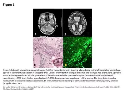
Figure 1 Figure 1. A) Magnetic resonance imaging (MRI) of the patient's brain showing a la
by angelina
Shirwadkar CG, Samant R, Sankhe M, Deshpande R, Ya...
SectionEditor
by reese
RESIDENT &FELLOW SECTION MitchellS.V.Elkind, MD,MS...
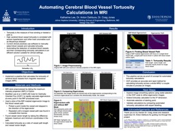
Automating Cerebral Blood Vessel Tortuosity Calculations in MRI
by emery
Katharine Lee, Dr. Anton . Dahbura. , Dr. Craig Jo...
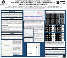
NEUROPSYCHOPHYSIOLOGICAL MAPPING:
by CountryBumpkin
CONCOMMITANT PSYCHOPHYSIOLOGICAL RECORDING . AND ....
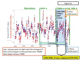
GPCP TRMM
by ButterflyKisses
La Nina:. Jul10+Jan11. July . /. January pairs at...
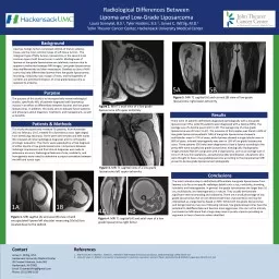
Radiological Differences Between
by Hardrocker
Lipoma. and Low-Grade . Liposarcoma. . Laura . S...
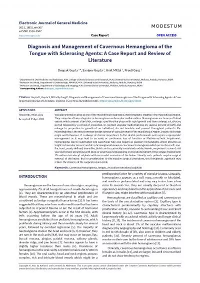
CopyrightbyAuthorsandLicensedbyModestumThisopenaccessarticledistribut
by priscilla
unrestricteduse,distribution,andreproductionanymed...
Medical Imaging By Anuja
by calandra-battersby
. Kulkarni. 1000722132. LIST OF ACRONYMS. CAT - ...
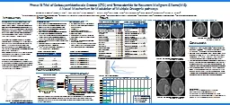
Phase IB Trial of Carboxyamidotriazole Orotate
by myesha-ticknor
(CTO) and . Temozolomide. for Recurrent Malignan...
Unusual case of a cystic pelvic mass in a male
by amelia
Dr Lorna Woodbridge (lorna.woodbridge@nhs.net). Dr...
M. AMOR, S. MAJDOUB, B. BEN SALAH, M. DHIFALLAH, H. ZAGHOUANI, T. RZIGA, H. AMARA, D. BAKIR, C. KRA
by SweetMelody
Radiology. service, . University Hospital . Farha...
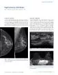
Figure 1 A and B
by fiona
. Left CC (A) and MLO (B) views from a digital mam...
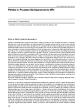
Pitfalls in Prostate Multiparametric MRI
by brown
128 Andrs Labra W.*, lvaroúñiga G.1. Radiologist...
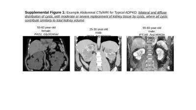
Supplemental Figure 1: Example Abdominal CTs/MRI for Typical ADPKD:
by helene
bilateral and diffuse distribution of cysts, with ...
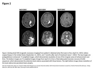
Figure 2 Figure 2. Axial MRI (magnetic resonance imaging) from patient 2 obtained when fir
by cora
Sohn AH, Probert WS, Glaser CA, Gupta N, Bollen AW...
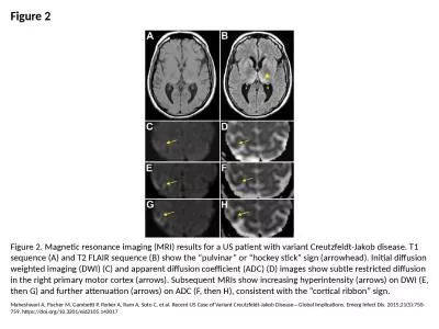
Figure 2 Figure 2. Magnetic resonance imaging (MRI) results for a US patient with variant Creutzfel
by carny
Maheshwari A, Fischer M, Gambetti P, Parker A, Ram...
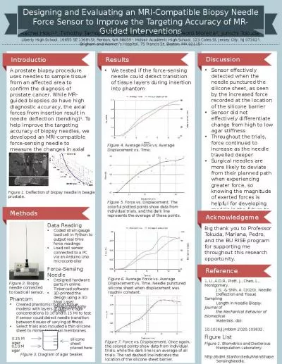
Designing and Evaluating an MRI-Compatible Biopsy Needle Force Sensor to Improve the Targeting Accu
by kareem
Phantom. Created phantoms (human tissue models) wi...
Figure 3 MRI T1W LS spine sagittal view Arrows show owing osteo
by delilah
367 Figure 4. MRI (T2W) L-S spine axial view. Arro...
suprascapular neuropathy electrophysiologic tests are usually perform
by elena
Figure 1.Infraspinatus damage due to suprascapular...
GASTROINTESTINAL
by jovita
57 57 Giant condyloma acuminatum (Buschke-Lowenste...
Article published online 20210730
by fauna
27 Bursae around the knee joints Priyank S Chatra...
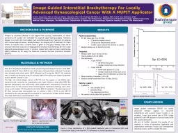
Image Guided Interstitial Brachytherapy For Locally Advanced
by hailey
Gynaecological. Cancer With A MUPIT Applicator . ...
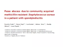
Psoas abscess due to community acquired methicillin-resistant
by sterialo
Staphylococcus aureus . in a patient with . spondy...
Load More...