
Endocrine and Hematologic Emergencies CHAPTER 21:












Description: Endocrine and Hematologic Emergencies CHAPTER 21: Focused Lecture National EMS Education Standard Competencies (1 of 3) Medicine Applies fundamental knowledge to provide basic and selected advanced emergency care and transportation based on
Related Topics
Download Presentation
"Endocrine and Hematologic Emergencies CHAPTER 21:" is the property of its rightful owner. Permission is granted to download and print the materials on this website for personal, non-commercial use only, and to display it on your personal computer provided you do not modify the materials and that you retain all copyright notices contained in the materials. By downloading content from our website, you accept the terms of this agreement.
Presentation Transcript
slide1. Endocrine and Hematologic Emergencies CHAPTER 21: Focused Lecture<br>
slide2. National EMS Education Standard Competencies (1 of 3) Medicine
Applies fundamental knowledge to provide basic and selected advanced emergency care and transportation based on assessment findings for an acutely ill patient.<br>
slide3. National EMS Education Standard Competencies (2 of 3) Endocrine Disorders
Awareness that
Diabetic emergencies cause altered mental status
Anatomy, physiology, pathophysiology, assessment, and management of
Acute diabetic emergencies<br>
slide4. National EMS Education Standard Competencies (3 of 3) Hematology
Anatomy, physiology, pathophysiology, assessment, and management of:
Sickle cell crisis
Clotting disorders<br>
slide5. Introduction Endocrine system influences every cell, organ, and body function.
Hematologic emergencies
Rare in most EMS systems
Difficult to assess and treat in the prehospital setting<br>
slide6. Anatomy and Physiology Review: The Endocrine System Network of glands that produce and secrete chemical messengers called hormones. © Jones & Bartlett Learning.<br>
slide7. Pituitary Gland Located at the base of the brain
Secretes hormones that stimulate other endocrine glands
Adrenocorticotropic hormone (ACTH)
Follicle-stimulating hormone
Growth hormone
Luteinizing hormone
Prolactin
Thyroid-stimulating hormone<br>
slide8. Thyroid Gland Secretes thyroxine when the body’s metabolic rate decreases
Thyroxine (T4) stimulates energy production in cells.
Secretes calcitonin, which helps maintain normal calcium levels in the blood<br>
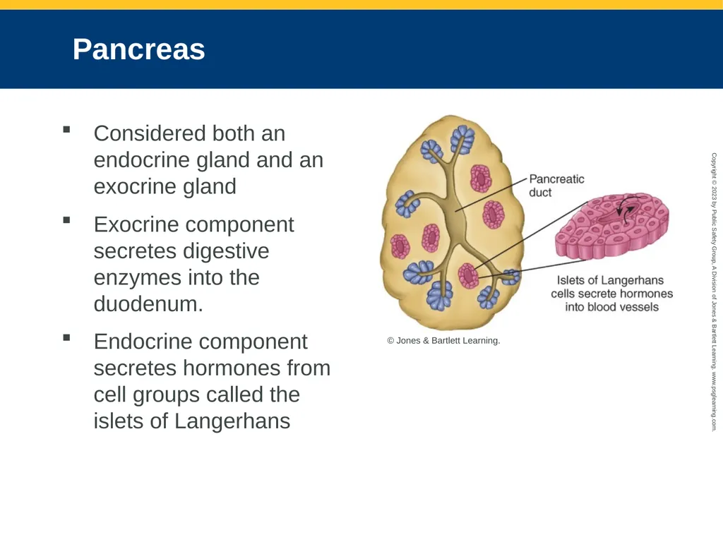
slide9. Pancreas Considered both an endocrine gland and an exocrine gland
Exocrine component secretes digestive enzymes into the duodenum.
Endocrine component secretes hormones from cell groups called the islets of Langerhans © Jones & Bartlett Learning.<br>
slide10. The Role of Glucose and Insulin (1 of 3) Glucose is the major source of energy for the body.
Without glucose, or with an extremely low level, brain cells rapidly suffer permanent damage.
Insulin is needed to allow glucose to enter individual body cells to fuel their functioning.<br>
slide11. The Role of Glucose and Insulin (2 of 3) © Jones & Bartlett Learning.<br>
slide12. The Role of Glucose and Insulin (3 of 3) When blood glucose levels are elevated
Insulin increases cell membrane permeability and mediates the transport of glucose across the cell membrane.
When the blood glucose level decreases
Glucagon enters the bloodstream and raises the blood glucose level.
Glucose is secreted into the bloodstream, where cells use it for energy.<br>
slide13. Pathophysiology of the Endocrine System Endocrine disorders can be caused by
Hypersecretion
Insufficient secretion of a gland
Hyperthyroidism increases metabolism.
Hypothyroidism decreases metabolism.<br>
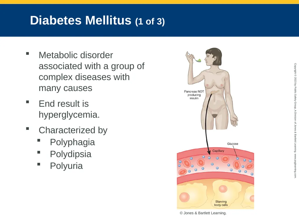
slide14. Diabetes Mellitus (1 of 3) Metabolic disorder associated with a group of complex diseases with many causes
End result is hyperglycemia.
Characterized by
Polyphagia
Polydipsia
Polyuria © Jones & Bartlett Learning.<br>
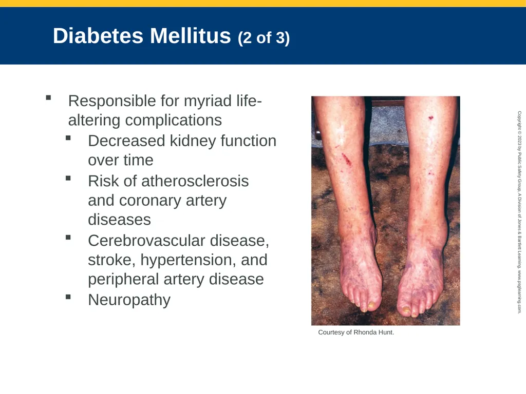
slide15. Diabetes Mellitus (2 of 3) Responsible for myriad life-altering complications
Decreased kidney function over time
Risk of atherosclerosis and coronary artery diseases
Cerebrovascular disease, stroke, hypertension, and peripheral artery disease
Neuropathy Courtesy of Rhonda Hunt.<br>
slide16. Diabetes Mellitus (3 of 3) Both chronic and acute complications are associated with diabetes mellitus.
Left untreated, results in organ system dysfunction, wasting of body tissues, and death.<br>
slide17. Type 1 Diabetes Mellitus (1 of 2) Insulin-dependent diabetes
Has a hereditary predisposition; environmental factors may play a role
In addition to daily insulin injections, strict dietary control must be observed.<br>
slide18. Type 1 Diabetes Mellitus (2 of 2) Always requires the use of insulin
Insulin pumps provide improved control of blood glucose levels. © Photographee.eu/Shutterstock.<br>
slide19. Type 2 Diabetes Mellitus (1 of 2) Most common form of diabetes
Blood glucose levels are elevated.
Typically develops in middle age
Symptoms of untreated type 2 diabetes
Fatigue, nausea, frequent urination, thirst, unexplained weight loss, blurred vision, slow healing of wounds, unresponsiveness, seizure<br>
slide20. Type 2 Diabetes Mellitus (2 of 2) Weight loss is an important factor in helping to control type 2 diabetes.
Metformin causes a decrease in glucose production. © Piotr Adamowicz/Shutterstock © Stockbyte/Thinkstock/Getty Images © instamatic/iStock/Getty Images.<br>
slide21. Prediabetes (1 of 2) Condition identified in people who have certain risk factors associated with type 2 diabetes
Exists when blood glucose levels or hemoglobin A1c levels are above normal<br>
slide22. Prediabetes (2 of 2) Risk factors include
Older than 45 years
Being overweight
Family history of diabetes
Losing 5% to 7% of the patient’s body weight and getting at least 150 minutes of physical activity per week can prevent or delay the onset.<br>
slide23. Gestational Diabetes Glucose intolerance that can occur during pregnancy
High levels of glucose enter the fetus, causing increased production of insulin by the fetus.
Management includes diet modification, exercise, and blood glucose testing.<br>
slide24. Hypoglycemia (1 of 2) Often the result of having taken too much insulin, too little food, or both
Counterregulation is the body’s natural defensive ability to maintain blood glucose.
First line of defense is reduced insulin production.
Second line of defense is secretion of catecholamines by the adrenal gland.<br>
slide25. Hypoglycemia (2 of 2) In patients with type 1 diabetes, the islets of Langerhans do not make insulin.
In patients with type 2 diabetes, the pancreas can generate insulin.
Patient will experience trembling; a rapid heart rate; rapid, shallow respirations; sweating; and a feeling of hunger.<br>
slide26. Hyperglycemia and Diabetic Ketoacidosis (1 of 4) Occurs when blood glucose levels remain elevated
Hyperglycemic crisis: State of unresponsiveness resulting from several problems
Common causes of DKA include infection, injury, alcohol use, emotional distress, and illness such as stroke or myocardial infarction<br>
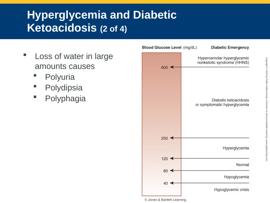
slide27. Hyperglycemia and Diabetic Ketoacidosis (2 of 4) Loss of water in large amounts causes
Polyuria
Polydipsia
Polyphagia © Jones & Bartlett Learning.<br>
slide28. Hyperglycemia and Diabetic Ketoacidosis (3 of 4) If proper fluid rehydration and insulin are not given, ketoacidosis will progress to unresponsiveness, diabetic coma, and, eventually, death.<br>
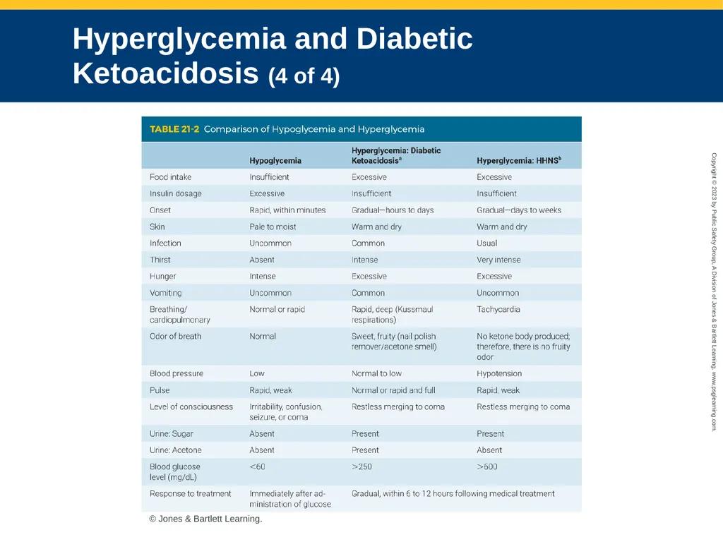
slide29. Hyperglycemia and Diabetic Ketoacidosis (4 of 4) © Jones & Bartlett Learning.<br>
slide30. Hyperosmolar Hyperglycemic Nonketotic Syndrome Occurs principally in patients with type 2 diabetes
Characterized by hyperglycemia, hyperosmolarity, and an absence of substantial ketosis
Signs and symptoms:
Hyperglycemia, seizures, partial paralysis
Altered mental status, drowsiness, and lethargy
Severe dehydration, thirst, and dark urine<br>
slide31. Patient Assessment of Endocrine Emergencies (1 of 5) Similar assessment as with any other medical patient
Scene size-up
Evaluate scene safety.
Ensure the personal protective equipment is readily available.
Question bystanders on events leading up to your arrival.<br>
slide32. Patient Assessment of Endocrine Emergencies (2 of 5) Primary survey
Form a general impression.
Determine level of consciousness using the AVPU scale.
Assess the patient’s airway and breathing.
A hyperglycemic patient may have rapid, deep respirations (Kussmaul respirations).
A hypoglycemic patient will have normal or shallow to rapid respirations.<br>
slide33. Patient Assessment of Endocrine Emergencies (3 of 5) History taking
Investigate the chief complaint.
Physical signs may guide you.
Obtain SAMPLE history.
Check for emergency medical identification device.<br>
slide34. Patient Assessment of Endocrine Emergencies (4 of 5) Secondary assessment
Assess unresponsive patients with a full-body exam.
Focus on the patient’s mental status and ability to swallow and protect the airway.
Obtain a complete set of vital signs.
Glucometers measure the glucose level in whole blood.<br>
slide35. Patient Assessment of Endocrine Emergencies (5 of 5) Reassessment
Monitor the airway carefully and be alert for potential vomiting.
Document each assessment, your findings, the time of the interventions, and any changes in the patient’s condition.
Base administration of glucose on serial readings.<br>
slide36. Emergency Medical Care of Endocrine Emergencies General management
Place the patient in a position of comfort.
If the patient has an altered mental status, establish an IV line.
Transport the unresponsive patient supine or in recovery position.
Keep the mouth and pharynx suctioned free of secretions, vomitus, and blood.<br>
slide37. Management of Hypoglycemia (1 of 3) Immediately increase blood glucose levels.
Do not give sugar-free drinks that are sweetened with saccharin.
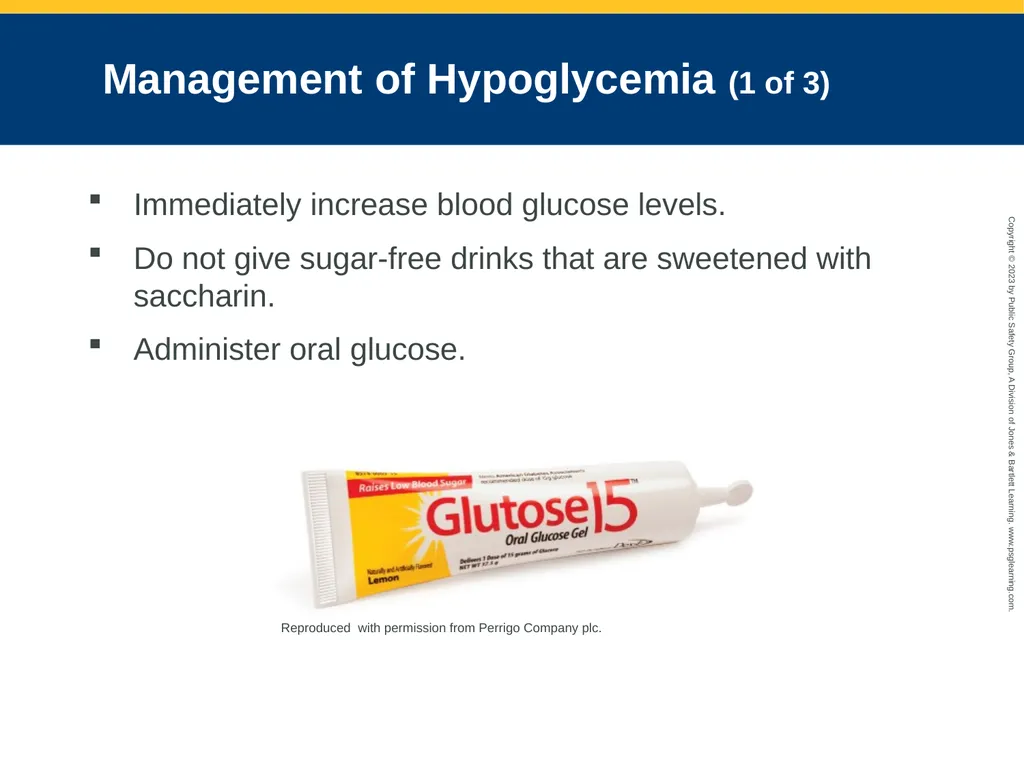
Administer oral glucose. Reproduced with permission from Perrigo Company plc.<br>
slide38. Management of Hypoglycemia (2 of 3) Administering intravenous dextrose
Administer if patient has an altered mental status or is unable to swallow.
Monitor for infiltration into local tissue.
Administer slowly to avoid rupturing the vein.<br>
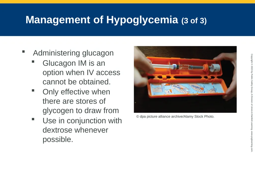
slide39. Management of Hypoglycemia (3 of 3) Administering glucagon
Glucagon IM is an option when IV access cannot be obtained.
Only effective when there are stores of glycogen to draw from
Use in conjunction with dextrose whenever possible. © dpa picture alliance archive/Alamy Stock Photo.<br>
slide40. Management of Hyperglycemia and DKA Treat for DKA if glucose level is more than 250 mg/dL.
Goals of prehospital treatment
Rehydration
Correct the patient’s electrolyte and acid-base abnormalities
Maintain the airway and administer oxygen.
Treat the patient symptomatically.<br>
slide41. Management of HHNS Follows pathway for dehydration and altered mental status
Complete advanced airway management as early as possible.
Cervical spine immobilization should be considered for all unresponsive patients.
Large-bore IV access (18 gauge) should be gained as soon as possible.<br>
slide42. Hematologic Emergencies (1 of 2) Hematology: Study of blood
Addresses the ways in which the constituent parts of blood are involved in health and disease
Blood components
Red blood cells (RBCs)
White blood cells (WBCs)
Platelets, proteins
Hematopoietic system<br>
slide43. Hematologic Emergencies (2 of 2) Hematologic disorder refers to any disorder of the blood.
Hemolytic disorders refer to disease processes that cause the breakdown of RBCs.
Hemostatic disorders refer to bleeding and clotting abnormalities.<br>
slide44. Anatomy and Physiology Review: Hematology (1 of 4) Blood and plasma
Functions of blood
Respiratory
Nutritional
Excretory
Regulatory
Defensive<br>
slide45. Anatomy and Physiology Review: Hematology (2 of 4) Components
Plasma
Transports all formed element components throughout the body
Red blood cells (RBCs)
Responsible for carrying oxygen to the tissues
White blood cells (WBCs)
Protect body against foreign invaders
Platelets<br>
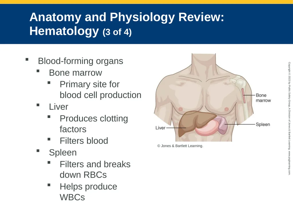
slide46. Anatomy and Physiology Review: Hematology (3 of 4) Blood-forming organs
Bone marrow
Primary site for blood cell production
Liver
Produces clotting factors
Filters blood
Spleen
Filters and breaks down RBCs
Helps produce WBCs © Jones & Bartlett Learning.<br>
slide47. Anatomy and Physiology Review: Hematology (4 of 4) Blood classifications
Ensure compatibility and prevent medical problems during blood component replacement
RBC classification types are O, A, B, and AB.
Transfusion reactions
Occur when patients receive a blood type different from their own<br>
slide48. Sickle Cell Disease (1 of 4) Most common inherited blood disorder
Starts with a genetic defect of the adult type hemoglobin (HbA)
Sickle cell crisis may manifest in several ways:
Vaso-occlusive crisis
Acute chest syndrome
Aplastic crisis
Hemolytic crisis
Splenic sequestration crisis<br>
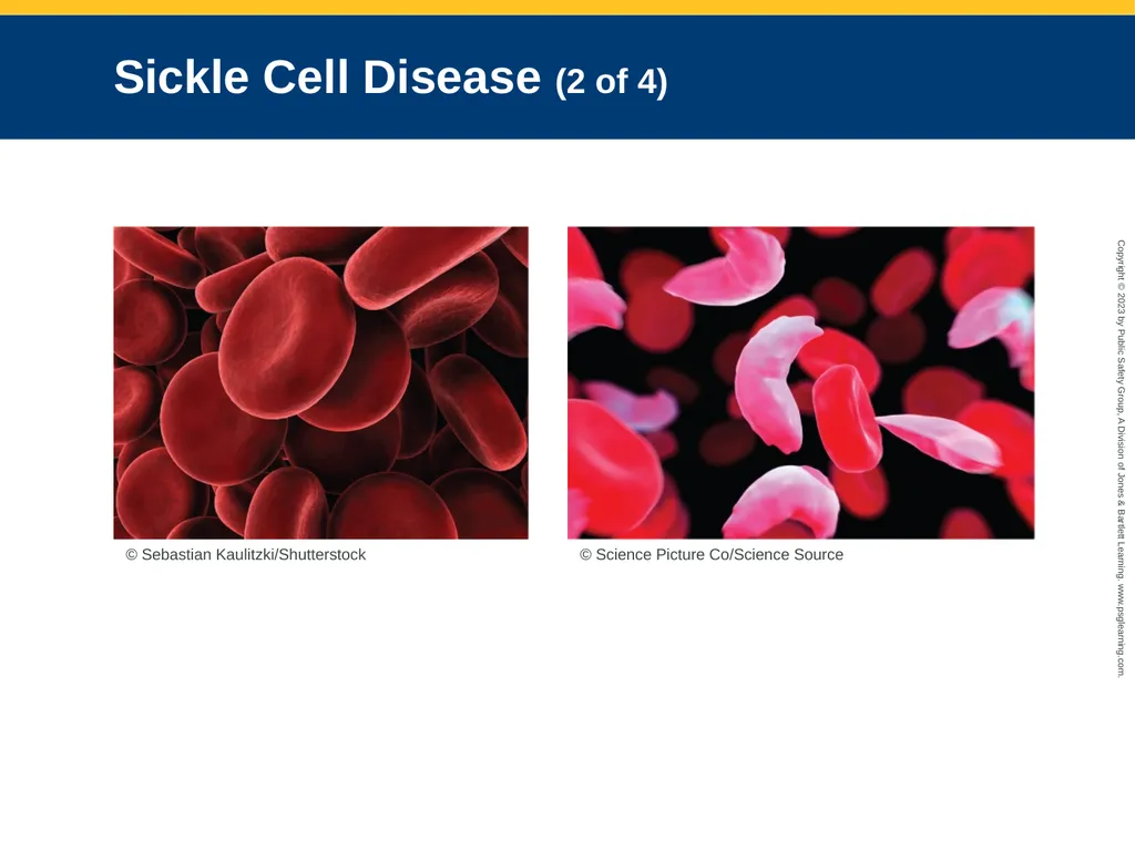
slide49. Sickle Cell Disease (2 of 4) © Sebastian Kaulitzki/Shutterstock © Science Picture Co/Science Source<br>
slide50. Sickle Cell Disease (3 of 4) In acute crises, patients may have substantial pain resulting from congested vessels.
Patients with chronic sickle cell attacks are susceptible to severe, life-threatening complications.<br>
slide51. Sickle Cell Disease (4 of 4) Potential complications
Cerebrovascular attack
Gallstones
Jaundice
Osteonecrosis
Splenic infections
Osteomyelitis (or septic arthritis)
Opiate tolerance
Leg ulcers
Retinopathy
Chronic pain
Pulmonary hypertension
Chronic renal failure<br>
slide52. Anemia (1 of 2) Defined as a hemoglobin or RBC level lower than normal
In autoimmune disorders, RBCs are destroyed by the body’s own antibodies.
Patients will report feeling worn down or having no energy.<br>
slide53. Anemia (2 of 2) Some patients may have angina-type chest pain related to decreased oxygen availability to the heart muscle.
Other conditions common in patients with anemia include leukopenia and thrombocytopenia.<br>
slide54. Clotting Disorders (1 of 4) Thrombosis: Blood clot in arterial or venous blood vessels
Thrombophilia: Tendency to develop blood clots
May manifest as the formation of a blood clot in a blood vessel or in one of the chambers of the heart<br>
slide55. Clotting Disorders (2 of 4) Risk factors:
Recent surgery
Impaired mobility
Heart failure
Cancer
Respiratory failure
Infectious diseases
Age older than 40 years
Overweight/obesity<br>
slide56. Clotting Disorders (3 of 4) Risk factors (cont’d):
Smoking
Oral contraceptive use
Recent trauma
Recent long-distance travel (multihour car or plane ride)<br>
slide57. Clotting Disorders (4 of 4) Hemophilia
Clotting does not occur or occurs insufficiently
Results in increased bleeding time
Classified as Hemophilia A or hemophilia B
Signs and symptoms are the same.<br>
slide58. Disseminated Intravascular Coagulation May result from any number of life-threatening conditions
Progresses in two stages
Identify signs and symptoms commonly associated with DIC or progression toward this coagulopathy.<br>
slide59. Patient Assessment of Hematologic Emergencies (1 of 7) Scene size-up
Consider MOI and/or NOI.
Determine the number of patients.
Assess for hazards and need for help.
Standard precautions should consist of gloves and eye protection at a minimum.
Evaluate each situation quickly.
Patients experiencing a vaso-occlusive crisis would benefit from administration of analgesics.<br>
slide60. Patient Assessment of Hematologic Emergencies (2 of 7) Primary survey
Always perform a thorough, careful primary survey.
Pay attention to ABCs and correct life threats.
Perform a rapid full-body scan.
Patients showing signs of inadequate breathing, an altered mental status, or SpO2 of 94% or less should receive high-flow oxygen.<br>
slide61. Patient Assessment of Hematologic Emergencies (3 of 7) An increased heart rate represents a compensatory mechanism.
In patients with suspected hemophilia, be alert for signs of acute blood loss.
Fluid may be necessary for these patients.
Rapid transportation will depend on the severity of the patient’s condition and wishes.<br>
slide62. Patient Assessment of Hematologic Emergencies (4 of 7) History taking
Investigate the chief complaint.
Obtain the patient’s history and SAMPLE history.
Physical signs include such as swelling of the fingers and toes, priapism, and jaundice.
Ascertain whether pain is isolated or felt throughout the body.
Look for changes in level of consciousness and symptoms.<br>
slide63. Patient Assessment of Hematologic Emergencies (5 of 7) In a patient with known sickle cell disease, ask:
Have you had a crisis before?
When was the last time you had a crisis?
How did your last crisis resolve?
Have you had any illness, unusual amount of activity, or stress lately?<br>
slide64. Patient Assessment of Hematologic Emergencies (6 of 7) Secondary assessment
May be performed on scene, en route to the emergency department, or not at all
Systematically examine the patient, focusing on major joints at which cells congregate.
Evaluate and document mental status using the AVPU scale.
Obtain a complete set of vital signs.
Use pulse oximetry, if available.<br>
slide65. Patient Assessment of Hematologic Emergencies (7 of 7) Reassessment
Reassess frequently.
Document each assessment, your findings, the time of the interventions, and any changes in the patient’s condition.<br>
slide66. Emergency Medical Care of Hematologic Emergencies (1 of 2) Emergency medical care should include:
Oxygen
Fluids
Transport
Pharmacology
Comfort and rest
Psychological support<br>
slide67. Emergency Medical Care of Hematologic Emergencies (2 of 2) Management of specific conditions
Care for sickle cell disease might include analgesics for pain, antibiotics to prevent infection, and blood transfusions as required.
In cases of anemia, check and monitor the airway and the patient’s breathing.
Identify DIC and establish treatment early.
Be optimistic but honest with patients and family.<br>
slide2. National EMS Education Standard Competencies (1 of 3) Medicine
Applies fundamental knowledge to provide basic and selected advanced emergency care and transportation based on assessment findings for an acutely ill patient.<br>
slide3. National EMS Education Standard Competencies (2 of 3) Endocrine Disorders
Awareness that
Diabetic emergencies cause altered mental status
Anatomy, physiology, pathophysiology, assessment, and management of
Acute diabetic emergencies<br>
slide4. National EMS Education Standard Competencies (3 of 3) Hematology
Anatomy, physiology, pathophysiology, assessment, and management of:
Sickle cell crisis
Clotting disorders<br>
slide5. Introduction Endocrine system influences every cell, organ, and body function.
Hematologic emergencies
Rare in most EMS systems
Difficult to assess and treat in the prehospital setting<br>
slide6. Anatomy and Physiology Review: The Endocrine System Network of glands that produce and secrete chemical messengers called hormones. © Jones & Bartlett Learning.<br>
slide7. Pituitary Gland Located at the base of the brain
Secretes hormones that stimulate other endocrine glands
Adrenocorticotropic hormone (ACTH)
Follicle-stimulating hormone
Growth hormone
Luteinizing hormone
Prolactin
Thyroid-stimulating hormone<br>
slide8. Thyroid Gland Secretes thyroxine when the body’s metabolic rate decreases
Thyroxine (T4) stimulates energy production in cells.
Secretes calcitonin, which helps maintain normal calcium levels in the blood<br>
slide9. Pancreas Considered both an endocrine gland and an exocrine gland
Exocrine component secretes digestive enzymes into the duodenum.
Endocrine component secretes hormones from cell groups called the islets of Langerhans © Jones & Bartlett Learning.<br>
slide10. The Role of Glucose and Insulin (1 of 3) Glucose is the major source of energy for the body.
Without glucose, or with an extremely low level, brain cells rapidly suffer permanent damage.
Insulin is needed to allow glucose to enter individual body cells to fuel their functioning.<br>
slide11. The Role of Glucose and Insulin (2 of 3) © Jones & Bartlett Learning.<br>
slide12. The Role of Glucose and Insulin (3 of 3) When blood glucose levels are elevated
Insulin increases cell membrane permeability and mediates the transport of glucose across the cell membrane.
When the blood glucose level decreases
Glucagon enters the bloodstream and raises the blood glucose level.
Glucose is secreted into the bloodstream, where cells use it for energy.<br>
slide13. Pathophysiology of the Endocrine System Endocrine disorders can be caused by
Hypersecretion
Insufficient secretion of a gland
Hyperthyroidism increases metabolism.
Hypothyroidism decreases metabolism.<br>
slide14. Diabetes Mellitus (1 of 3) Metabolic disorder associated with a group of complex diseases with many causes
End result is hyperglycemia.
Characterized by
Polyphagia
Polydipsia
Polyuria © Jones & Bartlett Learning.<br>
slide15. Diabetes Mellitus (2 of 3) Responsible for myriad life-altering complications
Decreased kidney function over time
Risk of atherosclerosis and coronary artery diseases
Cerebrovascular disease, stroke, hypertension, and peripheral artery disease
Neuropathy Courtesy of Rhonda Hunt.<br>
slide16. Diabetes Mellitus (3 of 3) Both chronic and acute complications are associated with diabetes mellitus.
Left untreated, results in organ system dysfunction, wasting of body tissues, and death.<br>
slide17. Type 1 Diabetes Mellitus (1 of 2) Insulin-dependent diabetes
Has a hereditary predisposition; environmental factors may play a role
In addition to daily insulin injections, strict dietary control must be observed.<br>
slide18. Type 1 Diabetes Mellitus (2 of 2) Always requires the use of insulin
Insulin pumps provide improved control of blood glucose levels. © Photographee.eu/Shutterstock.<br>
slide19. Type 2 Diabetes Mellitus (1 of 2) Most common form of diabetes
Blood glucose levels are elevated.
Typically develops in middle age
Symptoms of untreated type 2 diabetes
Fatigue, nausea, frequent urination, thirst, unexplained weight loss, blurred vision, slow healing of wounds, unresponsiveness, seizure<br>
slide20. Type 2 Diabetes Mellitus (2 of 2) Weight loss is an important factor in helping to control type 2 diabetes.
Metformin causes a decrease in glucose production. © Piotr Adamowicz/Shutterstock © Stockbyte/Thinkstock/Getty Images © instamatic/iStock/Getty Images.<br>
slide21. Prediabetes (1 of 2) Condition identified in people who have certain risk factors associated with type 2 diabetes
Exists when blood glucose levels or hemoglobin A1c levels are above normal<br>
slide22. Prediabetes (2 of 2) Risk factors include
Older than 45 years
Being overweight
Family history of diabetes
Losing 5% to 7% of the patient’s body weight and getting at least 150 minutes of physical activity per week can prevent or delay the onset.<br>
slide23. Gestational Diabetes Glucose intolerance that can occur during pregnancy
High levels of glucose enter the fetus, causing increased production of insulin by the fetus.
Management includes diet modification, exercise, and blood glucose testing.<br>
slide24. Hypoglycemia (1 of 2) Often the result of having taken too much insulin, too little food, or both
Counterregulation is the body’s natural defensive ability to maintain blood glucose.
First line of defense is reduced insulin production.
Second line of defense is secretion of catecholamines by the adrenal gland.<br>
slide25. Hypoglycemia (2 of 2) In patients with type 1 diabetes, the islets of Langerhans do not make insulin.
In patients with type 2 diabetes, the pancreas can generate insulin.
Patient will experience trembling; a rapid heart rate; rapid, shallow respirations; sweating; and a feeling of hunger.<br>
slide26. Hyperglycemia and Diabetic Ketoacidosis (1 of 4) Occurs when blood glucose levels remain elevated
Hyperglycemic crisis: State of unresponsiveness resulting from several problems
Common causes of DKA include infection, injury, alcohol use, emotional distress, and illness such as stroke or myocardial infarction<br>
slide27. Hyperglycemia and Diabetic Ketoacidosis (2 of 4) Loss of water in large amounts causes
Polyuria
Polydipsia
Polyphagia © Jones & Bartlett Learning.<br>
slide28. Hyperglycemia and Diabetic Ketoacidosis (3 of 4) If proper fluid rehydration and insulin are not given, ketoacidosis will progress to unresponsiveness, diabetic coma, and, eventually, death.<br>
slide29. Hyperglycemia and Diabetic Ketoacidosis (4 of 4) © Jones & Bartlett Learning.<br>
slide30. Hyperosmolar Hyperglycemic Nonketotic Syndrome Occurs principally in patients with type 2 diabetes
Characterized by hyperglycemia, hyperosmolarity, and an absence of substantial ketosis
Signs and symptoms:
Hyperglycemia, seizures, partial paralysis
Altered mental status, drowsiness, and lethargy
Severe dehydration, thirst, and dark urine<br>
slide31. Patient Assessment of Endocrine Emergencies (1 of 5) Similar assessment as with any other medical patient
Scene size-up
Evaluate scene safety.
Ensure the personal protective equipment is readily available.
Question bystanders on events leading up to your arrival.<br>
slide32. Patient Assessment of Endocrine Emergencies (2 of 5) Primary survey
Form a general impression.
Determine level of consciousness using the AVPU scale.
Assess the patient’s airway and breathing.
A hyperglycemic patient may have rapid, deep respirations (Kussmaul respirations).
A hypoglycemic patient will have normal or shallow to rapid respirations.<br>
slide33. Patient Assessment of Endocrine Emergencies (3 of 5) History taking
Investigate the chief complaint.
Physical signs may guide you.
Obtain SAMPLE history.
Check for emergency medical identification device.<br>
slide34. Patient Assessment of Endocrine Emergencies (4 of 5) Secondary assessment
Assess unresponsive patients with a full-body exam.
Focus on the patient’s mental status and ability to swallow and protect the airway.
Obtain a complete set of vital signs.
Glucometers measure the glucose level in whole blood.<br>
slide35. Patient Assessment of Endocrine Emergencies (5 of 5) Reassessment
Monitor the airway carefully and be alert for potential vomiting.
Document each assessment, your findings, the time of the interventions, and any changes in the patient’s condition.
Base administration of glucose on serial readings.<br>
slide36. Emergency Medical Care of Endocrine Emergencies General management
Place the patient in a position of comfort.
If the patient has an altered mental status, establish an IV line.
Transport the unresponsive patient supine or in recovery position.
Keep the mouth and pharynx suctioned free of secretions, vomitus, and blood.<br>
slide37. Management of Hypoglycemia (1 of 3) Immediately increase blood glucose levels.
Do not give sugar-free drinks that are sweetened with saccharin.
Administer oral glucose. Reproduced with permission from Perrigo Company plc.<br>
slide38. Management of Hypoglycemia (2 of 3) Administering intravenous dextrose
Administer if patient has an altered mental status or is unable to swallow.
Monitor for infiltration into local tissue.
Administer slowly to avoid rupturing the vein.<br>
slide39. Management of Hypoglycemia (3 of 3) Administering glucagon
Glucagon IM is an option when IV access cannot be obtained.
Only effective when there are stores of glycogen to draw from
Use in conjunction with dextrose whenever possible. © dpa picture alliance archive/Alamy Stock Photo.<br>
slide40. Management of Hyperglycemia and DKA Treat for DKA if glucose level is more than 250 mg/dL.
Goals of prehospital treatment
Rehydration
Correct the patient’s electrolyte and acid-base abnormalities
Maintain the airway and administer oxygen.
Treat the patient symptomatically.<br>
slide41. Management of HHNS Follows pathway for dehydration and altered mental status
Complete advanced airway management as early as possible.
Cervical spine immobilization should be considered for all unresponsive patients.
Large-bore IV access (18 gauge) should be gained as soon as possible.<br>
slide42. Hematologic Emergencies (1 of 2) Hematology: Study of blood
Addresses the ways in which the constituent parts of blood are involved in health and disease
Blood components
Red blood cells (RBCs)
White blood cells (WBCs)
Platelets, proteins
Hematopoietic system<br>
slide43. Hematologic Emergencies (2 of 2) Hematologic disorder refers to any disorder of the blood.
Hemolytic disorders refer to disease processes that cause the breakdown of RBCs.
Hemostatic disorders refer to bleeding and clotting abnormalities.<br>
slide44. Anatomy and Physiology Review: Hematology (1 of 4) Blood and plasma
Functions of blood
Respiratory
Nutritional
Excretory
Regulatory
Defensive<br>
slide45. Anatomy and Physiology Review: Hematology (2 of 4) Components
Plasma
Transports all formed element components throughout the body
Red blood cells (RBCs)
Responsible for carrying oxygen to the tissues
White blood cells (WBCs)
Protect body against foreign invaders
Platelets<br>
slide46. Anatomy and Physiology Review: Hematology (3 of 4) Blood-forming organs
Bone marrow
Primary site for blood cell production
Liver
Produces clotting factors
Filters blood
Spleen
Filters and breaks down RBCs
Helps produce WBCs © Jones & Bartlett Learning.<br>
slide47. Anatomy and Physiology Review: Hematology (4 of 4) Blood classifications
Ensure compatibility and prevent medical problems during blood component replacement
RBC classification types are O, A, B, and AB.
Transfusion reactions
Occur when patients receive a blood type different from their own<br>
slide48. Sickle Cell Disease (1 of 4) Most common inherited blood disorder
Starts with a genetic defect of the adult type hemoglobin (HbA)
Sickle cell crisis may manifest in several ways:
Vaso-occlusive crisis
Acute chest syndrome
Aplastic crisis
Hemolytic crisis
Splenic sequestration crisis<br>
slide49. Sickle Cell Disease (2 of 4) © Sebastian Kaulitzki/Shutterstock © Science Picture Co/Science Source<br>
slide50. Sickle Cell Disease (3 of 4) In acute crises, patients may have substantial pain resulting from congested vessels.
Patients with chronic sickle cell attacks are susceptible to severe, life-threatening complications.<br>
slide51. Sickle Cell Disease (4 of 4) Potential complications
Cerebrovascular attack
Gallstones
Jaundice
Osteonecrosis
Splenic infections
Osteomyelitis (or septic arthritis)
Opiate tolerance
Leg ulcers
Retinopathy
Chronic pain
Pulmonary hypertension
Chronic renal failure<br>
slide52. Anemia (1 of 2) Defined as a hemoglobin or RBC level lower than normal
In autoimmune disorders, RBCs are destroyed by the body’s own antibodies.
Patients will report feeling worn down or having no energy.<br>
slide53. Anemia (2 of 2) Some patients may have angina-type chest pain related to decreased oxygen availability to the heart muscle.
Other conditions common in patients with anemia include leukopenia and thrombocytopenia.<br>
slide54. Clotting Disorders (1 of 4) Thrombosis: Blood clot in arterial or venous blood vessels
Thrombophilia: Tendency to develop blood clots
May manifest as the formation of a blood clot in a blood vessel or in one of the chambers of the heart<br>
slide55. Clotting Disorders (2 of 4) Risk factors:
Recent surgery
Impaired mobility
Heart failure
Cancer
Respiratory failure
Infectious diseases
Age older than 40 years
Overweight/obesity<br>
slide56. Clotting Disorders (3 of 4) Risk factors (cont’d):
Smoking
Oral contraceptive use
Recent trauma
Recent long-distance travel (multihour car or plane ride)<br>
slide57. Clotting Disorders (4 of 4) Hemophilia
Clotting does not occur or occurs insufficiently
Results in increased bleeding time
Classified as Hemophilia A or hemophilia B
Signs and symptoms are the same.<br>
slide58. Disseminated Intravascular Coagulation May result from any number of life-threatening conditions
Progresses in two stages
Identify signs and symptoms commonly associated with DIC or progression toward this coagulopathy.<br>
slide59. Patient Assessment of Hematologic Emergencies (1 of 7) Scene size-up
Consider MOI and/or NOI.
Determine the number of patients.
Assess for hazards and need for help.
Standard precautions should consist of gloves and eye protection at a minimum.
Evaluate each situation quickly.
Patients experiencing a vaso-occlusive crisis would benefit from administration of analgesics.<br>
slide60. Patient Assessment of Hematologic Emergencies (2 of 7) Primary survey
Always perform a thorough, careful primary survey.
Pay attention to ABCs and correct life threats.
Perform a rapid full-body scan.
Patients showing signs of inadequate breathing, an altered mental status, or SpO2 of 94% or less should receive high-flow oxygen.<br>
slide61. Patient Assessment of Hematologic Emergencies (3 of 7) An increased heart rate represents a compensatory mechanism.
In patients with suspected hemophilia, be alert for signs of acute blood loss.
Fluid may be necessary for these patients.
Rapid transportation will depend on the severity of the patient’s condition and wishes.<br>
slide62. Patient Assessment of Hematologic Emergencies (4 of 7) History taking
Investigate the chief complaint.
Obtain the patient’s history and SAMPLE history.
Physical signs include such as swelling of the fingers and toes, priapism, and jaundice.
Ascertain whether pain is isolated or felt throughout the body.
Look for changes in level of consciousness and symptoms.<br>
slide63. Patient Assessment of Hematologic Emergencies (5 of 7) In a patient with known sickle cell disease, ask:
Have you had a crisis before?
When was the last time you had a crisis?
How did your last crisis resolve?
Have you had any illness, unusual amount of activity, or stress lately?<br>
slide64. Patient Assessment of Hematologic Emergencies (6 of 7) Secondary assessment
May be performed on scene, en route to the emergency department, or not at all
Systematically examine the patient, focusing on major joints at which cells congregate.
Evaluate and document mental status using the AVPU scale.
Obtain a complete set of vital signs.
Use pulse oximetry, if available.<br>
slide65. Patient Assessment of Hematologic Emergencies (7 of 7) Reassessment
Reassess frequently.
Document each assessment, your findings, the time of the interventions, and any changes in the patient’s condition.<br>
slide66. Emergency Medical Care of Hematologic Emergencies (1 of 2) Emergency medical care should include:
Oxygen
Fluids
Transport
Pharmacology
Comfort and rest
Psychological support<br>
slide67. Emergency Medical Care of Hematologic Emergencies (2 of 2) Management of specific conditions
Care for sickle cell disease might include analgesics for pain, antibiotics to prevent infection, and blood transfusions as required.
In cases of anemia, check and monitor the airway and the patient’s breathing.
Identify DIC and establish treatment early.
Be optimistic but honest with patients and family.<br>