Spore Stain: Spores represent a chemically and physically resistant form of the vegetative cell. Spore staining is applied to bacterial spore formers. After staining with simple or gram stain .The vegetative portion of the cell takes up the
"Spore Stain: Spores represent a chemically and" is the property of its rightful owner. Permission is granted to
download and print the materials on this website for personal, non-commercial use only, and to display it
on your personal computer provided you do not modify the materials and that you retain all copyright
notices contained in the materials. By downloading content from our website, you accept the terms of this
agreement.
Presentation Transcript
01
Spore Stain:
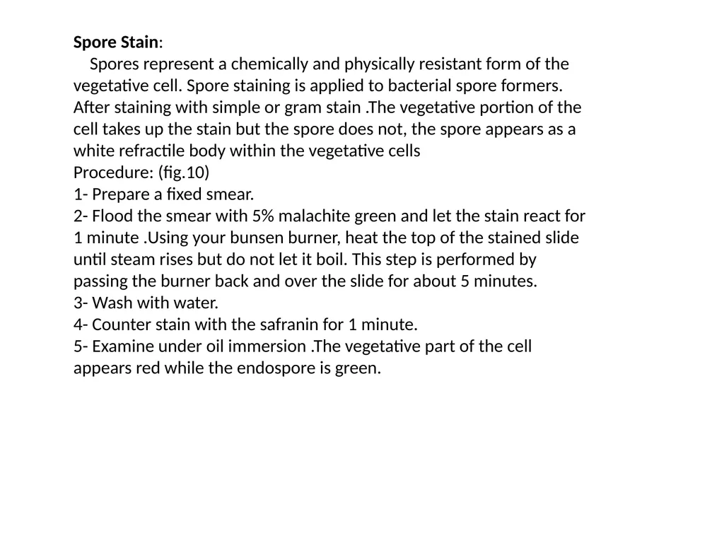
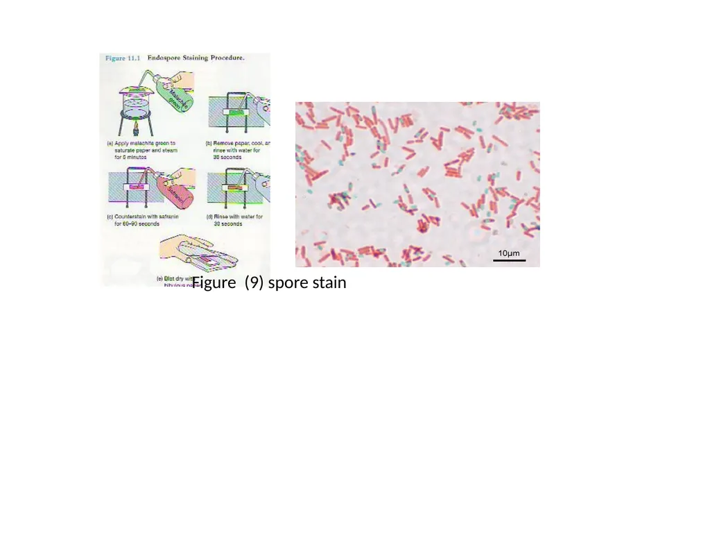
Spores represent a chemically and physically resistant form of the vegetative cell. Spore staining is applied to bacterial spore formers. After staining with simple or gram stain .The vegetative portion of the cell takes up the stain but the spore does not, the spore appears as a white refractile body within the vegetative cells
Procedure: (fig.10)
1- Prepare a fixed smear.
2- Flood the smear with 5% malachite green and let the stain react for 1 minute .Using your bunsen burner, heat the top of the stained slide until steam rises but do not let it boil. This step is performed by passing the burner back and over the slide for about 5 minutes.
3- Wash with water.
4- Counter stain with the safranin for 1 minute.
5- Examine under oil immersion .The vegetative part of the cell appears red while the endospore is green.<br>
02
Figure (9) spore stain<br>
03
Acid Fast Staining:
Acid – fast means the ability of a microorganism to resist decolarization by acid alcohol after primary staining. Acid alcohol resistant is due to the presence of a high lipid content (40-60%) in the cell envelope (gram negative bacteria have no more than 20% while gram positive bacteria have 1-4% lipid in their cell envelope).The acid – fast staining procedure also called the ziehl-neelson technique.
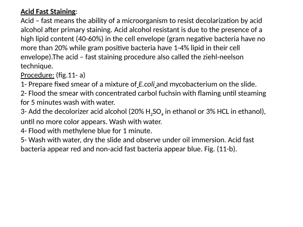
Procedure: (fig.11- a)
1- Prepare fixed smear of a mixture of E.coli and mycobacterium on the slide.
2- Flood the smear with concentrated carbol fuchsin with flaming until steaming for 5 minutes wash with water.
3- Add the decolorizer acid alcohol (20% H2SO4 in ethanol or 3% HCL in ethanol), until no more color appears. Wash with water.
4- Flood with methylene blue for 1 minute.
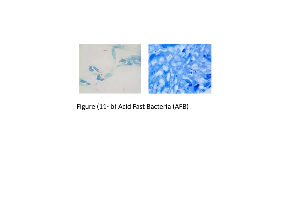
5- Wash with water, dry the slide and observe under oil immersion. Acid fast bacteria appear red and non-acid fast bacteria appear blue. Fig. (11-b).<br>
04
Figure (10-a) Acid-fast staining procedure<br>
05
Figure (11- b) Acid Fast Bacteria (AFB)<br>
06
Negative Staining:
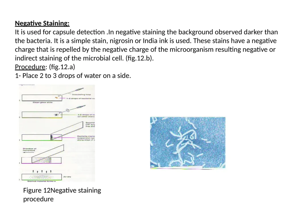
It is used for capsule detection .In negative staining the background observed darker than the bacteria. It is a simple stain, nigrosin or India ink is used. These stains have a negative charge that is repelled by the negative charge of the microorganism resulting negative or indirect staining of the microbial cell. (fig.12.b).
Procedure: (fig.12.a)
1- Place 2 to 3 drops of water on a side. Figure 12Negative staining procedure<br>