
StudyMafia.Org Submitted To: Submitted By:


Description: StudyMafia.Org Submitted To: Submitted By: Studymafia.org Studymafia.org Lungs Anatomy Table Contents Introduction Anatomical Position and Relations Lung Structure Vasculature Nerve Supply Lymphatic Drainage Conclusion 2 Introduction The
Related Topics
Download Presentation
"StudyMafia.Org Submitted To: Submitted By:" is the property of its rightful owner. Permission is granted to download and print the materials on this website for personal, non-commercial use only, and to display it on your personal computer provided you do not modify the materials and that you retain all copyright notices contained in the materials. By downloading content from our website, you accept the terms of this agreement.
Presentation Transcript
slide1. StudyMafia.Org Submitted To: Submitted By:
Studymafia.org Studymafia.org Lungs Anatomy<br>
slide2. Table Contents Introduction
Anatomical Position and Relations
Lung Structure
Vasculature
Nerve Supply
Lymphatic Drainage
Conclusion 2<br>
slide3. Introduction The lungs, which is the organ for respiration is a paired cone shaped organs lying in the thoracic cavity separated from each other by the heart and other structures in the mediastinum.
Each lung has a base resting on the diaphragm and an apex extending superiorly to a point approximately 2.5 cm superior to the clavicle. It also has a medial surface and with three borders- anterior, posterior and inferior. ●●●
3<br>
slide4. Introduction The lungs are the organs of respiration. They are located in the thorax, either side of the mediastinum.
The function of the lungs is to oxygenate blood. They achieve this by bringing inspired air into close contact with oxygen-poor blood in the pulmonary capillaries. ●●●
4<br>
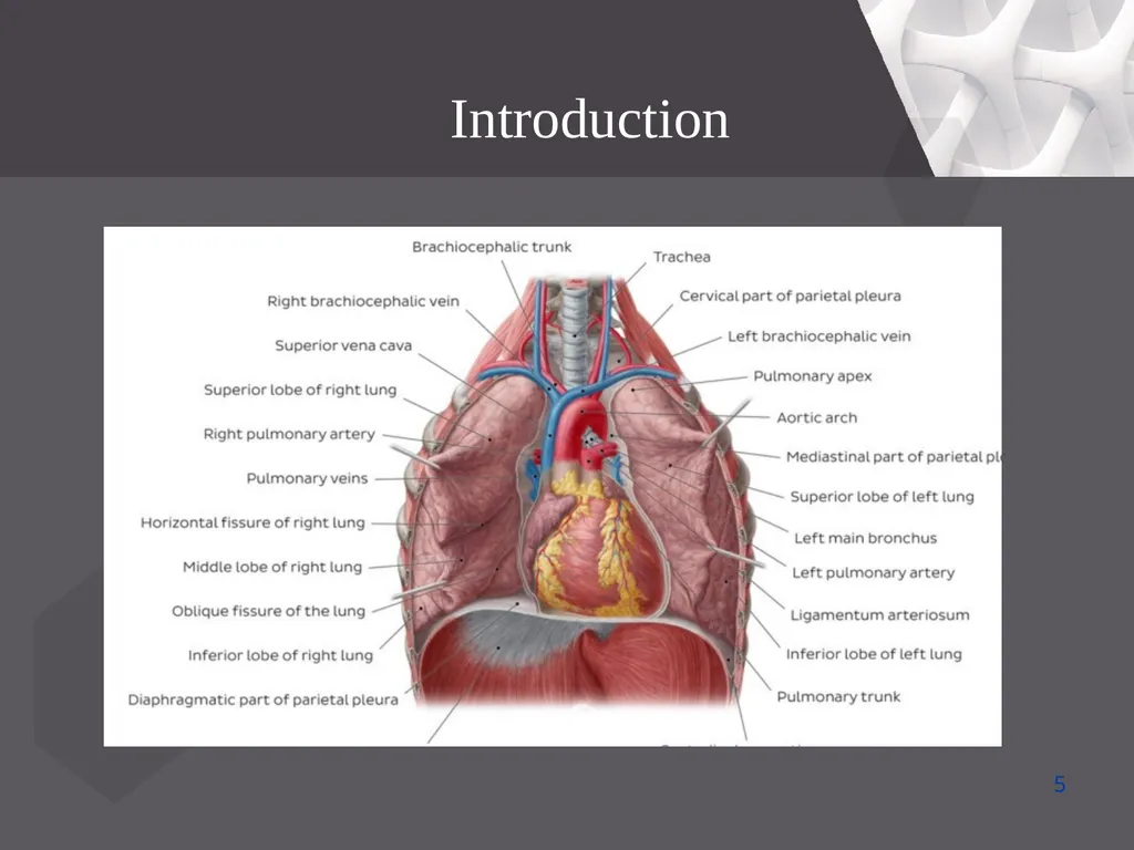
slide5. Introduction 5<br>
slide6. The lungs lie either side of the mediastinum, within the thoracic cavity. Each lung is surrounded by a pleural cavity, which is formed by the visceral and parietal pleura.
They are suspended from the mediastinum by the lung root – a collection of structures entering and leaving the lungs. The medial surfaces of both lungs lie in close proximity to several mediastinal structures. ●●●
6 Anatomical Position and Relations<br>
slide7. The lungs lie either side of the mediastinum, within the thoracic cavity. Each lung is surrounded by a pleural cavity, which is formed by the visceral and parietal pleura.
They are suspended from the mediastinum by the lung root – a collection of structures entering and leaving the lungs. The medial surfaces of both lungs lie in close proximity to several mediastinal structures. 7 Anatomical Position and Relations<br>
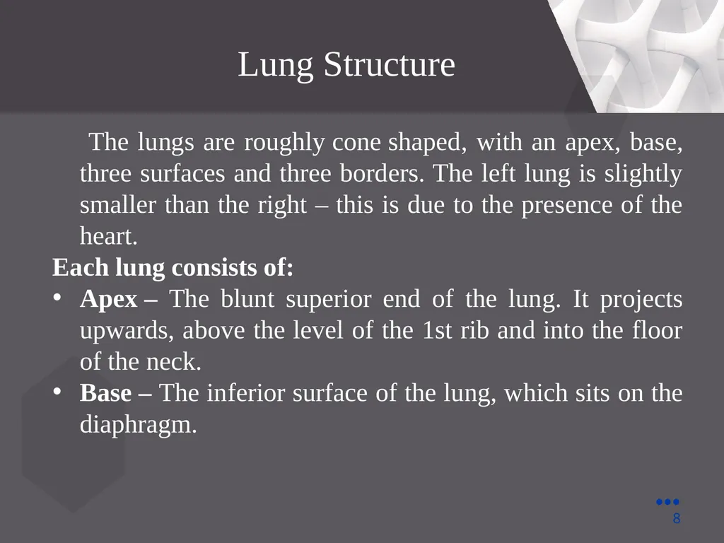
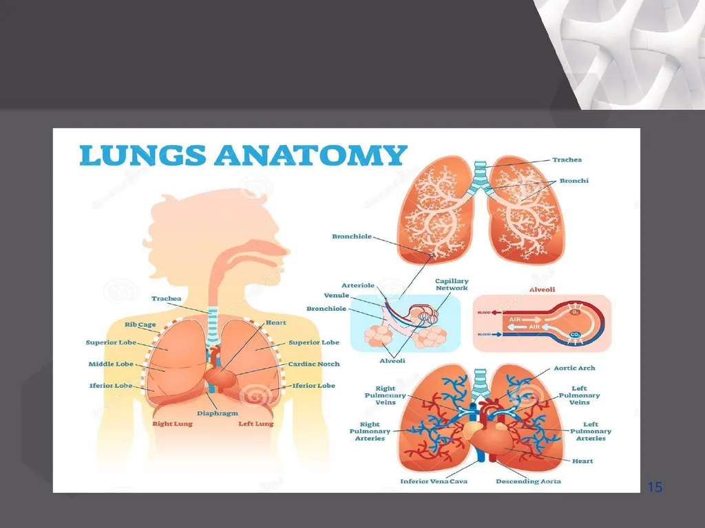
slide8. Lung Structure The lungs are roughly cone shaped, with an apex, base, three surfaces and three borders. The left lung is slightly smaller than the right – this is due to the presence of the heart.
Each lung consists of:
Apex – The blunt superior end of the lung. It projects upwards, above the level of the 1st rib and into the floor of the neck.
Base – The inferior surface of the lung, which sits on the diaphragm. ●●●
8<br>
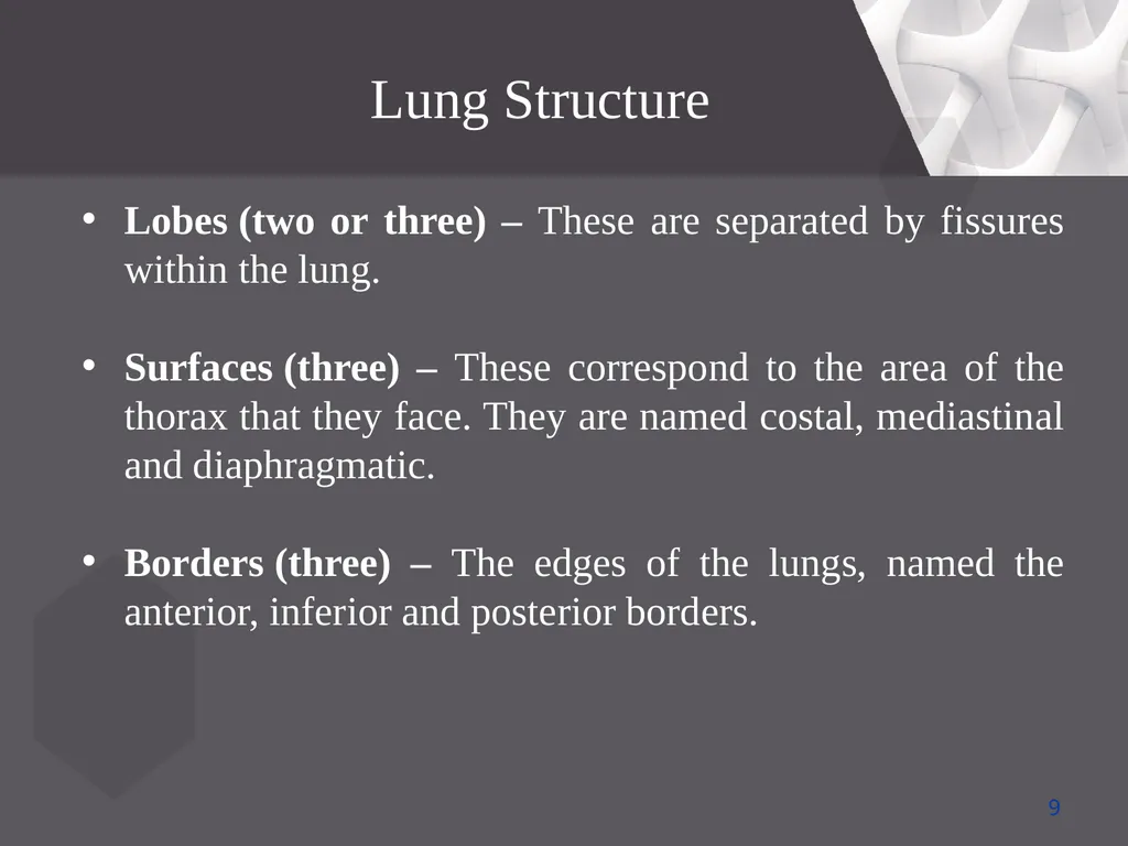
slide9. Lung Structure Lobes (two or three) – These are separated by fissures within the lung.
Surfaces (three) – These correspond to the area of the thorax that they face. They are named costal, mediastinal and diaphragmatic.
Borders (three) – The edges of the lungs, named the anterior, inferior and posterior borders. 9<br>
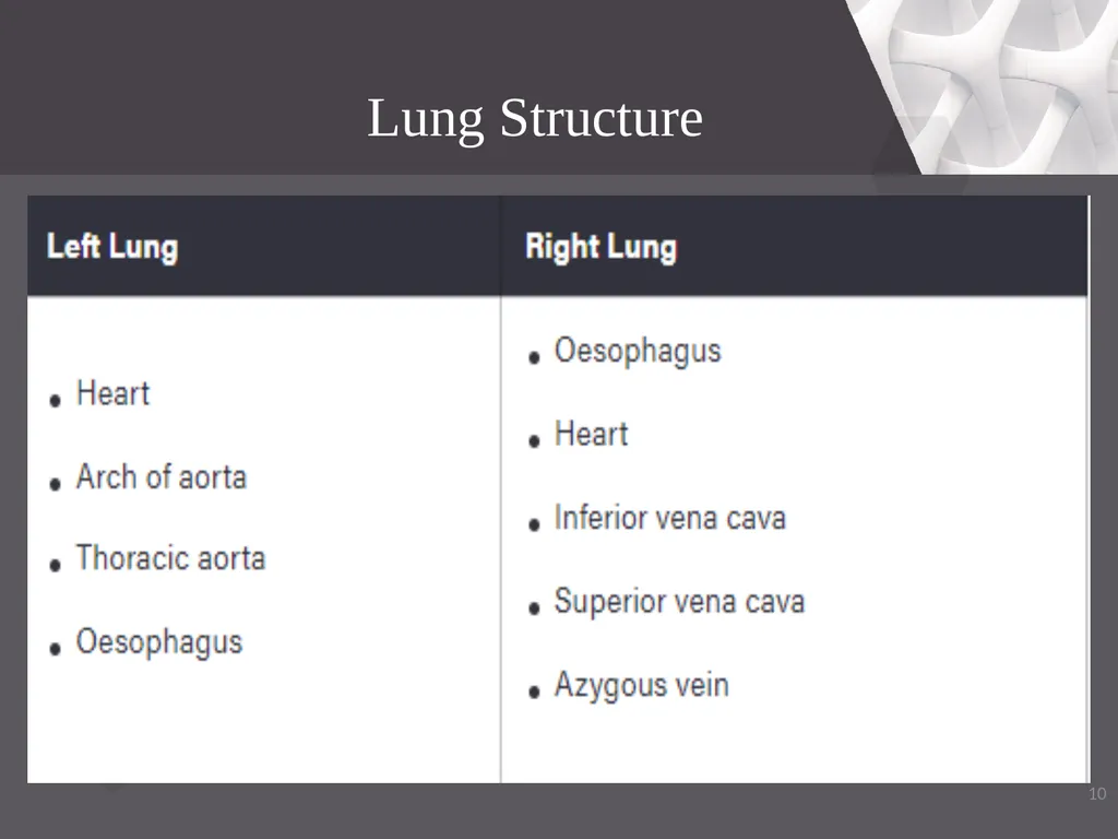
slide10. 10 Lung Structure<br>
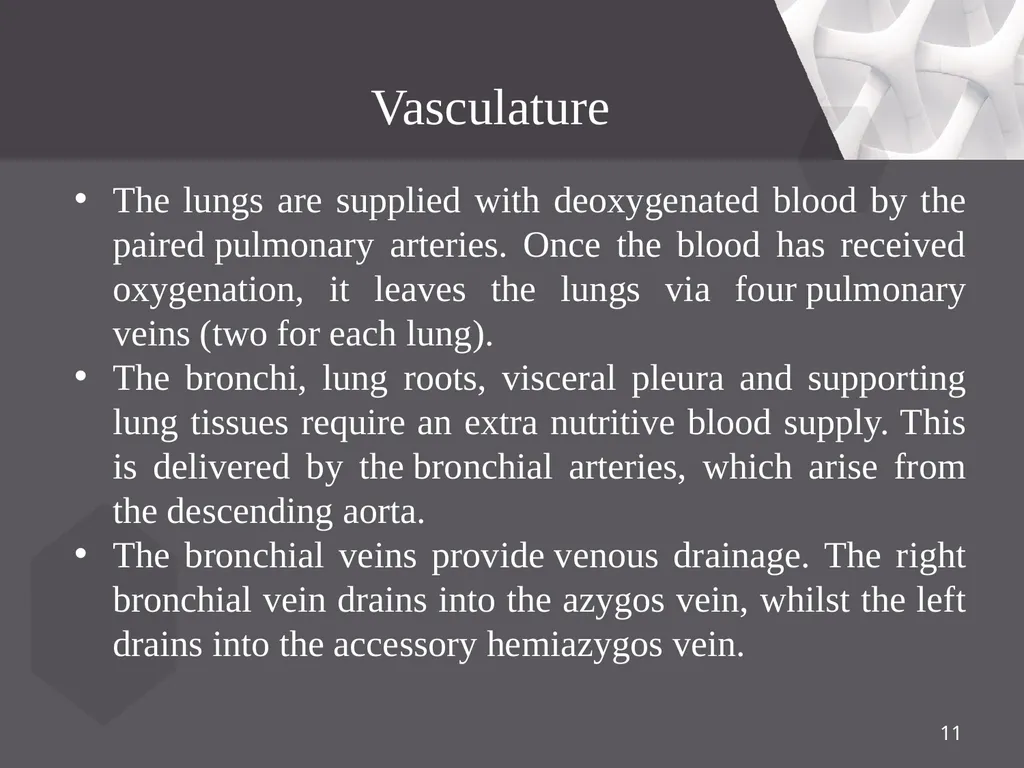
slide11. Vasculature The lungs are supplied with deoxygenated blood by the paired pulmonary arteries. Once the blood has received oxygenation, it leaves the lungs via four pulmonary veins (two for each lung).
The bronchi, lung roots, visceral pleura and supporting lung tissues require an extra nutritive blood supply. This is delivered by the bronchial arteries, which arise from the descending aorta.
The bronchial veins provide venous drainage. The right bronchial vein drains into the azygos vein, whilst the left drains into the accessory hemiazygos vein. 11<br>
slide12. Nerve Supply The nerves of the lungs are derived from the pulmonary plexuses. They feature sympathetic, parasympathetic and visceral afferent fibres:
Parasympathetic – derived from the vagus nerve. They stimulate secretion from the bronchial glands, contraction of the bronchial smooth muscle, and vasodilation of the pulmonary vessels. ●●●
12<br>
slide13. Nerve Supply Sympathetic – derived from the sympathetic trunks. They stimulate relaxation of the bronchial smooth muscle, and vasoconstriction of the pulmonary vessels.
Visceral afferent – conduct pain impulses to the sensory ganglion of the vagus nerve. 13<br>
slide14. The lymphatic vessels of the lung arise from two lymphatic plexuses:
Superficial (subpleural) – drains the lung parenchyma.
Deep – drains the structures of the lung root.
Both these plexuses empty into the trachebronchial nodes – located around the bifurcation of the trachea and the main bronchi. From here, lymph passes into the right and left bronchomediastinal trunks. 14 Lymphatic Drainage<br>
slide15. 15<br>
slide16. Conclusion The lungs are the major organs of the respiratory system, and are divided into sections, or lobes. The right lung has three lobes and is slightly larger than the left lung, which has two lobes. The lungs are separated by the mediastinum.
This area contains the heart, trachea, esophagus, and many lymph nodes. 16<br>
slide17. References Google.com
Wikipedia.org
Studymafia.org
Slidespanda.com<br>
slide18. Thanks To StudyMafia.org<br>
Studymafia.org Studymafia.org Lungs Anatomy<br>
slide2. Table Contents Introduction
Anatomical Position and Relations
Lung Structure
Vasculature
Nerve Supply
Lymphatic Drainage
Conclusion 2<br>
slide3. Introduction The lungs, which is the organ for respiration is a paired cone shaped organs lying in the thoracic cavity separated from each other by the heart and other structures in the mediastinum.
Each lung has a base resting on the diaphragm and an apex extending superiorly to a point approximately 2.5 cm superior to the clavicle. It also has a medial surface and with three borders- anterior, posterior and inferior. ●●●
3<br>
slide4. Introduction The lungs are the organs of respiration. They are located in the thorax, either side of the mediastinum.
The function of the lungs is to oxygenate blood. They achieve this by bringing inspired air into close contact with oxygen-poor blood in the pulmonary capillaries. ●●●
4<br>
slide5. Introduction 5<br>
slide6. The lungs lie either side of the mediastinum, within the thoracic cavity. Each lung is surrounded by a pleural cavity, which is formed by the visceral and parietal pleura.
They are suspended from the mediastinum by the lung root – a collection of structures entering and leaving the lungs. The medial surfaces of both lungs lie in close proximity to several mediastinal structures. ●●●
6 Anatomical Position and Relations<br>
slide7. The lungs lie either side of the mediastinum, within the thoracic cavity. Each lung is surrounded by a pleural cavity, which is formed by the visceral and parietal pleura.
They are suspended from the mediastinum by the lung root – a collection of structures entering and leaving the lungs. The medial surfaces of both lungs lie in close proximity to several mediastinal structures. 7 Anatomical Position and Relations<br>
slide8. Lung Structure The lungs are roughly cone shaped, with an apex, base, three surfaces and three borders. The left lung is slightly smaller than the right – this is due to the presence of the heart.
Each lung consists of:
Apex – The blunt superior end of the lung. It projects upwards, above the level of the 1st rib and into the floor of the neck.
Base – The inferior surface of the lung, which sits on the diaphragm. ●●●
8<br>
slide9. Lung Structure Lobes (two or three) – These are separated by fissures within the lung.
Surfaces (three) – These correspond to the area of the thorax that they face. They are named costal, mediastinal and diaphragmatic.
Borders (three) – The edges of the lungs, named the anterior, inferior and posterior borders. 9<br>
slide10. 10 Lung Structure<br>
slide11. Vasculature The lungs are supplied with deoxygenated blood by the paired pulmonary arteries. Once the blood has received oxygenation, it leaves the lungs via four pulmonary veins (two for each lung).
The bronchi, lung roots, visceral pleura and supporting lung tissues require an extra nutritive blood supply. This is delivered by the bronchial arteries, which arise from the descending aorta.
The bronchial veins provide venous drainage. The right bronchial vein drains into the azygos vein, whilst the left drains into the accessory hemiazygos vein. 11<br>
slide12. Nerve Supply The nerves of the lungs are derived from the pulmonary plexuses. They feature sympathetic, parasympathetic and visceral afferent fibres:
Parasympathetic – derived from the vagus nerve. They stimulate secretion from the bronchial glands, contraction of the bronchial smooth muscle, and vasodilation of the pulmonary vessels. ●●●
12<br>
slide13. Nerve Supply Sympathetic – derived from the sympathetic trunks. They stimulate relaxation of the bronchial smooth muscle, and vasoconstriction of the pulmonary vessels.
Visceral afferent – conduct pain impulses to the sensory ganglion of the vagus nerve. 13<br>
slide14. The lymphatic vessels of the lung arise from two lymphatic plexuses:
Superficial (subpleural) – drains the lung parenchyma.
Deep – drains the structures of the lung root.
Both these plexuses empty into the trachebronchial nodes – located around the bifurcation of the trachea and the main bronchi. From here, lymph passes into the right and left bronchomediastinal trunks. 14 Lymphatic Drainage<br>
slide15. 15<br>
slide16. Conclusion The lungs are the major organs of the respiratory system, and are divided into sections, or lobes. The right lung has three lobes and is slightly larger than the left lung, which has two lobes. The lungs are separated by the mediastinum.
This area contains the heart, trachea, esophagus, and many lymph nodes. 16<br>
slide17. References Google.com
Wikipedia.org
Studymafia.org
Slidespanda.com<br>
slide18. Thanks To StudyMafia.org<br>