/
Color Tests for Proteins and Amino Acids Color Tests for Proteins and Amino Acids

Color Tests for Proteins and Amino Acids - PowerPoint Presentation

paige
paige . @paige
Follow
66 views
Uploaded On 2023-08-31

Color Tests for Proteins and Amino Acids - PPT Presentation

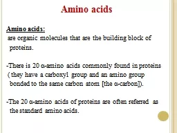
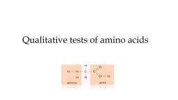
Test for Specific Amino Acids CLS281 Exp2 Done by Daheeya Alenazi Haifa Altwaijry 1 Millons Test Used for detecting the presence of monohydroxybenzene derivatives ID: 1014905

ml1 acid test ml2 acid ml1 ml2 test ml5 color ml3 tube amino group ring drops3 arginine procedure protein

Share:

Link:

Embed:

Download Presentation from below link

Download Presentation The PPT/PDF document "Color Tests for Proteins and Amino Acids" is the property of its rightful owner. Permission is granted to download and print the materials on this web site for personal, non-commercial use only, and to display it on your personal computer provided you do not modify the materials and that you retain all copyright notices contained in the materials. By downloading content from our website, you accept the terms of this agreement.


Presentation Transcript

1. Color Tests for Proteins and Amino Acids(Test for Specific Amino Acids)CLS281Exp.:2 Done by: Daheeya Alenazi Haifa Altwaijry

2. 1/Millon's Test:Used for detecting the presence of monohydroxybenzene derivatives (e.g: Tyrosine, Tyrosine derivatives, Phenol).*Millon's reagent is a solution of mercuric and mercurous ions in nitric and nitrous acids.*Phenolic group of tyrosine reacts with Millon's reagent and give a red color, which is due to a mercury salt of nitrated tyrosine.

3. Materials:WaterEgg albumin: protein contains tyrosine Salicylic acid:is a monohydroxybenzoic acid, a type of phenolic acid TyrosineGelatin: is a translucent, colorless, flavorless solid substance, derived from collagen. It contains tyrosinePhenylalanine : a.aPhenol

4. Procedure:Tube 1Tube 2 Tube 3 Tube 4Tube 5Tube 6 Tube7 0.02% phenol0.02% phenylalanine1% gelatin0.02% tyrosine0.02% salicylic acid1% egg albuminH2O2 ml2 ml2 ml2 ml2 ml2 ml2 mlMillon's Reagent3-5 drops3-5 drops3-5 drops3-5 drops3-5 drops3-5 drops3-5 dropsIncubate in boiling water bath for 2 min. Note the color formed

5. Results:

6. 2/Hopkins-Cole Test:The hopkins-cole test is used to determine the presence of the amino acid tryptophan.Tryptophan has an indole nucleus which is responsible for the violet ring found at the junction between the two layersTryptophan + Hopkins-Cole reagent purple ring*Hopkins-Cole reagent contain magnesium glyoxylate (Magnesium powder and Oxalic acid).Indole

7. Purple ring is formed by the reaction of glyoxylic acid ( CHO-COOH ) with the indole ring of tryptophan in presence of sulfuric acid (H2SO4)glyoxylic acid + indole ring purple ring sulfuric acid

8. Materials:TyrosineTryptophanCasein: protein contains tryptophanGelatin: incomplete protein, because it lacks the essential amino acid tryptophan Egg albumin: protein contains tryptophanWater : as control

9. Procedure: 1234560.02% tyrosine0.02% tryptophan1% casein1% gelatin1%egg albuminH2O2 ml2 ml2 ml2 ml2 ml2 mlHopkins-Cole Reagent3 ml3 ml3 ml3 ml3 ml3 ml*Concentrated H2SO45 ml5 ml5 ml5 ml5 ml5 ml*Carefully add 5 ml of con. H2SO4 from buret by touching the tip of the stopcock to the inside wall of the test tube and allowing the acid to slowly drain down the tube so that the two liquids form separate layers. Add 5 ml of con. H2SO4 to each tube. Observe the color at the zone of contact of the two fluids. If no color appears, swirl the tube gently, but do not mix.

10. Observe the color formed at the junction of the two liquids.If necessary, gently rotate the tube to develop the colored ring.Results:

11. 3/Lead Acetate Test:*Used for detecting the presence of Sulfur in Cystine , free cysteine and methionine…*Proteins or peptides containing these amino acid heated with NaOH to split the sulfide group from amino acids.*Then, lead acetate reacts with free sulfide ions resulting in the formation of lead sulfide ( pbs ) a brown to black color.

12. Procedure:12340.02% methionine0.02% cystine1% egg albuminH2O2 ml2 ml2 ml2 ml5% NaOH5 ml5 ml5 ml5 mlLead AcetateFew crystalsFew crystalsFew crystalsFew crystalsIncubate in boiling water bath for 10 minutes with occasional mixing.Describe the color changes in each test tubes.

13. Results:

14. 4/Sakaguchi Test:*This test is specific for the guanidine group of arginine (H2N-C-NH), but a non-amino acid like creatine which contains this group will also answer this test.*This test positive for all proteins containing arginine and for arginine itself. *When arginine reacts with α-Naphthol and Sodium hypobromite (NaOBr) a red color result.arginineguanidine group

15. Red color is due to a reaction between the hypobromite and the –NH2 group of the guanidino part of arginine

16. Materials:Urea: is an organic compound Creatine: contains guanidine group ArginineGelatin: contains arginine Water: as control

17. Procedure:0.02% urea0.02% creatine0.02% arginine0.1% gelatinH2O5 ml5 ml5 ml5 ml5 ml10% NaOH1 ml1 ml1 ml1 ml1 ml0.02% α-Naphthol1 ml1 ml1 ml1 ml1 mlMix, incubate at room temperature for 3 minutesNaOBr4 drops4 drops4 drops4 drops4 dropsNote the color formed

18. 5/Folin's Test:*This test for free cystine, therefore protein or peptide must be first hydrolyzed by sodium carbonate and sodium sulfite.*Then, Folin's uric acid reagent (phosphotungstic acid) is added which is reduced to tungsten blue.*This can be used for the quantitative determination of cystine.

19. Procedure:12340.1% methionine1% egg albumin0.1% cystineH2O1 ml1 ml1 ml1 mlSodium Carbonate (saturated)7 ml7 ml7 ml7 ml20% Sodium Sulfite3 ml3 ml3 ml3 ml Incubate at R.T for 5 min.Uric acid reagent1 ml1 ml1 ml1 mlNote the color formed