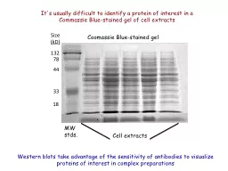
/
It's usually difficult to identify a protein of interest in It's usually difficult to identify a protein of interest in

It's usually difficult to identify a protein of interest in - PowerPoint Presentation

pasty-toler
pasty-toler . @pasty-toler
Follow
410 views
Uploaded On 2016-02-22

It's usually difficult to identify a protein of interest in - PPT Presentation

Coomassie Bluestained gel MW stds Cell extracts 33 44 78 18 132 Size kD Western blots take advantage of the sensitivity of antibodies to visualize proteins of interest in complex preparations ID: 226245

antibody proteins protein epitope proteins antibody epitope protein hrp pbg1805 gel pyes2 western encoded epitopes domain blots binding antibodies

Share:

Link:

Embed:

Download Presentation from below link

Download Presentation The PPT/PDF document "It's usually difficult to identify a pro..." is the property of its rightful owner. Permission is granted to download and print the materials on this web site for personal, non-commercial use only, and to display it on your personal computer provided you do not modify the materials and that you retain all copyright notices contained in the materials. By downloading content from our website, you accept the terms of this agreement.


Presentation Transcript

Slide1

It's usually difficult to identify a protein of interest in a Commassie Blue-stained gel of cell extracts

Coomassie Blue-stained gel

MW stds.

Cell extracts

33

44

78

18

132

Size

(kD)

Western blots take advantage of the sensitivity of antibodies to visualize proteins of interest in complex preparationsSlide2

How are epitope tags used to locate proteins on western blots?

What steps are involved in western blots? Slide3

Proteins are expressed from plasmids that encode epitopes in the same reading frame with the cloned sequence

Protein of interest

Epitope added by plasmid sequences

Antibody that binds the epitope

Epitopes add antibody binding sites to proteinsSlide4

9 AA

ORF

His

6

HA

antibody binding domain of

S. aureus

protein A

14 AA

ORF

His

6

V5

pBG1805-encoded proteins

have multiple epitopes, which add 19 kDa to the protein’s MW

pYES2.1-encoded proteins

have two epitopes, which add ~5 kDa to the protein’s MW Slide5

Epitope-tagged proteins on gel

1. Primary antibody binds proteins (stoichiometric)

2. Secondary antibodies recognize multiple sites on primary antibody

3. Enzyme or fluorochrome amplifies the signal

Western blots

typically use multiple antibodies

to visualize epitope-tagged proteinsSlide6

HRP

HRP

Horseradish peroxidase (HRP)

40,000 MW enzyme

Catalyzes reaction that produces a colored product

Blue reaction product

3,3'5,5'-tetramethyl benzidine

+ H

2

O

2

HRP-catalyzed reaction amplifies the signal and generates a visible productSlide7

14 AA

ORF

His

6

V5

pYES2.1

9 AA

ORF

His

6

HA

IgG binding domain

pBG1805

Problem

: How can proteins encoded by pBG1805 and pYES2.1 plasmids be visualized on the same gel?

Sensitive (and expensive) monoclonal antibodies are available for the viral HA and V5 epitopes

The His

6 tag is useful for purifying proteins, but it is a poor antigenSlide8

Staphylococcus aureus

Protein A

A solution:

Use an alternative strategy to identify proteins encoded by pBG1805, based on the IgG binding domain of

S. aureus

protein A

Protein A is a transmembrane protein with multiple binding sites for the F

c

portion of IgGs

Helps bacterium to evade the host’s immune system during infectionsSlide9

Different strategies will be used to detect pBG1805- and pYES2.1-encoded proteins

pYES2.1 -encoded proteins on gel

Mouse monoclonal Ab detects V5 antigen

HRP

HRP

HRP

pBG1805-encoded proteins on gel

HRP

HRP

HRP-conjugated rabbit anti-mouse IgG polyclonal antibodySlide10

How are epitope tags used to locate proteins on western blots?

What steps are involved in western blots? Slide11

A multi-layer "sandwich" of filter papers and sponges encloses the gel and the PVDF membrane within a transfer cassetteSlide12

The cassette with its "sandwich" is inserted into the transfer apparatus

Be mindful of the correct polarity!

Black plate of sandwich toward black side of cassette holder

The electric current transfers proteins from the gel to the membraneSlide13

SDS-PAGE gel

PVDF membranes are hydrophobic. To bind proteins, they must first be wetted with methanol, followed by water and transfer buffer

Proteins are electrophoretically transferred to a PVDF (essentially

Teflon

) membraneSlide14

Blocking step

PVDF membranes will bind proteins nonspecifically. Milk contains high concentrations of casein proteins, which bind and saturate these low affinity, nonspecific sites on the membranes.

Epitope-tagged proteins (invisible)

MW standards

Membranes are incubated with 5% nonfat milk

MW standardsSlide15

Primary antibody binding step

Primary antibody is a mouse monoclonal antibody that recognizes the V5 epitope

1 2 3

1 – MW standards

2 – proteins with V5 epitope tag (pYES2.1)

3 – proteins with protein A domain (pBG1805)

Antibodies bind with high affinity to V5 epitopesSlide16

1 – MW standards2 – proteins with V5 epitope tag (pYES2.1)

3 – proteins with protein A domain (pBG1805)

Secondary antibody is a rabbit polyclonal antibody that:

recognizes the constant region of the mouse

IgGs

(lane 2)

binds to

S.

aureus

Protein A (lane 3)

1 2 3Slide17

1 – MW standards2 – proteins with V5 epitope tag (pYES2.1)

3 – proteins with protein A domain (pBG1805)

Detection

Blot is incubated with substrates for HRP

1 2 3

Blot is incubated until reaction products appearSlide18

Blue-black bands indicate the position of epitope-tagged proteins

Western blot done by BC studentsSlide19

Process begins with the electrophoretic transfer of proteins to a membrane

Blocking

Membrane replica

Primary antibody

Secondary antibody

Enzymatic detection

Final blot