Uploads
Contact
/
Login
Upload
Search Results for 'Figure 2 Figure 2 Magnetic Resonance Imaging Scans Showing Brain Abnormalities In A Previously Hea'
Figure 3. – A linear fit to observed magnetic fields extrapolated
jane-oiler
Statistical
celsa-spraggs
1 Electron Spin Resonance Spectroscopy
celsa-spraggs
amplification efficiency with hairpin stem length, loop size,important
conchita-marotz
Figure 1.
karlyn-bohler
On The Feasibility Of Magneto-Thermo-Acoustic Imaging Using
karlyn-bohler
3-Axis Magnetometer Construction and Data
myesha-ticknor
A cerebral aneurysm is a weak area in a blood vessel, w
danika-pritchard
FIBER OPTIC MAGNETIC FIELD SENSOR
phoebe-click
Comparing Transcranial Magnetic Stimulation & Direct Electric Stimulation:
debby-jeon
Intro to Map and Compass
min-jolicoeur
Six-figure grid references
aaron
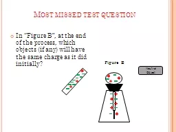
In “Figure B”, at the end of the process, which objects (if any) will have the same
danika-pritchard
PHYSICS OF MAGNETIC RESONANCE
alexa-scheidler
Physics of Magnetic Resonance
mitsue-stanley
Adaptation of the PRETOR Code to Stellarator Simulations.Application t
tatyana-admore
air products and chemicals inc 2005 magnetic field and c
danika-pritchard
Nuclear magnetic resonance spectroscopy
briana-ranney
Figure 26.0-1 Why Reproduction Matters
phoebe-click
Muscle Diagrams Figure 10.10a
giovanna-bartolotta
Figure 17-5b The Sectional Anatomy of the Eye.
olivia-moreira
Review Unit 3 and 4 Figure 6.8a
olivia-moreira
Figure 2.26 Some examples of alternative RNA splicing
test
Figure
calandra-battersby
1
2
3
4
5
6
7
8
9