Explore
Featured
Recent
Articles
Topics
Login
Upload
Featured
Recent
Articles
Topics
Login
Upload
Search Results for 'Objective-Magnification'
Objective-Magnification published presentations and documents on DocSlides.
magnification
by stefany-barnette
50X. magnification. 300X. magnification. 1000X. m...
the Microscope Types of Microscopes
by iris
- Compound microscope. - Dissecting microscope. - ...
Proper use and care of microscope
by stella
The Compound Microscope. Parts of the Microscope. ...
also uses electrons but instead of scanning the surface as with SEMs e
by amelia
and 100X depending on the objective in use The len...
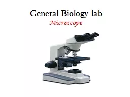
General Biology
by debby-jeon
lab. Microscope. Microscope. . is an instrument ...
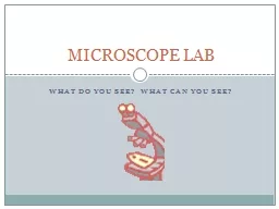
WHAT DO YOU SEE? WHAT CAN YOU SEE?
by briana-ranney
MICROSCOPE LAB. Parts of the Microscope. Fill and...
Standard 7-1.1
by tawny-fly
Microscopes. Microscopes. A microscope is a tool ...
Microscope Structure
by lindy-dunigan
From its humble beginnings the microscope has und...
Microscope Basics Parts of the Microscope
by isabella
1 . Eyepiece : the part th. a. t you look through ...
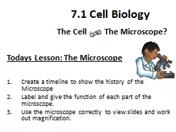
Todays Lesson: The Microscope
by anderson
Learning Objectives…... Create a timeline to sho...
r.zammit@stmonicas-epping.com
by trish-goza
www.year8sciencewithmisszammit.weebly.com. Line u...
Compound Light Microscope
by cheryl-pisano
TOTAL MAGNIFICATION. Power of the. . eyepiece. ...
Types of Microscopes Light Microscope
by calandra-battersby
- . the models found in most schools, use com...
1 Microscopy and
by phoebe-click
Specimen Preparation. Microscope. Basics...
Microscopes Moisey (2009),
by liane-varnes
Optical Microscope. [photograph]. . Retrieved fr...
Cell Theory
by liane-varnes
The History Behind Cells. Where does life come fr...
Microscope
by tatiana-dople
Basics. Nosepiece. Objectives. Stage Clips. Light...
1.1 Intro to the microscope and Calculating cell size
by liane-varnes
Introduction to the Microscope. Types of . Micros...
Microscope
by stefany-barnette
Basics. By: Kaitlyn Schroeder; ETEAMS 2014. Modif...
Bellwork
by ellena-manuel
Why do scientists use Microscopes?. Microscopes. ...
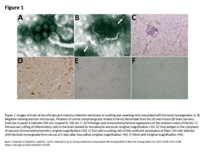
Figure 1 Figure 1. Images of brain of fox with group A rotavirus infection and brains of suckling a
by alonzo
Busi C, Martella V, Papetti A, Sabelli C, Lelli D,...
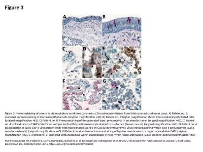
Figure 3 Figure 3. Immunostaining of severe acute respiratory syndrome coronavirus 2 in pulmonary t
by alan
Martines RB, Ritter JM, Matkovic E, Gary J, Bollwe...
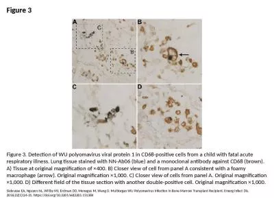
Figure 3 Figure 3. Detection of WU polyomavirus viral protein 1 in CD68-positive cells from a child
by tate
Siebrasse EA, Nguyen NL, Willby MJ, Erdman DD, Men...
The Thin Lens Equation The Thin Lens Equation
by melody
Let’s us predict mathematically the properties o...
Year 8 Lesson 8 – cells
by delilah
Science. Learning intention. To know how microscop...
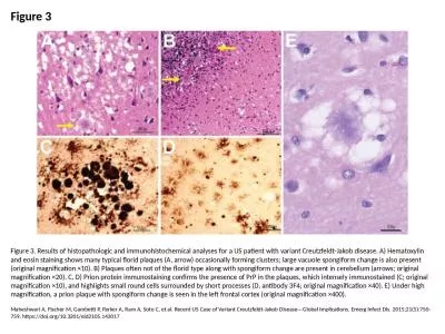
Figure 3 Figure 3. Results of histopathologic and immunohistochemical analyses for a US patient wit
by badra
Maheshwari A, Fischer M, Gambetti P, Parker A, Ram...
Microscope quiz Review Mrs. Barnes
by mary
Biology I Honors. Which statement BEST describes a...
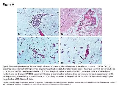
Figure 6 Figure 6. Representative histopathologic changes of brains of infected equines. A
by ida
Gonzalez D, Estrada-Franco JG, Carrara A, Aronson ...
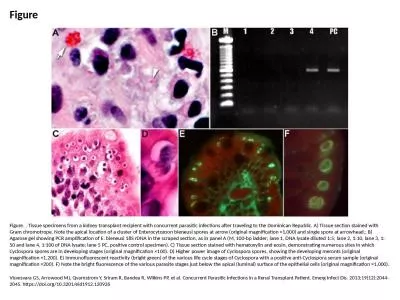
Figure Figure. . Tissue specimens from a kidney transplant recipient with concurrent parasitic infe
by belinda
Visvesvara GS, Arrowood MJ, Qvarnstrom Y, Sriram R...
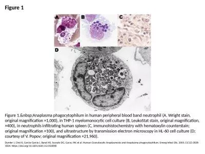
Figure 1 Figure 1. Anaplasma phagocytophilum in human peripheral blood band neutrophil (A.
by skylar
Dumler J, Choi K, Garcia-Garcia J, Barat NS, Scorp...
Microscopes magnifications, resolutions and calculations
by sophie
Light . microscope- uses light and lenses to enlar...
Telescopes faint objects
by deborah
– gathers light. small apparent size – magnifi...
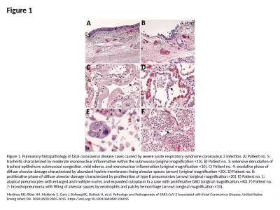
Figure 1 Figure 1. Pulmonary histopathology in fatal coronavirus disease cases caused by severe acu
by piper
Martines RB, Ritter JM, Matkovic E, Gary J, Bollwe...
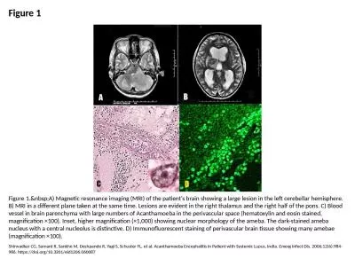
Figure 1 Figure 1. A) Magnetic resonance imaging (MRI) of the patient's brain showing a la
by angelina
Shirwadkar CG, Samant R, Sankhe M, Deshpande R, Ya...
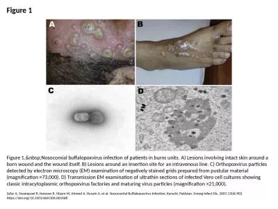
Figure 1 Figure 1. Nosocomial buffalopoxvirus infection of patients in burns units. A) Les
by lauren
Zafar A, Swanepoel R, Hewson R, Nizam M, Ahmed A, ...
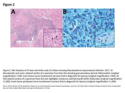
Figure 2 Figure 2. Skin biopsies of 59-year-old white male US citizen showing Mycobacterium leproma
by olivia
Virk A, Pritt B, Patel R, Uhl JR, Bezalel SA, Gibs...
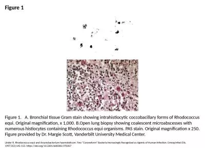
Figure 1 Figure 1. A. Bronchial tissue Gram stain showing intrahistiocytic coccobacillary forms o
by scarlett
Linder R. Rhodococcus equi and Arcanobacterium hae...
Supplement 1: A) HE staining of healthy bone of humanized mice. Asterisks (*) indicate human immun
by bety
) HE staining of healthy bone of . a humanized mou...
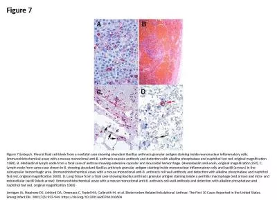
Figure 7 Figure 7. A. Pleural fluid cell block from a nonfatal case showing abundant Bacil
by morton
Jernigan JA, Stephens DS, Ashford DA, Omenaca C, T...
Figure Figure. Acute form of African swine fever in wild boars. A) Petechial and larger ec
by dorothy
Rahimi P, Sohrabi A, Ashrafihelan J, Edalat R, Ala...
Load More...