
DNA Structure, Replication, and Repair in






Description: DNA Structure, Replication, and Repair in prokaryote Ali I.A.Gharip, B.Sc Biotechnology TA, Omdurman Islamic University dedication To spirit of my mother OVERVIEW Nucleic acids are required for the storage and expression of genetic
Related Topics
Download Presentation
"DNA Structure, Replication, and Repair in" is the property of its rightful owner. Permission is granted to download and print the materials on this website for personal, non-commercial use only, and to display it on your personal computer provided you do not modify the materials and that you retain all copyright notices contained in the materials. By downloading content from our website, you accept the terms of this agreement.
Presentation Transcript
slide1. DNA Structure, Replication,
and Repair in prokaryote Ali I.A.Gharip, B.Sc
Biotechnology TA,
Omdurman Islamic University<br>
slide2. dedication To spirit of my mother<br>
slide3. OVERVIEW Nucleic acids are required for the storage and expression of genetic information. There are two chemically distinct types of nucleic acids:
deoxyribonucleic acid (DNA)
and ribonucleic acid (RNA)
DNA, the repository of genetic information, is present not only in chromosomes in the nucleus of eukaryotic organisms, but also in mitochondria and the chloroplasts of plants.
Prokaryotic cells, which lack nuclei, have a single chromosome, but may also contain non-chromosomal DNA in the form of plasmids.<br>
slide4. The genetic information found in DNA is copied and transmitted to daughter cells through DNA replication.
The DNA contained in a fertilized egg encodes the information that directs the development of an organism.<br>
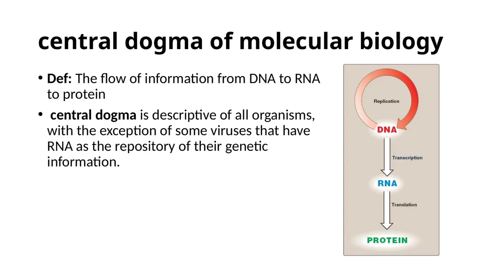
slide5. central dogma of molecular biology Def: The flow of information from DNA to RNA to protein
central dogma is descriptive of all organisms, with the exception of some viruses that have RNA as the repository of their genetic information.<br>
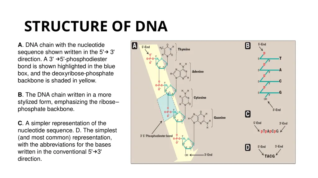
slide6. STRUCTURE OF DNA A. DNA chain with the nucleotide sequence shown written in the 5'→ 3' direction. A 3' →5'-phosphodiester
bond is shown highlighted in the blue box, and the deoxyribose-phosphate backbone is shaded in yellow.
B. The DNA chain written in a more stylized form, emphasizing the ribose–phosphate backbone.
C. A simpler representation of the nucleotide sequence. D. The simplest (and most common) representation,
with the abbreviations for the bases written in the conventional 5'→3' direction.<br>
slide7. DNA: is a polymer of deoxyribonucleoside monophosphates covalently linked by 3'→5'–phosphodiester bonds.
With the exception of a few viruses that contain single-stranded (ss) DNA, DNA exists as a doublestranded (ds) molecule, in which the two strands wind around each other, forming a double helix.
In eukaryotic cells, DNA is found associated with various types of proteins (known collectively as nucleoprotein) present in the nucleus, whereas in prokaryotes, the protein–DNA complex is present in a non-membrane-bound region known as the nucleoid.<br>
slide8. 3'→5'-Phosphodiester bonds Phosphodiester bonds join the 3'-hydroxyl group of the deoxy pentose of one nucleotide to the 5'-hydroxyl group of the deoxy pentose of an adjacent nucleotide through a phosphate group.
3'-end: (the end with the free hydroxyl) that are not attached to other nucleotides.
5'-end: (the end with the free phosphate) that are not attached to other nucleotides.
The bases located along the resulting deoxy ribose–phosphate backbone are, by convention, always written in sequence from the 5'-end of the chain to the 3'-end. For example, the sequence of bases in the DNA shown.<br>
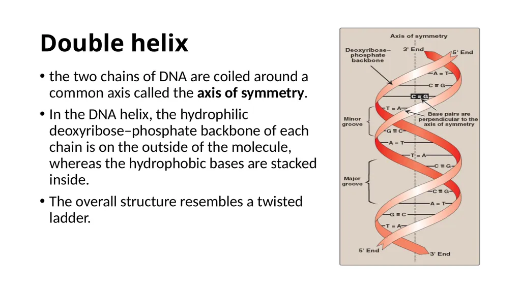
slide9. Double helix the two chains of DNA are coiled around a common axis called the axis of symmetry.
In the DNA helix, the hydrophilic deoxyribose–phosphate backbone of each chain is on the outside of the molecule, whereas the hydrophobic bases are stacked inside.
The overall structure resembles a twisted ladder.<br>
slide10. grooves provide access for the binding of regulatory proteins to their specific recognition sequences along the DNA chain.
Certain anticancer drugs, such as dactinomycin (actinomycin D), exert their cytotoxic effect by intercalating into the narrow groove of the DNA double helix, thus interfering with DNA and RNA synthesis.
Base pairing: The bases of one strand of DNA are paired with the bases of the second strand, so that an adenine is always paired with a thymine and a cytosine is always paired with a guanine.
one polynucleotide chain of the DNA double helix is always the complement of the other.<br>
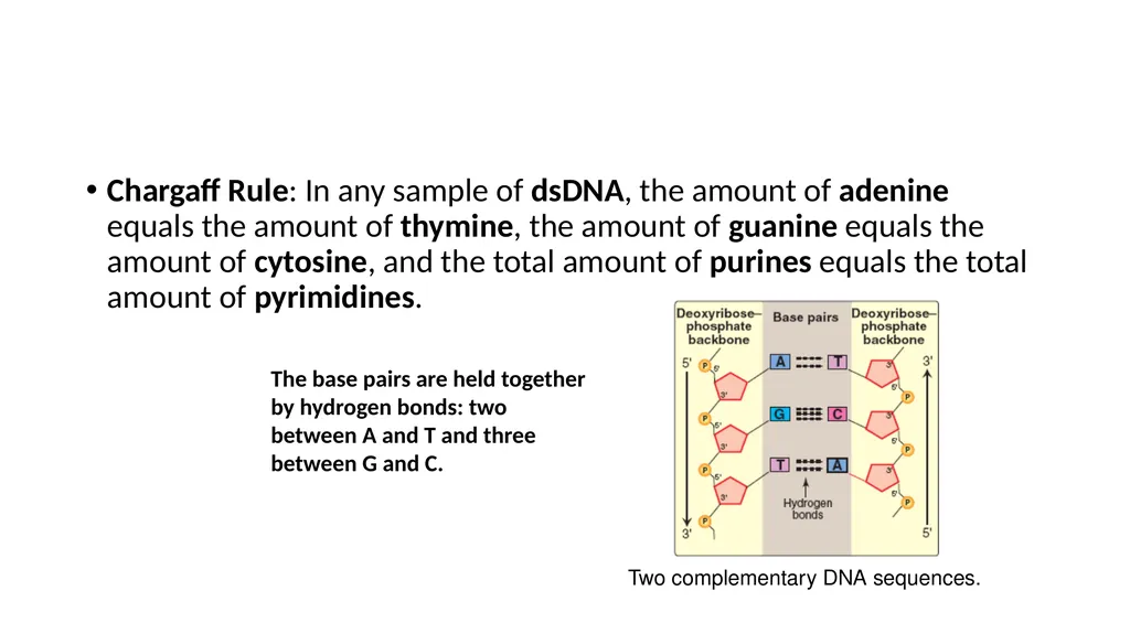
slide11. Chargaff Rule: In any sample of dsDNA, the amount of adenine equals the amount of thymine, the amount of guanine equals the amount of cytosine, and the total amount of purines equals the total amount of pyrimidines. The base pairs are held together by hydrogen bonds: two between A and T and three between G and C. Two complementary DNA sequences.<br>
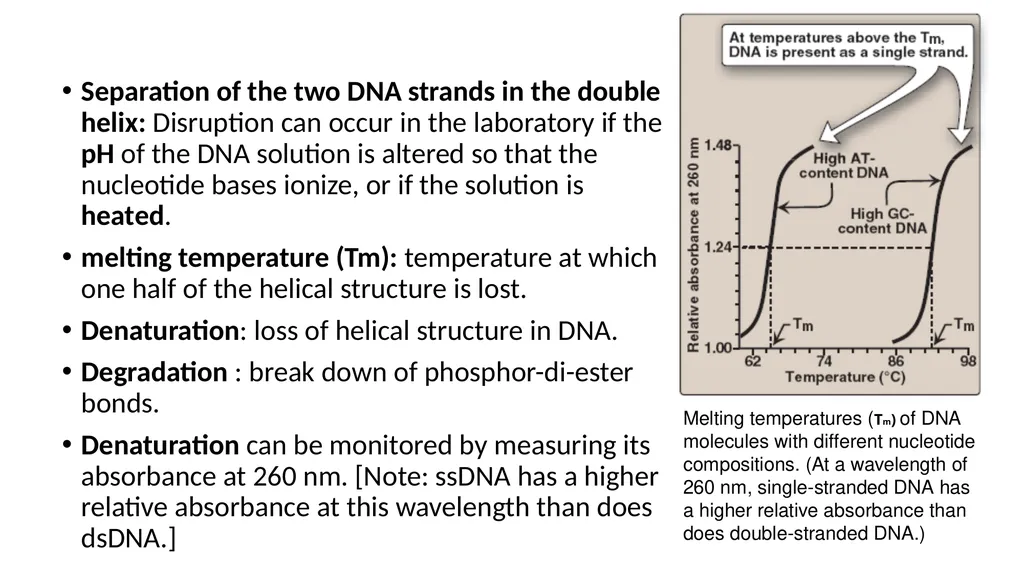
slide12. Separation of the two DNA strands in the double helix: Disruption can occur in the laboratory if the pH of the DNA solution is altered so that the nucleotide bases ionize, or if the solution is heated.
melting temperature (Tm): temperature at which one half of the helical structure is lost.
Denaturation: loss of helical structure in DNA.
Degradation : break down of phosphor-di-ester bonds.
Denaturation can be monitored by measuring its absorbance at 260 nm. [Note: ssDNA has a higher relative absorbance at this wavelength than does dsDNA.] Melting temperatures (Tm) of DNA
molecules with different nucleotide
compositions. (At a wavelength of
260 nm, single-stranded DNA has
a higher relative absorbance than
does double-stranded DNA.)<br>
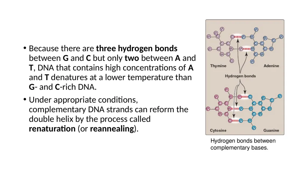
slide13. Because there are three hydrogen bonds between G and C but only two between A and T, DNA that contains high concentrations of A and T denatures at a lower temperature than G- and C-rich DNA.
Under appropriate conditions, complementary DNA strands can reform the double helix by the process called renaturation (or reannealing). Hydrogen bonds between
complementary bases.<br>
slide14. Structural forms of the double helix: There are three major structural forms of DNA:
the B form, described by Watson and Crick in 1953,
the A form,
The Z form.
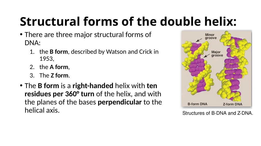
The B form is a right-handed helix with ten residues per 360° turn of the helix, and with the planes of the bases perpendicular to the helical axis. Structures of B-DNA and Z-DNA.<br>
slide15. The A form is produced by moderately dehydrating the B form. It is also a right-handed helix, but there are 11 base pairs per turn, and the planes of the base pairs are tilted 20° away from the perpendicular to the helical axis.
The conformation found in DNA–RNA hybrids or RNA–RNA double-stranded regions is probably very close to the A form.
Z-DNA is a left-handed helix that contains about 12 base pairs per turn [Note: The deoxyribose –phosphate backbone “zigzags,” hence, the name “Z”-DNA.]<br>
slide16. Stretches of Z-DNA can occur naturally in regions of DNA that have a sequence of alternating purines and pyrimidines, for example, poly GC.
Transitions between the B and Z helical forms of DNA may play a role in regulating gene expression.<br>
slide17. Linear and circular DNA molecules Each chromosome in the nucleus of a eukaryote contains one long, linear molecule of dsDNA, which is bound to a complex mixture of proteins (histone and non-histone) to form chromatin.
Eukaryotes have closed, circular DNA molecules in their mitochondria, as do plant chloroplasts. A prokaryotic organism typically contains a single, double-stranded, supercoiled, circular chromosome.
Each prokaryotic chromosome is associated with non-histone proteins that can condense the DNA to form a nucleoid. In addition, most species of bacteria also contain small, circular, extrachromosomal DNA molecules called plasmids.<br>
slide18. Plasmid DNA carries genetic information, and undergoes replication that may or may not be synchronized to chromosomal division.
Plasmids may carry genes that convey antibiotic resistance to the host bacterium, and may facilitate the transfer of genetic information from one bacterium to another.<br>
slide19. STEPS IN PROKARYOTIC DNA SYNTHESIS When the two strands of the DNA double helix are separated, each can serve as a template for the replication of a new complementary strand.
This produces two daughter molecules, each of which contains two DNA strands with an antiparallel orientation .This process is called semiconservative replication because, although the parental duplex is separated into two halves (and, therefore, is not “conserved” as an entity), each of the individual parental strands remains intact in one of the two new duplexes . Semiconservative replication of DNA.<br>
slide20. The enzymes involved in the DNA replication process are template-directed polymerases that can synthesize the complementary sequence of each strand with extraordinary fidelity.
The reactions described in this section were first known from studies of the bacterium Escherichia coli (E. coli), and the description given below refers to the process in prokaryotes.
DNA synthesis in higher organisms is less well understood, but involves the same types of mechanisms. In either case, initiation of DNA replication commits the cell to continue the process until the entire genome has been replicated.<br>
slide21. The steps of DNA synthesis consists of :
Separation of the two complementary DNA strands
Formation of the replication fork
RNA primer
Chain elongation
Excision of RNA primers and their replacement by DNA
DNA ligase<br>
slide22. A. Separation of the two complementary DNA strands In order for the two strands of the parental double helical DNA to be replicated, they must first separate (or “melt”) over a small region, because the polymerases use only ssDNA as a template.
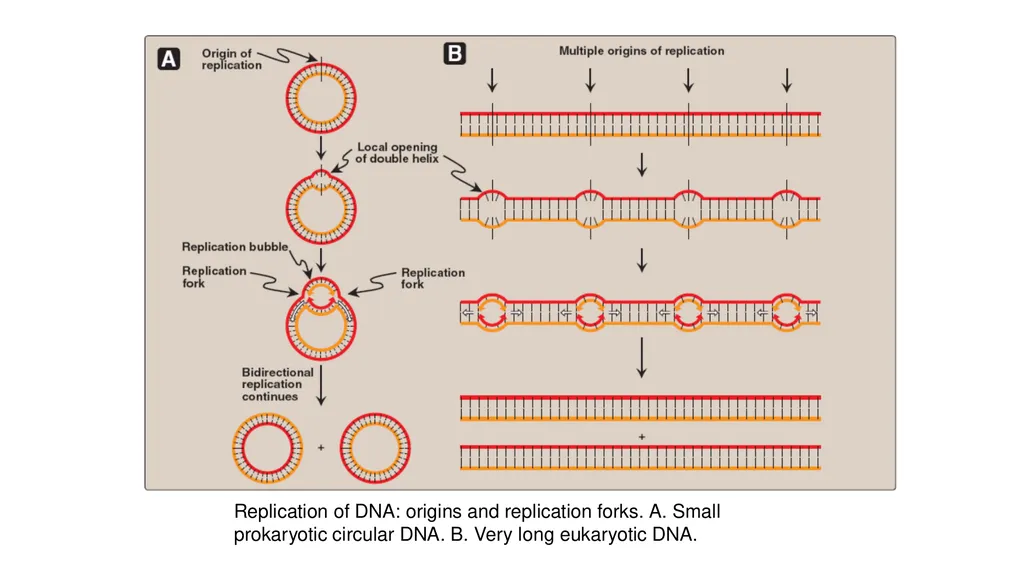
In prokaryotic organisms, DNA replication begins at a single, unique nucleotide sequence—a site called the origin of replication.
[Note: This is referred to as a consensus sequence, because the order of nucleotides is essentially the same at each site.] This site includes a short sequence composed almost exclusively of AT base pairs that facilitate melting.
In eukaryotes, replication begins at multiple sites along the DNA helix .<br>
slide23. Having multiple origins of replication provides a mechanism for rapidly replicating the great length of the eukaryotic DNA molecules.<br>
slide24. B. Formation of the replication fork As the two strands unwind and separate, they form a “V” where active synthesis occurs. This region is called the replication fork. It moves along the DNA molecule as synthesis occurs.
Replication of dsDNA is bidirectional—that is, the replication forks move in opposite directions from the origin, generating a replication bubble.<br>
slide25. Replication of DNA: origins and replication forks. A. Small prokaryotic circular DNA. B. Very long eukaryotic DNA.<br>
slide26. 1. Proteins required for DNA strand separation: Initiation of DNA replication requires the recognition of the origin of replication by a group of proteins that form the prepriming complex. These proteins are responsible for maintaining the separation of the parental strands, and for unwinding the double helix ahead of the advancing replication fork.
These proteins include the following:
DnaA protein
DNA helicases
Single-stranded DNA-binding (SSB) proteins<br>
slide27. DnaA protein: protein binds to specific nucleotide sequences at the origin of replication, causing short, tandemly arranged (one after the other) AT-rich regions in the origin to melt.
Melting is ATP-dependent, and results in strand separation with the formation of localized regions of ssDNA.
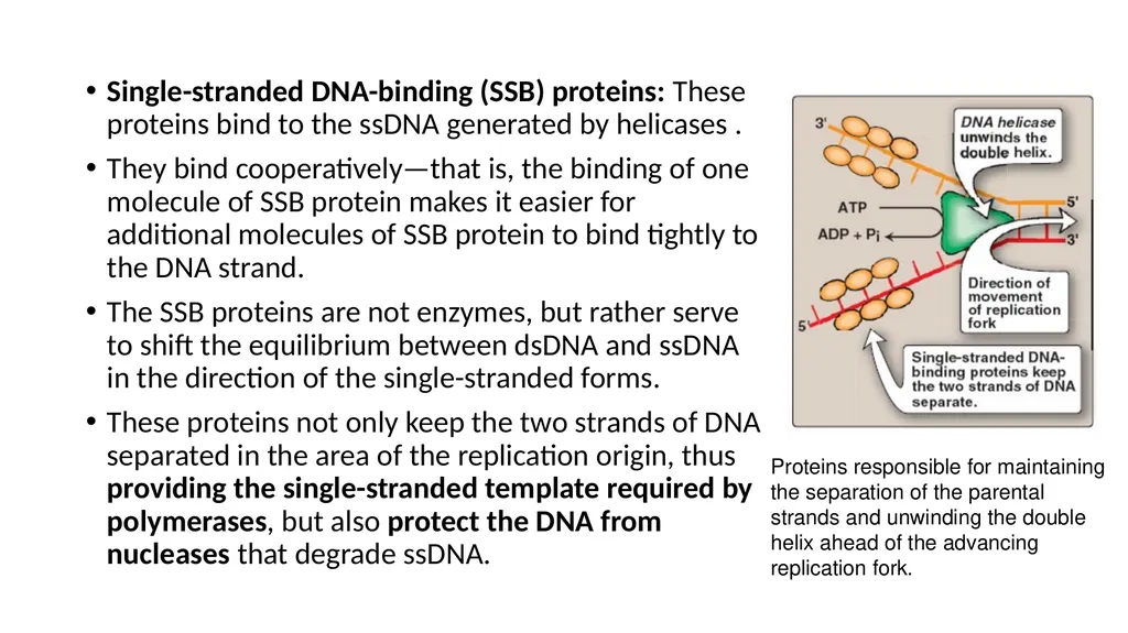
DNA helicases: These enzymes bind to ssDNA near the replication fork, and then move into the neighboring doublestranded region, forcing the strands apart—in effect, unwinding the double helix.
Helicases require energy provided by ATP .
[Note: DnaB is the principal helicase of replication in E. coli. Its binding to DNA requires DnaC.]<br>
slide28. Single-stranded DNA-binding (SSB) proteins: These proteins bind to the ssDNA generated by helicases .
They bind cooperatively—that is, the binding of one molecule of SSB protein makes it easier for additional molecules of SSB protein to bind tightly to the DNA strand.
The SSB proteins are not enzymes, but rather serve to shift the equilibrium between dsDNA and ssDNA in the direction of the single-stranded forms.
These proteins not only keep the two strands of DNA separated in the area of the replication origin, thus providing the single-stranded template required by polymerases, but also protect the DNA from nucleases that degrade ssDNA. Proteins responsible for maintaining
the separation of the parental
strands and unwinding the double
helix ahead of the advancing
replication fork.<br>
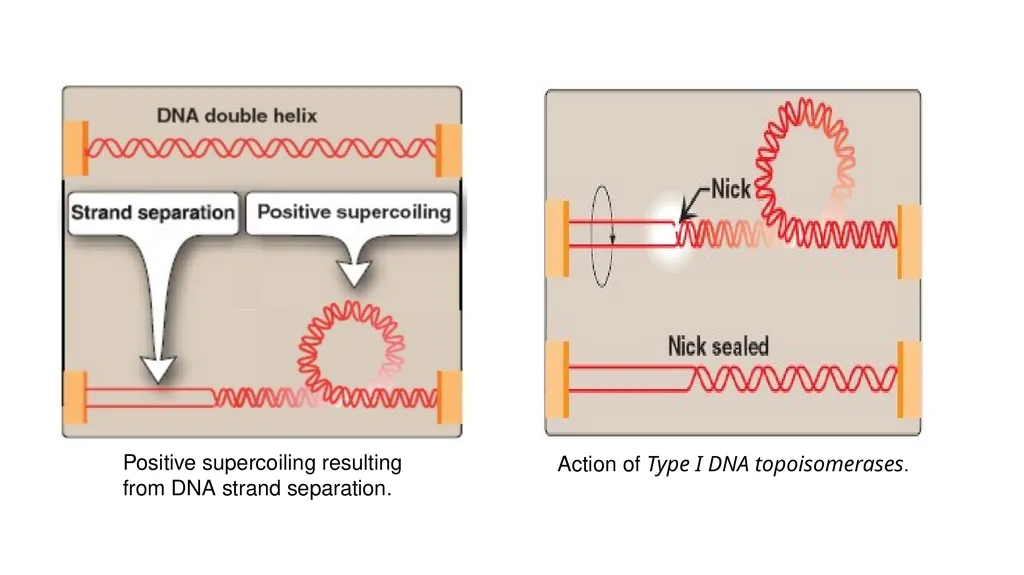
slide29. 2. Solving the problem of supercoils: As the two strands of the double helix are separated, a problem is encountered, namely, the appearance of positive supercoils (also called supertwists) in the region of DNA ahead of the replication fork .
The accumulating positive supercoils interfere with further unwinding of the double helix.
To solve this problem, there is a group of enzymes called DNA topoisomerases, which are responsible for removing supercoils in the helix.
Type I DNA topoisomerases
Type II DNA topoisomerases<br>
slide30. Type I DNA topoisomerases: These enzymes reversibly cut one strand of the double helix. They have both nuclease (strand-cutting) and ligase (strand-resealing) activities.
They do not require ATP, but rather appear to store the energy from the phosphodiester bond they cleave, reusing the energy to reseal the strand .
Each time a transient “nick” is created in one DNA strand, the intact DNA strand is passed through the break before it is resealed, thus relieving (“relaxing”) accumulated supercoils.
Type I topoisomerases relax negative supercoils (that is, those that contain fewer turns of the helix than relaxed DNA) in E. coli, and both negative and positive supercoils (that is, those that contain fewer or more turns of the helix than relaxed DNA) in eukaryotic cells.<br>
slide31. Positive supercoiling resulting
from DNA strand separation. Action of Type I DNA topoisomerases.<br>
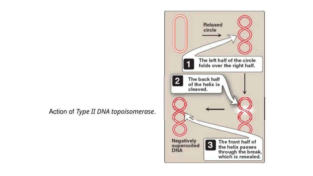
slide32. Type II DNA topoisomerases: These enzymes bind tightly to the DNA double helix and make transient breaks in both strands.
The enzyme then causes a second stretch of the DNA double helix to pass through the break and, finally, reseals the break .
As a result, both negative and positive supercoils can be relieved by this ATP-requiring process.
Type II DNA topoisomerases are also required in both prokaryotes and eukaryotes for the separation of interlocked molecules of DNA following chromosomal replication.<br>
slide33. DNA gyrase, a Type II topoisomerase found in bacteria and plants, has the unusual property of being able to introduce negative supercoils into relaxed circular DNA using energy from the hydrolysis of ATP. This facilitates the future replication of DNA because the negative supercoils neutralize the positive supercoils introduced during opening of the double helix. It also aids in the transient strand separation required during transcription .
Anticancer agents, such as etoposide, target human topoisomerase II.
Bacterial DNA gyrase is a unique target of a group of anti microbial agents called quinolones, for example, cipro - floxacin.<br>
slide34. Action of Type II DNA topoisomerase.<br>
slide35. Direction of DNA replication The DNA polymerases responsible for copying the DNA templates are only able to “read” the parental nucleotide sequences in the 3'→5' direction, and they synthesize the new DNA strands only in the 5'→3' (antiparallel) direction.
Therefore, beginning with one parental double helix, the two newly synthesized stretches of nucleotide chains must grow in opposite directions—one in the 5'→3' direction toward the replication fork (Leading strand) and one in the 5'→3' direction away from the replication fork (Lagging strand).
This feat is accomplished by a slightly different mechanism on each strand.<br>
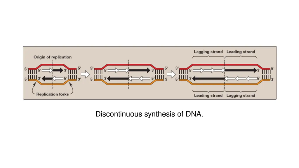
slide36. Leading strand: The strand that is being copied in the direction of the advancing replication fork and is synthesized continuously.
Lagging strand: The strand that is being copied in the direction away from the replication fork is synthesized discontinuously, with small fragments of DNA being copied near the replication fork.
These short stretches of discontinuous DNA, termed Okazaki fragments, are eventually joined (ligated) to become a single, continuous strand.
The new strand of DNA produced by this mechanism is termed the lagging strand.<br>
slide37. Discontinuous synthesis of DNA.<br>
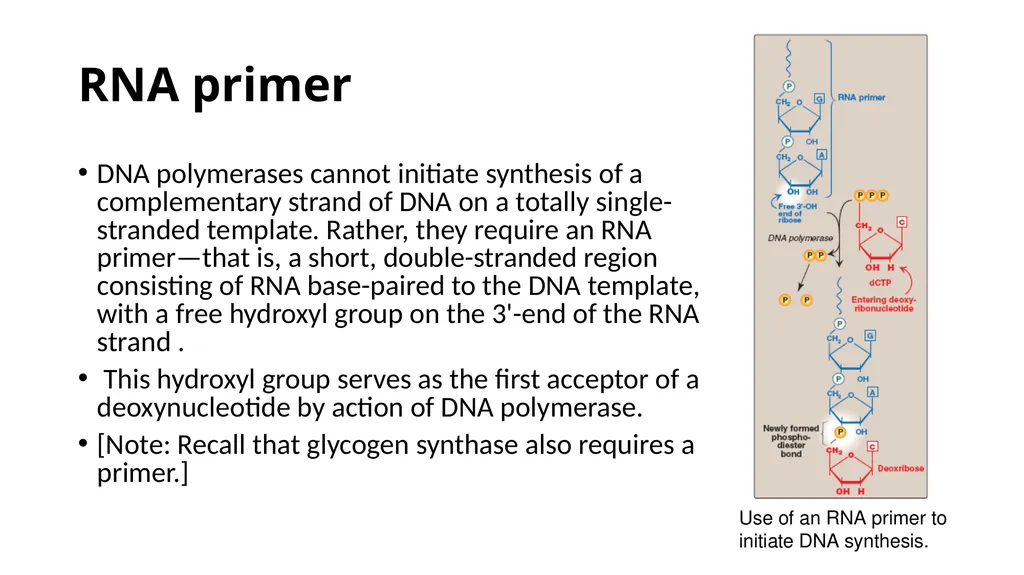
slide38. RNA primer DNA polymerases cannot initiate synthesis of a complementary strand of DNA on a totally single-stranded template. Rather, they require an RNA primer—that is, a short, double-stranded region consisting of RNA base-paired to the DNA template, with a free hydroxyl group on the 3'-end of the RNA strand .
This hydroxyl group serves as the first acceptor of a deoxynucleotide by action of DNA polymerase.
[Note: Recall that glycogen synthase also requires a primer.] Use of an RNA primer to initiate DNA synthesis.<br>
slide39. Primase: A specific RNA polymerase, called primase (DnaG), synthesizes the short stretches of RNA (approximately ten nucleotides long) that are complementary and antiparallel to the DNA template. In the resulting hybrid duplex, the U in RNA pairs with A in DNA, these short RNA sequences are constantly being synthesized at the replication fork on the lagging strand, but only one RNA sequence at the origin of replication is required on the leading strand.
The substrates for this process are 5'-ribonucleoside triphosphates, and pyrophosphate is released as each ribonucleoside monophosphate is added through formation of a 3'→5' phosphodiester bond.
[Note: The RNA primer is later removed.]<br>
slide40. Primosome: The addition of primase converts the prepriming complex of proteins required for DNA strand separation to a primosome.
The primosome makes the RNA primer required for leading strand synthesis, and initiates Okazaki fragment formation in lagging strand synthesis.
As with DNA synthesis, the direction of synthesis of the primer is 5'→3'.<br>
slide41. Chain elongation Prokaryotic (and eukaryotic) DNA polymerases elongate a new DNA strand by adding deoxyribonucleotides, one at a time, to the 3'- end of the growing chain .
The sequence of nucleotides that are added is dictated by the base sequence of the template strand with which the incoming nucleotides are paired.
DNA polymerase III: DNA chain elongation is catalyzed by DNA polymerase III. Using the 3'-hydroxyl group of the RNA primer as the acceptor of the first deoxyribonucleotide, DNA polymerase III begins to add nucleotides along the single-stranded template that specifies the sequence of bases in the newly synthesized chain.<br>
slide42. DNA polymerase III is a highly “processive” enzyme—that is, it remains bound to the template strand as it moves along, and does not diffuse away and then rebind before adding each new nucleotide. The processivity of DNA polymerase III is the result of its β subunit forming a ring that encircles and moves along the template strand of the DNA, thus serving as a sliding DNA clamp.
The new strand grows in the 5'→3' direction, antiparallel to the parental strand .
The nucleotide substrates are 5'-deoxy ribo nucleoside triphosphates.
Pyrophosphate (PPi) is released when each new deoxynucleoside monophosphate is added to the growing chain .
Hydrolysis of PPi to 2Pi means that a total of two high-energy bonds are used to drive the addition of each deoxynucleotide.<br>
slide43. Elongation of the leading and lagging strands.<br>
slide44. All four deoxyribonucleoside triphosphates (dATP, dTTP, dCTP, and dGTP) must be present for DNA elongation to occur. If one of the four is in short supply, DNA synthesis stops when that nucleotide is depleted.
Proofreading of newly synthesized DNA: It is highly important for the survival of an organism that the nucleotide sequence of DNA be replicated with as few errors as possible.
Misreading of the template sequence could result in deleterious, perhaps lethal, mutations.
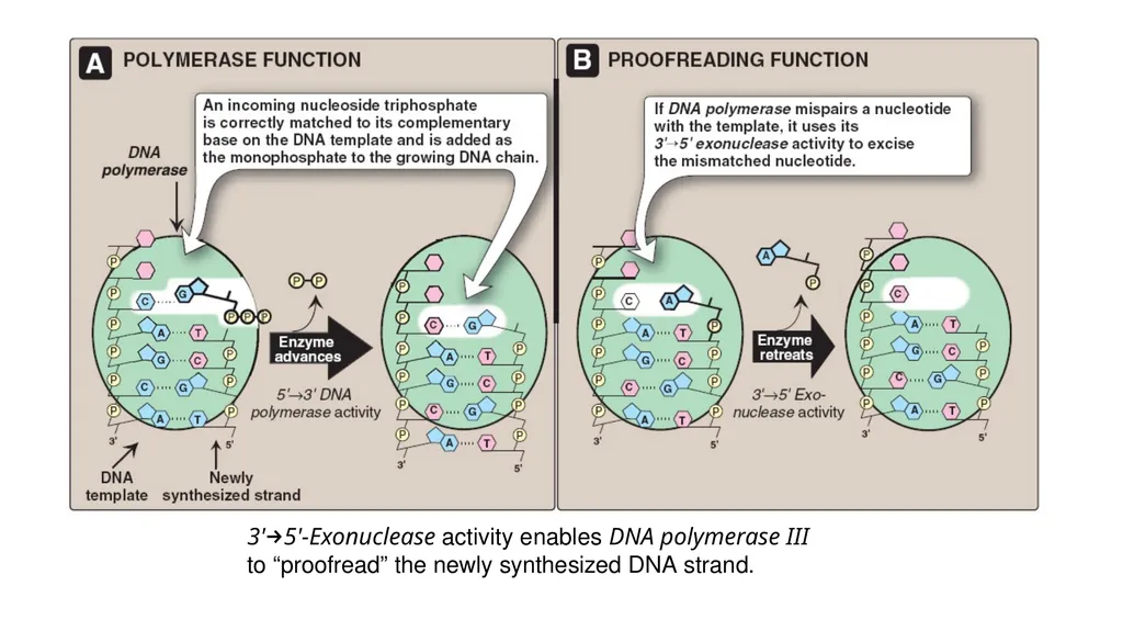
To ensure replication fidelity, DNA polymerase III has, in addition to its 5'→3' polymerase activity, a “proofreading” activity (3'→5' exonuclease.<br>
slide45. As each nucleotide is added to the chain, DNA polymerase III checks to make certain the added nucleotide is, in fact, correctly matched to its complementary base on the template. If it is not, the 3'→5' exonuclease activity corrects the mistake.
[Note: The enzyme requires an improperly basepaired 3'-hydroxy terminus and, therefore, does not degrade correctly paired nucleotide sequences.] For example, if the template base is cytosine and the enzyme mistakenly inserts an adenine instead of a guanine into the new chain, the 3'→5' exonuclease activity hydrolytically removes the misplaced nucleotide.
The 5' →3‘polymerase activity then replaces it with the correct nucleotide containing guanine.<br>
slide46. [Note: The proofreading exonuclease activity requires movement in the 3'→5' direction, not 5' →3' like the polymerase activity. This is because the excision must be done in the reverse direction from that of synthesis.]<br>
slide47. 3'→5'-Exonuclease activity enables DNA polymerase III to “proofread” the newly synthesized DNA strand.<br>
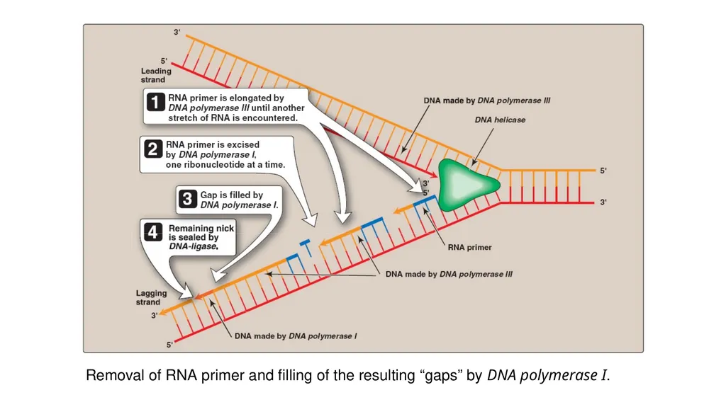
slide48. Excision of RNA primers and their replacement by DNA DNA polymerase III continues to synthesize DNA on the lagging strand until it is blocked by proximity to an RNA primer. When this occurs, the RNA is excised and the gap filled by DNA polymerase I.
5'→3' Exonuclease activity : In addition to having the 5'→3' polymerase activity that synthesizes DNA, and the 3' →5' exonuclease activity that proofreads the newly synthesized DNA chain like DNA polymerase III, DNA polymerase I also has a 5'→3' exonuclease activity that is able to hydrolytically remove the RNA primer.<br>
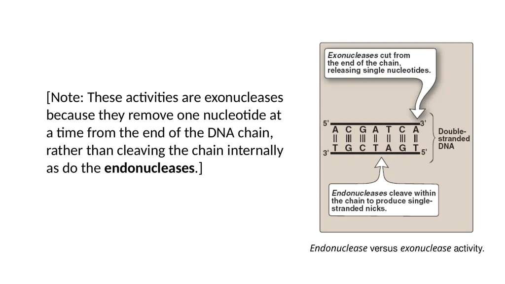
slide49. Endonuclease versus exonuclease activity. [Note: These activities are exonucleases because they remove one nucleotide at a time from the end of the DNA chain, rather than cleaving the chain internally as do the endonucleases.]<br>
slide50. First, DNA polymerase I locates the space (“nick”) between the 3'-end of the DNA newly synthesized by DNA polymerase III and the 5'-end of the adjacent RNA primer.
Next, DNA polymerase I hydrolytically removes the RNA nucleotides “ahead” of itself, moving in the 5'→3' direction (5'→3' exonuclease activity).
As it removes the RNA, DNA polymerase I replaces it with deoxyribonucleotides, synthesizing DNA in the 5'→3' direction (5'→3' polymerase activity). As it synthesizes the DNA, it also “proofreads” the new chain using 3'→5' exonuclease activity.<br>
slide51. Differences between 5'→3' and 3'→5' exonucleases:
The 5'→3‘ exonuclease activity of DNA polymerase I differs from the 3'→5‘ exonuclease used by both DNA polymerase I and III in two important ways:
First, 5'→3' exonuclease can remove one nucleotide at a time from a region of DNA that is properly base-paired. The nucleotides it removes can be either ribonucleotides or deoxyribonucleotides.
Second, 5'→3' exonuclease can also remove groups of altered nucleotides in the 5'→3' direction, removing from one to ten nucleotides at a time. This ability is important in the repair of some types of damaged DNA.<br>
slide52. Removal of RNA primer and filling of the resulting “gaps” by DNA polymerase I.<br>
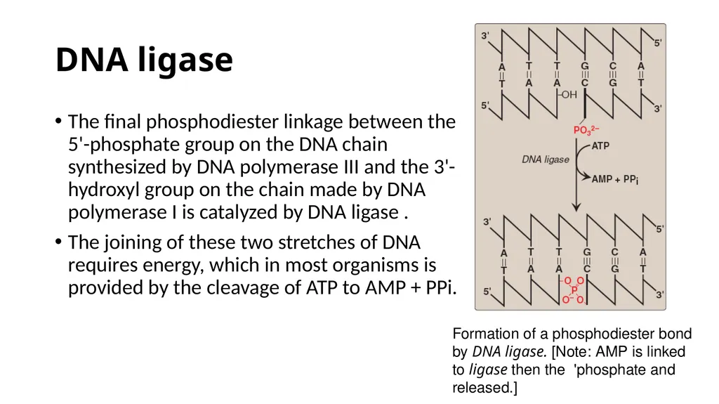
slide53. DNA ligase The final phosphodiester linkage between the 5'-phosphate group on the DNA chain synthesized by DNA polymerase III and the 3'- hydroxyl group on the chain made by DNA polymerase I is catalyzed by DNA ligase .
The joining of these two stretches of DNA requires energy, which in most organisms is provided by the cleavage of ATP to AMP + PPi. Formation of a phosphodiester bond by DNA ligase. [Note: AMP is linked to ligase then the 'phosphate and released.]<br>
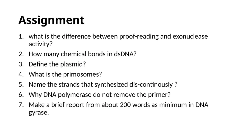
slide54. Assignment what is the difference between proof-reading and exonuclease activity?
How many chemical bonds in dsDNA?
Define the plasmid?
What is the primosomes?
Name the strands that synthesized dis-continously ?
Why DNA polymerase do not remove the primer?
Make a brief report from about 200 words as minimum in DNA gyrase.<br>
Biotechnology TA,
Omdurman Islamic University<br>
slide2. dedication To spirit of my mother<br>
slide3. OVERVIEW Nucleic acids are required for the storage and expression of genetic information. There are two chemically distinct types of nucleic acids:
deoxyribonucleic acid (DNA)
and ribonucleic acid (RNA)
DNA, the repository of genetic information, is present not only in chromosomes in the nucleus of eukaryotic organisms, but also in mitochondria and the chloroplasts of plants.
Prokaryotic cells, which lack nuclei, have a single chromosome, but may also contain non-chromosomal DNA in the form of plasmids.<br>
slide4. The genetic information found in DNA is copied and transmitted to daughter cells through DNA replication.
The DNA contained in a fertilized egg encodes the information that directs the development of an organism.<br>
slide5. central dogma of molecular biology Def: The flow of information from DNA to RNA to protein
central dogma is descriptive of all organisms, with the exception of some viruses that have RNA as the repository of their genetic information.<br>
slide6. STRUCTURE OF DNA A. DNA chain with the nucleotide sequence shown written in the 5'→ 3' direction. A 3' →5'-phosphodiester
bond is shown highlighted in the blue box, and the deoxyribose-phosphate backbone is shaded in yellow.
B. The DNA chain written in a more stylized form, emphasizing the ribose–phosphate backbone.
C. A simpler representation of the nucleotide sequence. D. The simplest (and most common) representation,
with the abbreviations for the bases written in the conventional 5'→3' direction.<br>
slide7. DNA: is a polymer of deoxyribonucleoside monophosphates covalently linked by 3'→5'–phosphodiester bonds.
With the exception of a few viruses that contain single-stranded (ss) DNA, DNA exists as a doublestranded (ds) molecule, in which the two strands wind around each other, forming a double helix.
In eukaryotic cells, DNA is found associated with various types of proteins (known collectively as nucleoprotein) present in the nucleus, whereas in prokaryotes, the protein–DNA complex is present in a non-membrane-bound region known as the nucleoid.<br>
slide8. 3'→5'-Phosphodiester bonds Phosphodiester bonds join the 3'-hydroxyl group of the deoxy pentose of one nucleotide to the 5'-hydroxyl group of the deoxy pentose of an adjacent nucleotide through a phosphate group.
3'-end: (the end with the free hydroxyl) that are not attached to other nucleotides.
5'-end: (the end with the free phosphate) that are not attached to other nucleotides.
The bases located along the resulting deoxy ribose–phosphate backbone are, by convention, always written in sequence from the 5'-end of the chain to the 3'-end. For example, the sequence of bases in the DNA shown.<br>
slide9. Double helix the two chains of DNA are coiled around a common axis called the axis of symmetry.
In the DNA helix, the hydrophilic deoxyribose–phosphate backbone of each chain is on the outside of the molecule, whereas the hydrophobic bases are stacked inside.
The overall structure resembles a twisted ladder.<br>
slide10. grooves provide access for the binding of regulatory proteins to their specific recognition sequences along the DNA chain.
Certain anticancer drugs, such as dactinomycin (actinomycin D), exert their cytotoxic effect by intercalating into the narrow groove of the DNA double helix, thus interfering with DNA and RNA synthesis.
Base pairing: The bases of one strand of DNA are paired with the bases of the second strand, so that an adenine is always paired with a thymine and a cytosine is always paired with a guanine.
one polynucleotide chain of the DNA double helix is always the complement of the other.<br>
slide11. Chargaff Rule: In any sample of dsDNA, the amount of adenine equals the amount of thymine, the amount of guanine equals the amount of cytosine, and the total amount of purines equals the total amount of pyrimidines. The base pairs are held together by hydrogen bonds: two between A and T and three between G and C. Two complementary DNA sequences.<br>
slide12. Separation of the two DNA strands in the double helix: Disruption can occur in the laboratory if the pH of the DNA solution is altered so that the nucleotide bases ionize, or if the solution is heated.
melting temperature (Tm): temperature at which one half of the helical structure is lost.
Denaturation: loss of helical structure in DNA.
Degradation : break down of phosphor-di-ester bonds.
Denaturation can be monitored by measuring its absorbance at 260 nm. [Note: ssDNA has a higher relative absorbance at this wavelength than does dsDNA.] Melting temperatures (Tm) of DNA
molecules with different nucleotide
compositions. (At a wavelength of
260 nm, single-stranded DNA has
a higher relative absorbance than
does double-stranded DNA.)<br>
slide13. Because there are three hydrogen bonds between G and C but only two between A and T, DNA that contains high concentrations of A and T denatures at a lower temperature than G- and C-rich DNA.
Under appropriate conditions, complementary DNA strands can reform the double helix by the process called renaturation (or reannealing). Hydrogen bonds between
complementary bases.<br>
slide14. Structural forms of the double helix: There are three major structural forms of DNA:
the B form, described by Watson and Crick in 1953,
the A form,
The Z form.
The B form is a right-handed helix with ten residues per 360° turn of the helix, and with the planes of the bases perpendicular to the helical axis. Structures of B-DNA and Z-DNA.<br>
slide15. The A form is produced by moderately dehydrating the B form. It is also a right-handed helix, but there are 11 base pairs per turn, and the planes of the base pairs are tilted 20° away from the perpendicular to the helical axis.
The conformation found in DNA–RNA hybrids or RNA–RNA double-stranded regions is probably very close to the A form.
Z-DNA is a left-handed helix that contains about 12 base pairs per turn [Note: The deoxyribose –phosphate backbone “zigzags,” hence, the name “Z”-DNA.]<br>
slide16. Stretches of Z-DNA can occur naturally in regions of DNA that have a sequence of alternating purines and pyrimidines, for example, poly GC.
Transitions between the B and Z helical forms of DNA may play a role in regulating gene expression.<br>
slide17. Linear and circular DNA molecules Each chromosome in the nucleus of a eukaryote contains one long, linear molecule of dsDNA, which is bound to a complex mixture of proteins (histone and non-histone) to form chromatin.
Eukaryotes have closed, circular DNA molecules in their mitochondria, as do plant chloroplasts. A prokaryotic organism typically contains a single, double-stranded, supercoiled, circular chromosome.
Each prokaryotic chromosome is associated with non-histone proteins that can condense the DNA to form a nucleoid. In addition, most species of bacteria also contain small, circular, extrachromosomal DNA molecules called plasmids.<br>
slide18. Plasmid DNA carries genetic information, and undergoes replication that may or may not be synchronized to chromosomal division.
Plasmids may carry genes that convey antibiotic resistance to the host bacterium, and may facilitate the transfer of genetic information from one bacterium to another.<br>
slide19. STEPS IN PROKARYOTIC DNA SYNTHESIS When the two strands of the DNA double helix are separated, each can serve as a template for the replication of a new complementary strand.
This produces two daughter molecules, each of which contains two DNA strands with an antiparallel orientation .This process is called semiconservative replication because, although the parental duplex is separated into two halves (and, therefore, is not “conserved” as an entity), each of the individual parental strands remains intact in one of the two new duplexes . Semiconservative replication of DNA.<br>
slide20. The enzymes involved in the DNA replication process are template-directed polymerases that can synthesize the complementary sequence of each strand with extraordinary fidelity.
The reactions described in this section were first known from studies of the bacterium Escherichia coli (E. coli), and the description given below refers to the process in prokaryotes.
DNA synthesis in higher organisms is less well understood, but involves the same types of mechanisms. In either case, initiation of DNA replication commits the cell to continue the process until the entire genome has been replicated.<br>
slide21. The steps of DNA synthesis consists of :
Separation of the two complementary DNA strands
Formation of the replication fork
RNA primer
Chain elongation
Excision of RNA primers and their replacement by DNA
DNA ligase<br>
slide22. A. Separation of the two complementary DNA strands In order for the two strands of the parental double helical DNA to be replicated, they must first separate (or “melt”) over a small region, because the polymerases use only ssDNA as a template.
In prokaryotic organisms, DNA replication begins at a single, unique nucleotide sequence—a site called the origin of replication.
[Note: This is referred to as a consensus sequence, because the order of nucleotides is essentially the same at each site.] This site includes a short sequence composed almost exclusively of AT base pairs that facilitate melting.
In eukaryotes, replication begins at multiple sites along the DNA helix .<br>
slide23. Having multiple origins of replication provides a mechanism for rapidly replicating the great length of the eukaryotic DNA molecules.<br>
slide24. B. Formation of the replication fork As the two strands unwind and separate, they form a “V” where active synthesis occurs. This region is called the replication fork. It moves along the DNA molecule as synthesis occurs.
Replication of dsDNA is bidirectional—that is, the replication forks move in opposite directions from the origin, generating a replication bubble.<br>
slide25. Replication of DNA: origins and replication forks. A. Small prokaryotic circular DNA. B. Very long eukaryotic DNA.<br>
slide26. 1. Proteins required for DNA strand separation: Initiation of DNA replication requires the recognition of the origin of replication by a group of proteins that form the prepriming complex. These proteins are responsible for maintaining the separation of the parental strands, and for unwinding the double helix ahead of the advancing replication fork.
These proteins include the following:
DnaA protein
DNA helicases
Single-stranded DNA-binding (SSB) proteins<br>
slide27. DnaA protein: protein binds to specific nucleotide sequences at the origin of replication, causing short, tandemly arranged (one after the other) AT-rich regions in the origin to melt.
Melting is ATP-dependent, and results in strand separation with the formation of localized regions of ssDNA.
DNA helicases: These enzymes bind to ssDNA near the replication fork, and then move into the neighboring doublestranded region, forcing the strands apart—in effect, unwinding the double helix.
Helicases require energy provided by ATP .
[Note: DnaB is the principal helicase of replication in E. coli. Its binding to DNA requires DnaC.]<br>
slide28. Single-stranded DNA-binding (SSB) proteins: These proteins bind to the ssDNA generated by helicases .
They bind cooperatively—that is, the binding of one molecule of SSB protein makes it easier for additional molecules of SSB protein to bind tightly to the DNA strand.
The SSB proteins are not enzymes, but rather serve to shift the equilibrium between dsDNA and ssDNA in the direction of the single-stranded forms.
These proteins not only keep the two strands of DNA separated in the area of the replication origin, thus providing the single-stranded template required by polymerases, but also protect the DNA from nucleases that degrade ssDNA. Proteins responsible for maintaining
the separation of the parental
strands and unwinding the double
helix ahead of the advancing
replication fork.<br>
slide29. 2. Solving the problem of supercoils: As the two strands of the double helix are separated, a problem is encountered, namely, the appearance of positive supercoils (also called supertwists) in the region of DNA ahead of the replication fork .
The accumulating positive supercoils interfere with further unwinding of the double helix.
To solve this problem, there is a group of enzymes called DNA topoisomerases, which are responsible for removing supercoils in the helix.
Type I DNA topoisomerases
Type II DNA topoisomerases<br>
slide30. Type I DNA topoisomerases: These enzymes reversibly cut one strand of the double helix. They have both nuclease (strand-cutting) and ligase (strand-resealing) activities.
They do not require ATP, but rather appear to store the energy from the phosphodiester bond they cleave, reusing the energy to reseal the strand .
Each time a transient “nick” is created in one DNA strand, the intact DNA strand is passed through the break before it is resealed, thus relieving (“relaxing”) accumulated supercoils.
Type I topoisomerases relax negative supercoils (that is, those that contain fewer turns of the helix than relaxed DNA) in E. coli, and both negative and positive supercoils (that is, those that contain fewer or more turns of the helix than relaxed DNA) in eukaryotic cells.<br>
slide31. Positive supercoiling resulting
from DNA strand separation. Action of Type I DNA topoisomerases.<br>
slide32. Type II DNA topoisomerases: These enzymes bind tightly to the DNA double helix and make transient breaks in both strands.
The enzyme then causes a second stretch of the DNA double helix to pass through the break and, finally, reseals the break .
As a result, both negative and positive supercoils can be relieved by this ATP-requiring process.
Type II DNA topoisomerases are also required in both prokaryotes and eukaryotes for the separation of interlocked molecules of DNA following chromosomal replication.<br>
slide33. DNA gyrase, a Type II topoisomerase found in bacteria and plants, has the unusual property of being able to introduce negative supercoils into relaxed circular DNA using energy from the hydrolysis of ATP. This facilitates the future replication of DNA because the negative supercoils neutralize the positive supercoils introduced during opening of the double helix. It also aids in the transient strand separation required during transcription .
Anticancer agents, such as etoposide, target human topoisomerase II.
Bacterial DNA gyrase is a unique target of a group of anti microbial agents called quinolones, for example, cipro - floxacin.<br>
slide34. Action of Type II DNA topoisomerase.<br>
slide35. Direction of DNA replication The DNA polymerases responsible for copying the DNA templates are only able to “read” the parental nucleotide sequences in the 3'→5' direction, and they synthesize the new DNA strands only in the 5'→3' (antiparallel) direction.
Therefore, beginning with one parental double helix, the two newly synthesized stretches of nucleotide chains must grow in opposite directions—one in the 5'→3' direction toward the replication fork (Leading strand) and one in the 5'→3' direction away from the replication fork (Lagging strand).
This feat is accomplished by a slightly different mechanism on each strand.<br>
slide36. Leading strand: The strand that is being copied in the direction of the advancing replication fork and is synthesized continuously.
Lagging strand: The strand that is being copied in the direction away from the replication fork is synthesized discontinuously, with small fragments of DNA being copied near the replication fork.
These short stretches of discontinuous DNA, termed Okazaki fragments, are eventually joined (ligated) to become a single, continuous strand.
The new strand of DNA produced by this mechanism is termed the lagging strand.<br>
slide37. Discontinuous synthesis of DNA.<br>
slide38. RNA primer DNA polymerases cannot initiate synthesis of a complementary strand of DNA on a totally single-stranded template. Rather, they require an RNA primer—that is, a short, double-stranded region consisting of RNA base-paired to the DNA template, with a free hydroxyl group on the 3'-end of the RNA strand .
This hydroxyl group serves as the first acceptor of a deoxynucleotide by action of DNA polymerase.
[Note: Recall that glycogen synthase also requires a primer.] Use of an RNA primer to initiate DNA synthesis.<br>
slide39. Primase: A specific RNA polymerase, called primase (DnaG), synthesizes the short stretches of RNA (approximately ten nucleotides long) that are complementary and antiparallel to the DNA template. In the resulting hybrid duplex, the U in RNA pairs with A in DNA, these short RNA sequences are constantly being synthesized at the replication fork on the lagging strand, but only one RNA sequence at the origin of replication is required on the leading strand.
The substrates for this process are 5'-ribonucleoside triphosphates, and pyrophosphate is released as each ribonucleoside monophosphate is added through formation of a 3'→5' phosphodiester bond.
[Note: The RNA primer is later removed.]<br>
slide40. Primosome: The addition of primase converts the prepriming complex of proteins required for DNA strand separation to a primosome.
The primosome makes the RNA primer required for leading strand synthesis, and initiates Okazaki fragment formation in lagging strand synthesis.
As with DNA synthesis, the direction of synthesis of the primer is 5'→3'.<br>
slide41. Chain elongation Prokaryotic (and eukaryotic) DNA polymerases elongate a new DNA strand by adding deoxyribonucleotides, one at a time, to the 3'- end of the growing chain .
The sequence of nucleotides that are added is dictated by the base sequence of the template strand with which the incoming nucleotides are paired.
DNA polymerase III: DNA chain elongation is catalyzed by DNA polymerase III. Using the 3'-hydroxyl group of the RNA primer as the acceptor of the first deoxyribonucleotide, DNA polymerase III begins to add nucleotides along the single-stranded template that specifies the sequence of bases in the newly synthesized chain.<br>
slide42. DNA polymerase III is a highly “processive” enzyme—that is, it remains bound to the template strand as it moves along, and does not diffuse away and then rebind before adding each new nucleotide. The processivity of DNA polymerase III is the result of its β subunit forming a ring that encircles and moves along the template strand of the DNA, thus serving as a sliding DNA clamp.
The new strand grows in the 5'→3' direction, antiparallel to the parental strand .
The nucleotide substrates are 5'-deoxy ribo nucleoside triphosphates.
Pyrophosphate (PPi) is released when each new deoxynucleoside monophosphate is added to the growing chain .
Hydrolysis of PPi to 2Pi means that a total of two high-energy bonds are used to drive the addition of each deoxynucleotide.<br>
slide43. Elongation of the leading and lagging strands.<br>
slide44. All four deoxyribonucleoside triphosphates (dATP, dTTP, dCTP, and dGTP) must be present for DNA elongation to occur. If one of the four is in short supply, DNA synthesis stops when that nucleotide is depleted.
Proofreading of newly synthesized DNA: It is highly important for the survival of an organism that the nucleotide sequence of DNA be replicated with as few errors as possible.
Misreading of the template sequence could result in deleterious, perhaps lethal, mutations.
To ensure replication fidelity, DNA polymerase III has, in addition to its 5'→3' polymerase activity, a “proofreading” activity (3'→5' exonuclease.<br>
slide45. As each nucleotide is added to the chain, DNA polymerase III checks to make certain the added nucleotide is, in fact, correctly matched to its complementary base on the template. If it is not, the 3'→5' exonuclease activity corrects the mistake.
[Note: The enzyme requires an improperly basepaired 3'-hydroxy terminus and, therefore, does not degrade correctly paired nucleotide sequences.] For example, if the template base is cytosine and the enzyme mistakenly inserts an adenine instead of a guanine into the new chain, the 3'→5' exonuclease activity hydrolytically removes the misplaced nucleotide.
The 5' →3‘polymerase activity then replaces it with the correct nucleotide containing guanine.<br>
slide46. [Note: The proofreading exonuclease activity requires movement in the 3'→5' direction, not 5' →3' like the polymerase activity. This is because the excision must be done in the reverse direction from that of synthesis.]<br>
slide47. 3'→5'-Exonuclease activity enables DNA polymerase III to “proofread” the newly synthesized DNA strand.<br>
slide48. Excision of RNA primers and their replacement by DNA DNA polymerase III continues to synthesize DNA on the lagging strand until it is blocked by proximity to an RNA primer. When this occurs, the RNA is excised and the gap filled by DNA polymerase I.
5'→3' Exonuclease activity : In addition to having the 5'→3' polymerase activity that synthesizes DNA, and the 3' →5' exonuclease activity that proofreads the newly synthesized DNA chain like DNA polymerase III, DNA polymerase I also has a 5'→3' exonuclease activity that is able to hydrolytically remove the RNA primer.<br>
slide49. Endonuclease versus exonuclease activity. [Note: These activities are exonucleases because they remove one nucleotide at a time from the end of the DNA chain, rather than cleaving the chain internally as do the endonucleases.]<br>
slide50. First, DNA polymerase I locates the space (“nick”) between the 3'-end of the DNA newly synthesized by DNA polymerase III and the 5'-end of the adjacent RNA primer.
Next, DNA polymerase I hydrolytically removes the RNA nucleotides “ahead” of itself, moving in the 5'→3' direction (5'→3' exonuclease activity).
As it removes the RNA, DNA polymerase I replaces it with deoxyribonucleotides, synthesizing DNA in the 5'→3' direction (5'→3' polymerase activity). As it synthesizes the DNA, it also “proofreads” the new chain using 3'→5' exonuclease activity.<br>
slide51. Differences between 5'→3' and 3'→5' exonucleases:
The 5'→3‘ exonuclease activity of DNA polymerase I differs from the 3'→5‘ exonuclease used by both DNA polymerase I and III in two important ways:
First, 5'→3' exonuclease can remove one nucleotide at a time from a region of DNA that is properly base-paired. The nucleotides it removes can be either ribonucleotides or deoxyribonucleotides.
Second, 5'→3' exonuclease can also remove groups of altered nucleotides in the 5'→3' direction, removing from one to ten nucleotides at a time. This ability is important in the repair of some types of damaged DNA.<br>
slide52. Removal of RNA primer and filling of the resulting “gaps” by DNA polymerase I.<br>
slide53. DNA ligase The final phosphodiester linkage between the 5'-phosphate group on the DNA chain synthesized by DNA polymerase III and the 3'- hydroxyl group on the chain made by DNA polymerase I is catalyzed by DNA ligase .
The joining of these two stretches of DNA requires energy, which in most organisms is provided by the cleavage of ATP to AMP + PPi. Formation of a phosphodiester bond by DNA ligase. [Note: AMP is linked to ligase then the 'phosphate and released.]<br>
slide54. Assignment what is the difference between proof-reading and exonuclease activity?
How many chemical bonds in dsDNA?
Define the plasmid?
What is the primosomes?
Name the strands that synthesized dis-continously ?
Why DNA polymerase do not remove the primer?
Make a brief report from about 200 words as minimum in DNA gyrase.<br>