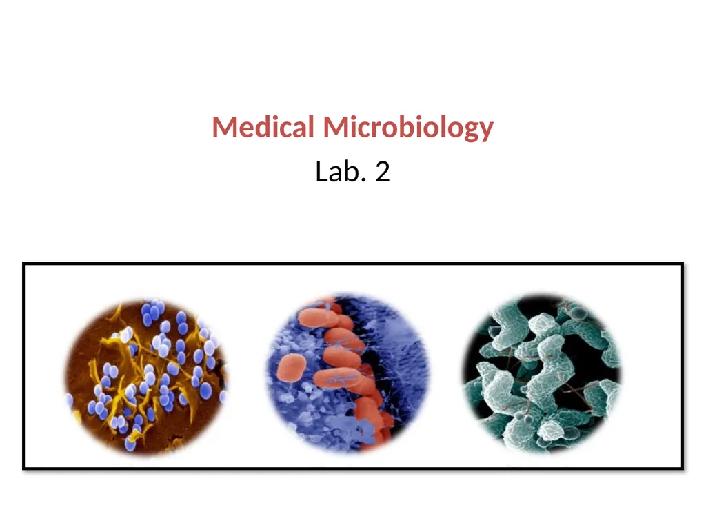
Medical Microbiology Lab. 2 Bacterial Morphology Bacteria: are unicellular free living organisms without chlorophil having doth DNA and RNA Bacterial morphology Can be grouped into three types 1- Spherical (cocci) 2- Rod shaped (bacili) 3
"Medical Microbiology Lab. 2 Bacterial Morphology" is the property of its rightful owner. Permission is granted to
download and print the materials on this website for personal, non-commercial use only, and to display it
on your personal computer provided you do not modify the materials and that you retain all copyright
notices contained in the materials. By downloading content from our website, you accept the terms of this
agreement.
Presentation Transcript
01
Medical Microbiology
Lab. 2<br>
02
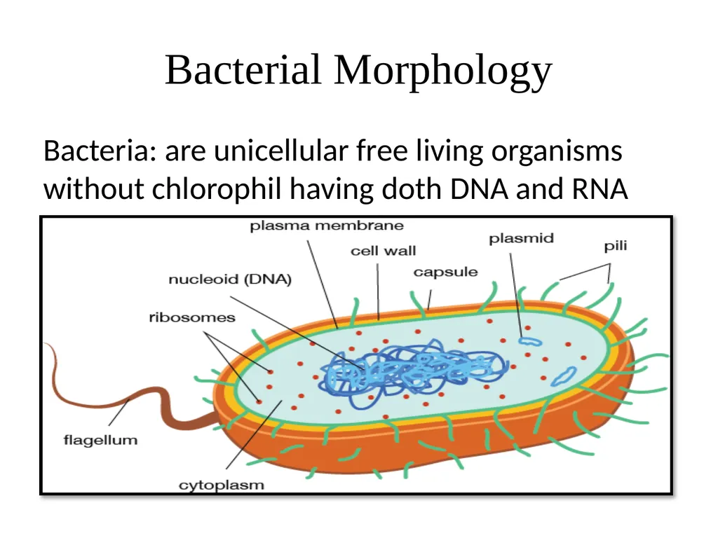
Bacterial Morphology Bacteria: are unicellular free living organisms without chlorophil having doth DNA and RNA<br>
03
Bacterial morphology
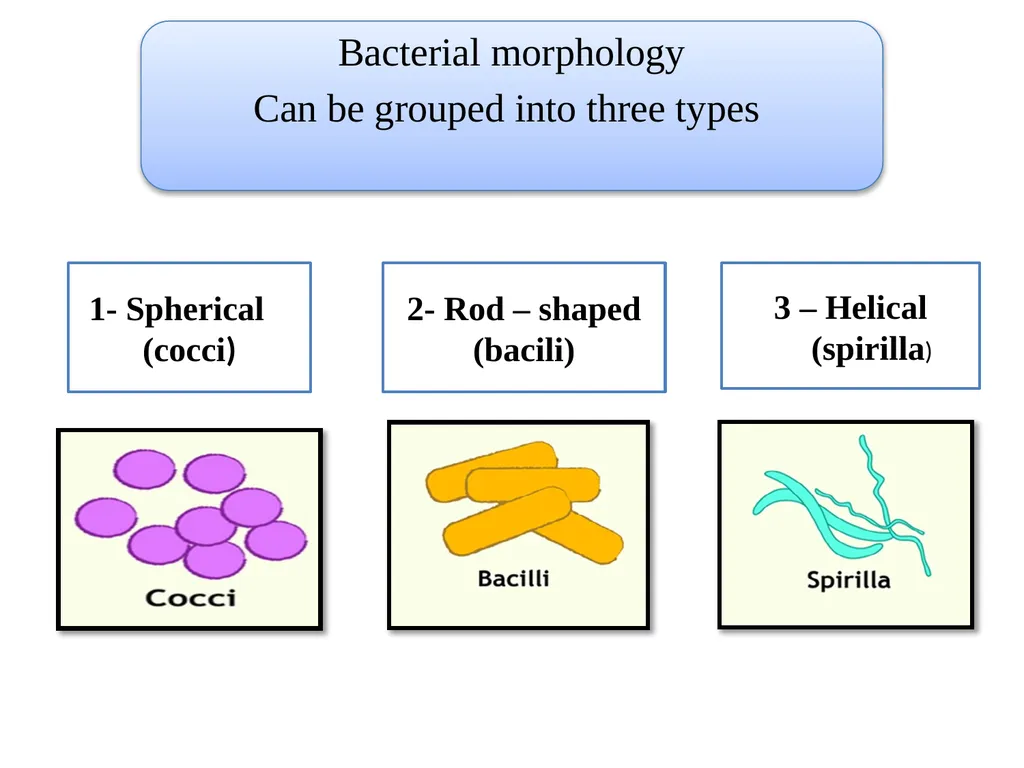
Can be grouped into three types 1- Spherical (cocci) 2- Rod – shaped
(bacili) 3 – Helical
(spirilla)<br>
04
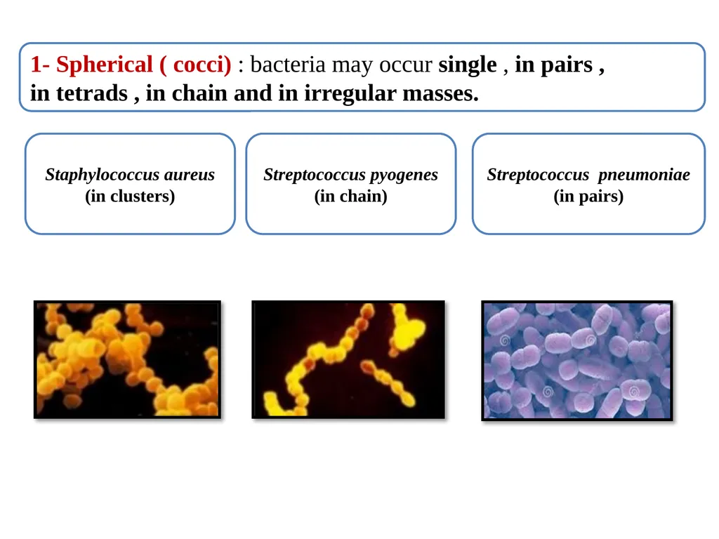
1- Spherical ( cocci) : bacteria may occur single , in pairs ,
in tetrads , in chain and in irregular masses. Staphylococcus aureus
(in clusters) Streptococcus pyogenes
(in chain) Streptococcus pneumoniae
(in pairs)<br>
05
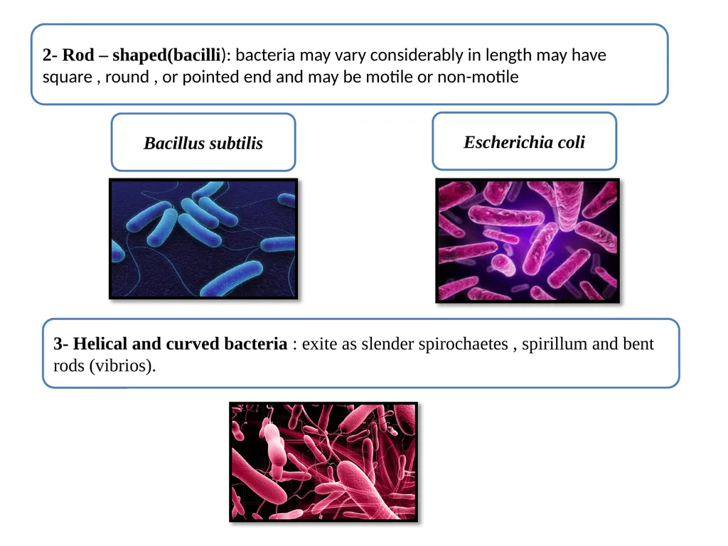
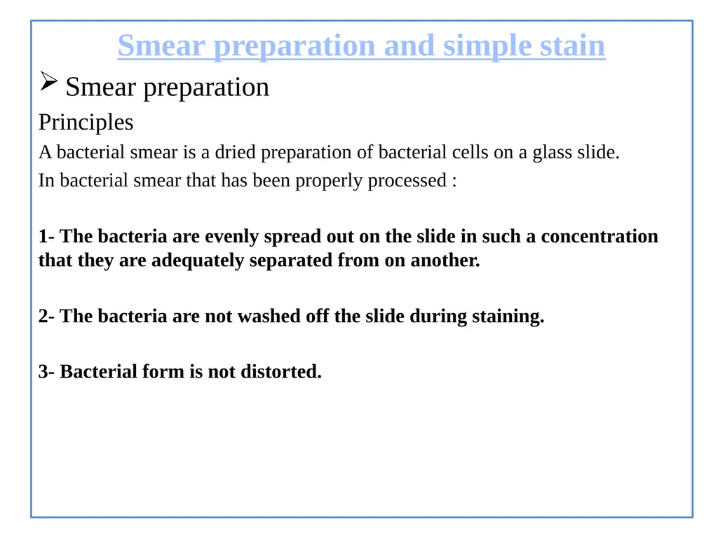
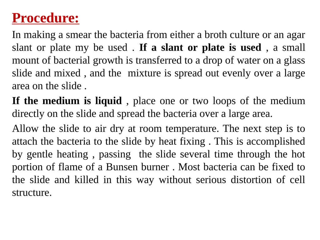
2- Rod – shaped(bacilli): bacteria may vary considerably in length may have square , round , or pointed end and may be motile or non-motile Bacillus subtilis Escherichia coli 3- Helical and curved bacteria : exite as slender spirochaetes , spirillum and bent rods (vibrios).<br>
06
Smear preparation and simple stain
Smear preparation
Principles
A bacterial smear is a dried preparation of bacterial cells on a glass slide.
In bacterial smear that has been properly processed :
1- The bacteria are evenly spread out on the slide in such a concentration that they are adequately separated from on another.
2- The bacteria are not washed off the slide during staining.
3- Bacterial form is not distorted.<br>
07
Procedure:
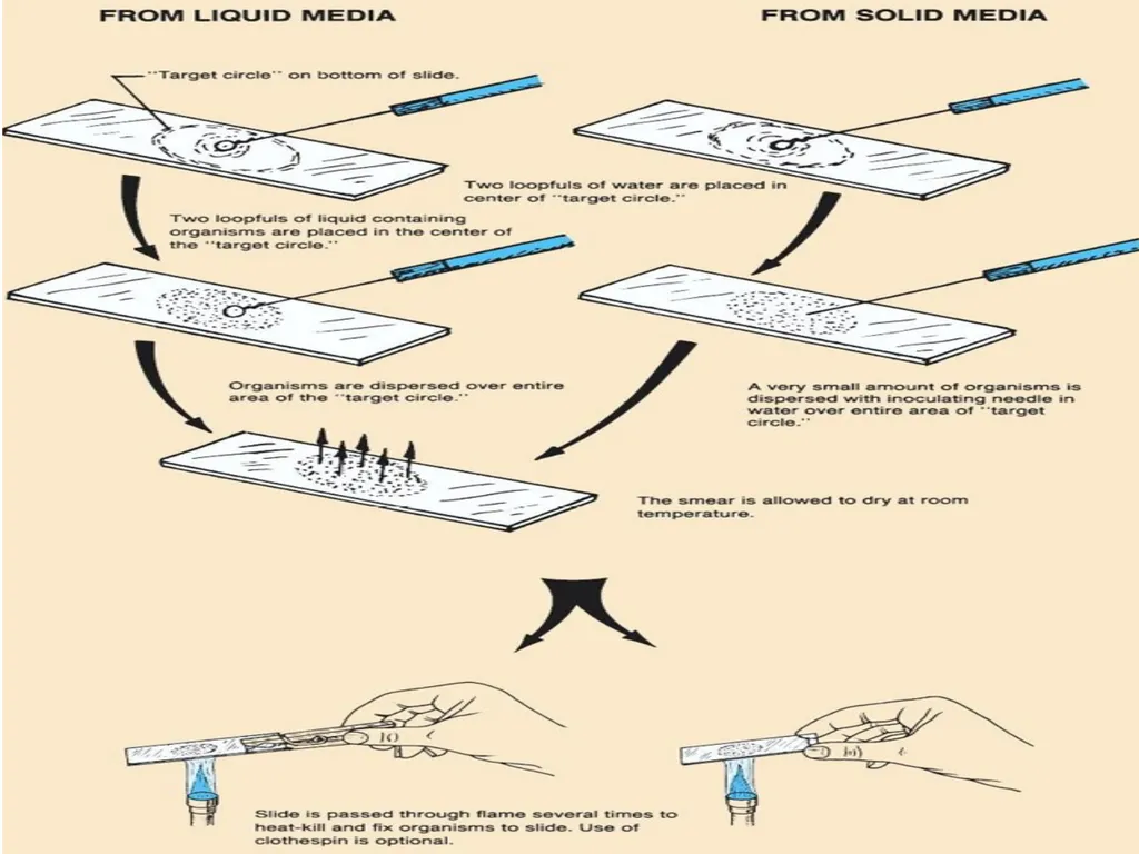
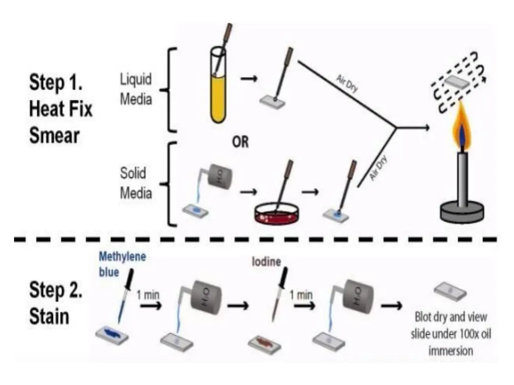
In making a smear the bacteria from either a broth culture or an agar slant or plate my be used . If a slant or plate is used , a small mount of bacterial growth is transferred to a drop of water on a glass slide and mixed , and the mixture is spread out evenly over a large area on the slide .
If the medium is liquid , place one or two loops of the medium directly on the slide and spread the bacteria over a large area.
Allow the slide to air dry at room temperature. The next step is to attach the bacteria to the slide by heat fixing . This is accomplished by gentle heating , passing the slide several time through the hot portion of flame of a Bunsen burner . Most bacteria can be fixed to the slide and killed in this way without serious distortion of cell structure.<br>
08
Simple stain
Principles
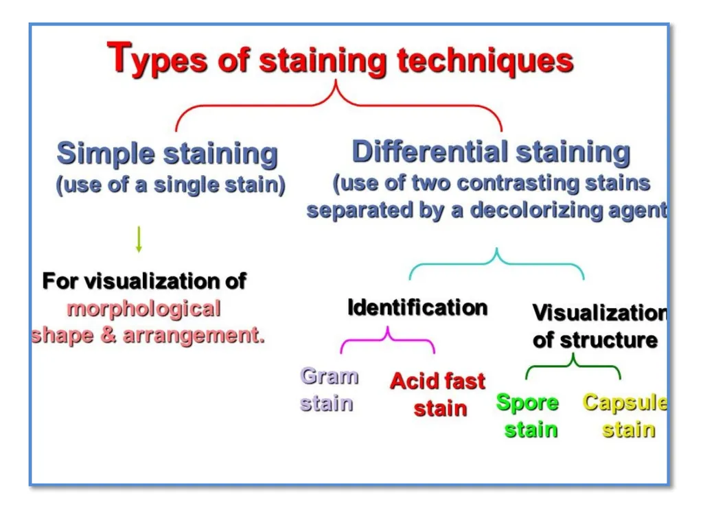
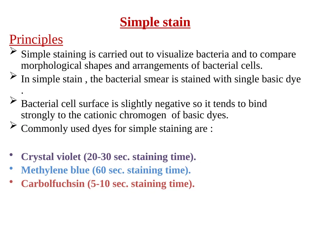
Simple staining is carried out to visualize bacteria and to compare morphological shapes and arrangements of bacterial cells.
In simple stain , the bacterial smear is stained with single basic dye .
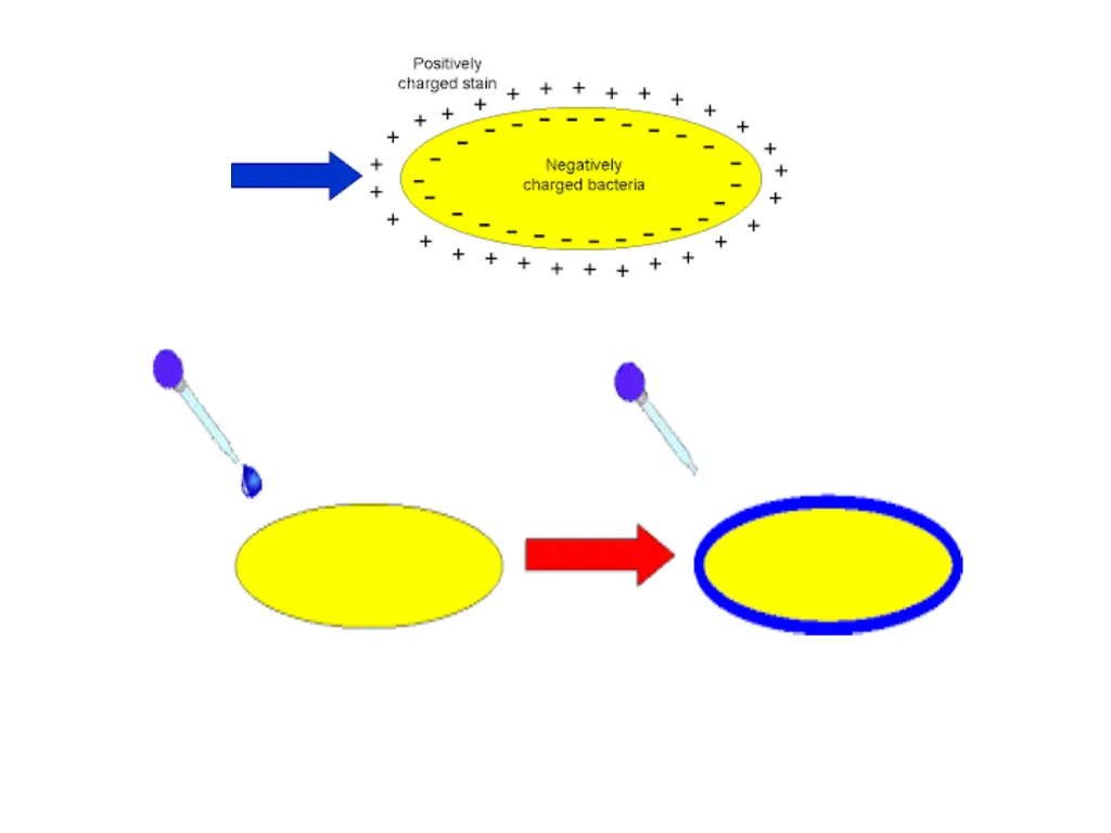
Bacterial cell surface is slightly negative so it tends to bind strongly to the cationic chromogen of basic dyes.
Commonly used dyes for simple staining are :
Crystal violet (20-30 sec. staining time).
Methylene blue (60 sec. staining time).
Carbolfuchsin (5-10 sec. staining time).<br>