How to look at bacteria Gram Staining There are a couple pieces of data to collect from bacteria: 1. how many bacteria grew on your plate (count colonies) 2. surface area of plate covered with growth (estimate using graph paper) 3. colony
"How to look at bacteria & Gram Staining There are" is the property of its rightful owner. Permission is granted to
download and print the materials on this website for personal, non-commercial use only, and to display it
on your personal computer provided you do not modify the materials and that you retain all copyright
notices contained in the materials. By downloading content from our website, you accept the terms of this
agreement.
Presentation Transcript
01
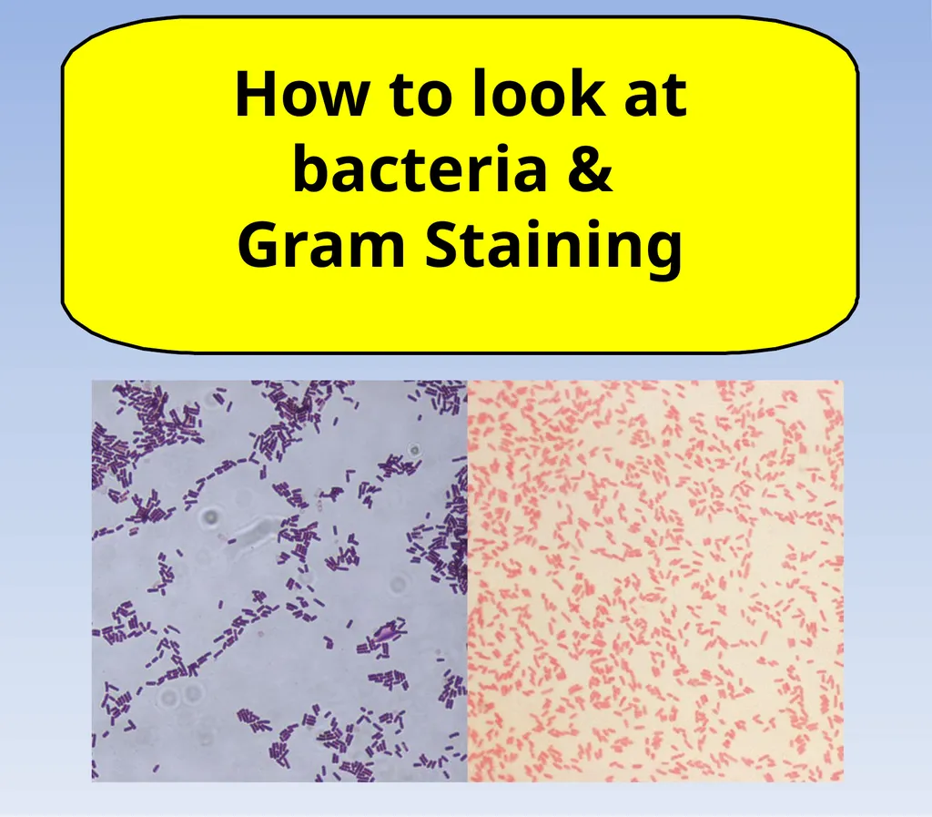
How to look at bacteria &
Gram Staining<br>
02
There are a couple pieces of data to collect from bacteria:
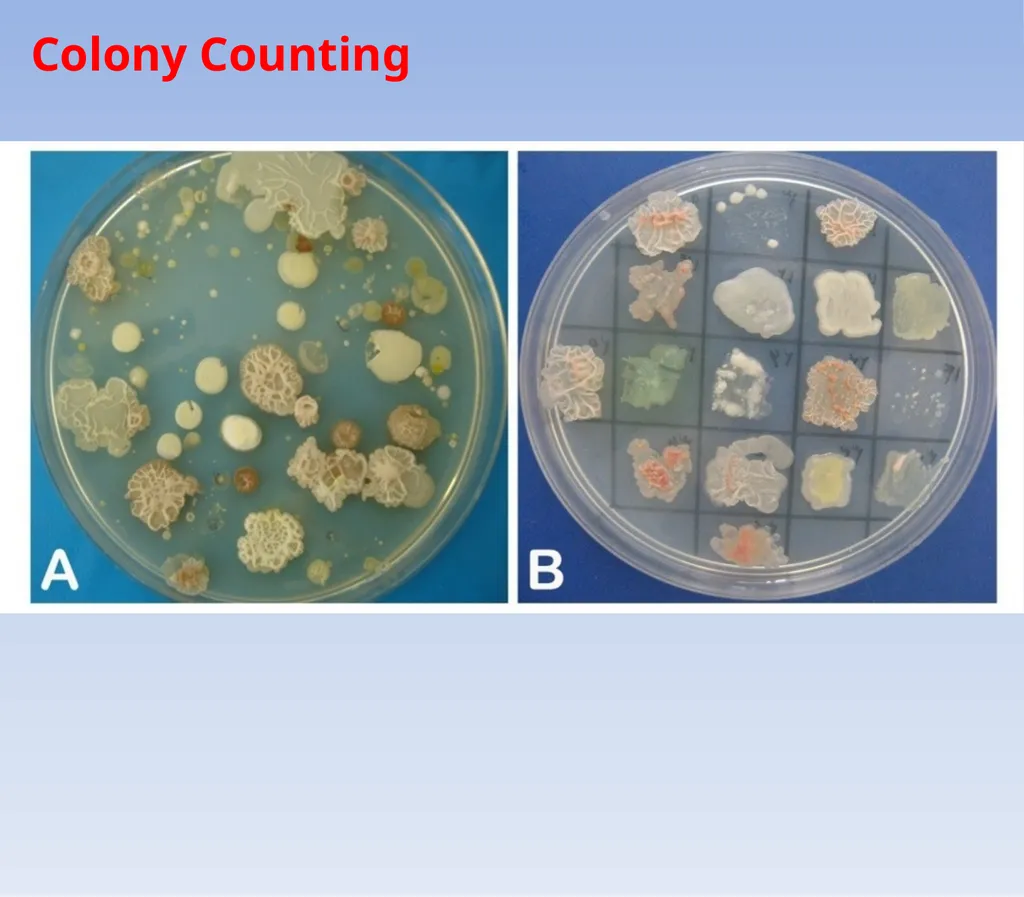
1. how many bacteria grew on your plate (count colonies)
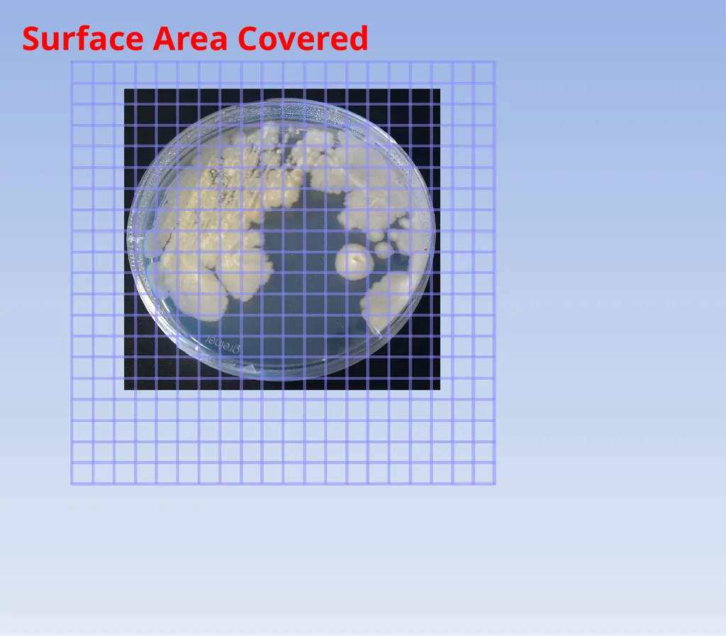
2. surface area of plate covered with growth (estimate using graph paper)
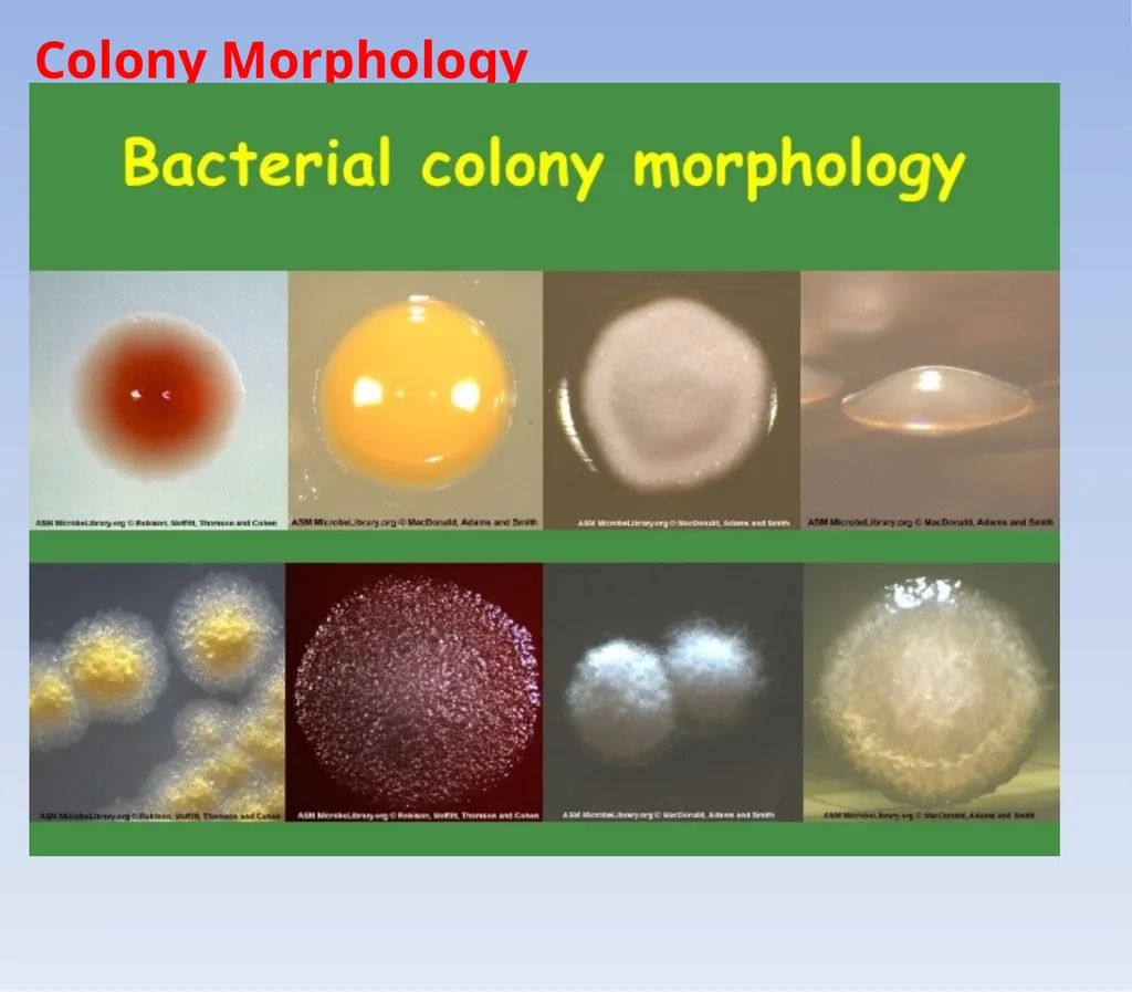
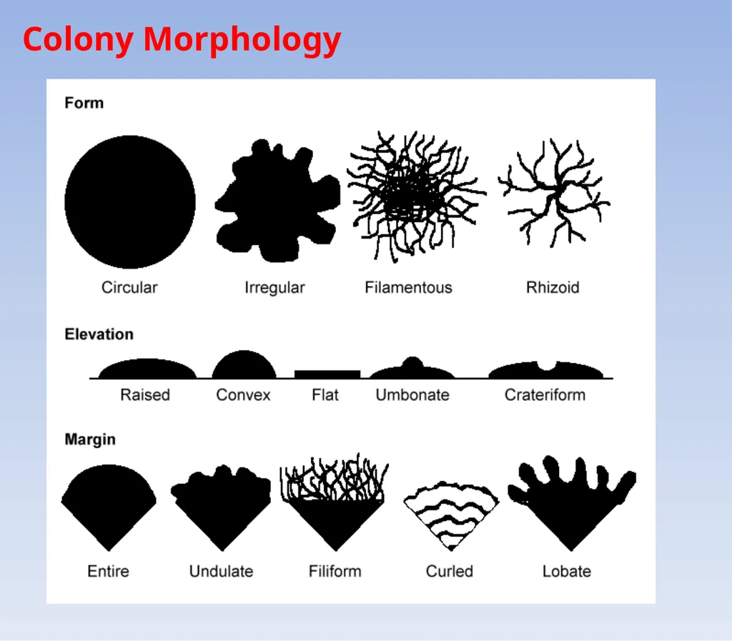
3. colony morphology (see hand out)
4. gram staining<br>
03
Colony Counting<br>
04
Surface Area Covered<br>
05
Colony Morphology<br>
06
Colony Morphology<br>
07
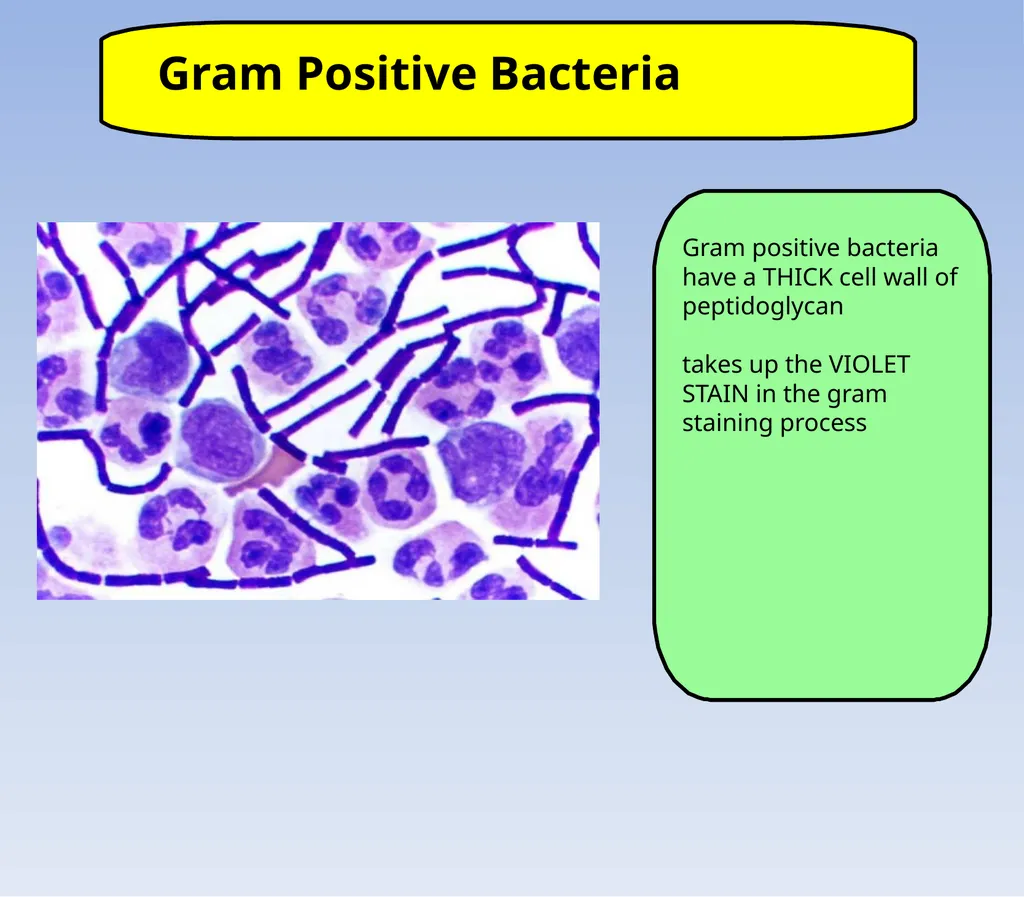
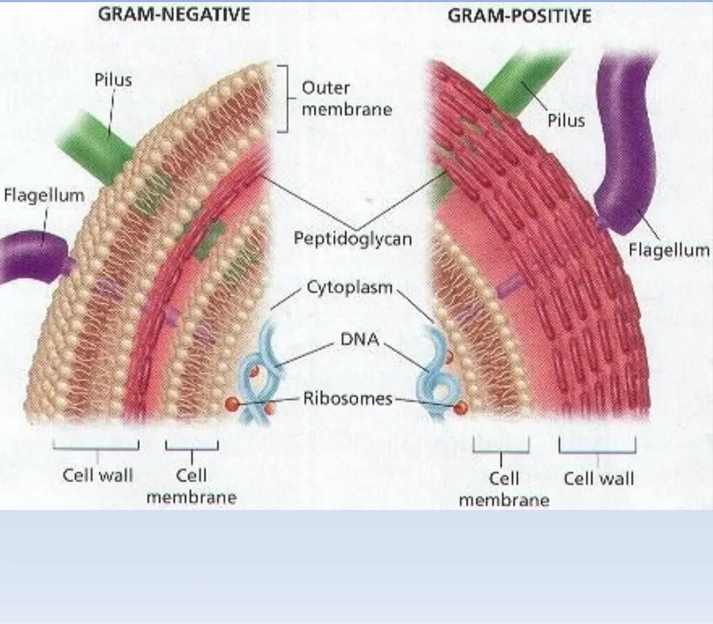
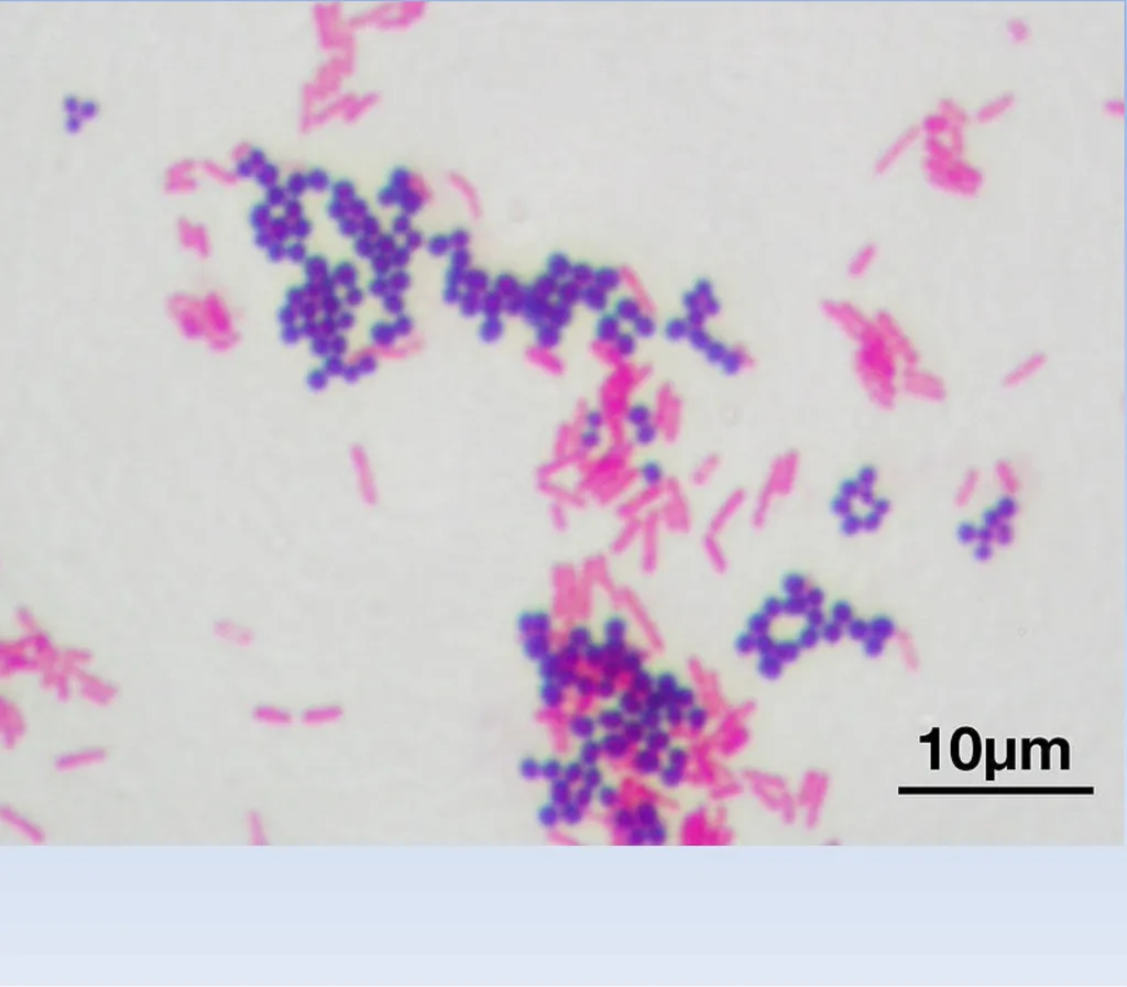
Gram Positive Bacteria Gram positive bacteria have a THICK cell wall of peptidoglycan
takes up the VIOLET STAIN in the gram staining process<br>
08
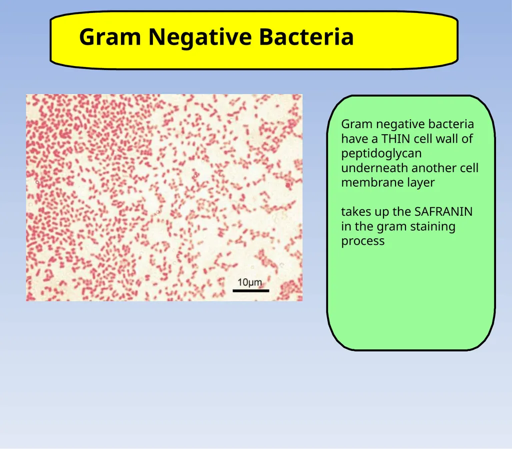
Gram Negative Bacteria Gram negative bacteria have a THIN cell wall of peptidoglycan underneath another cell membrane layer
takes up the SAFRANIN in the gram staining process<br>
09
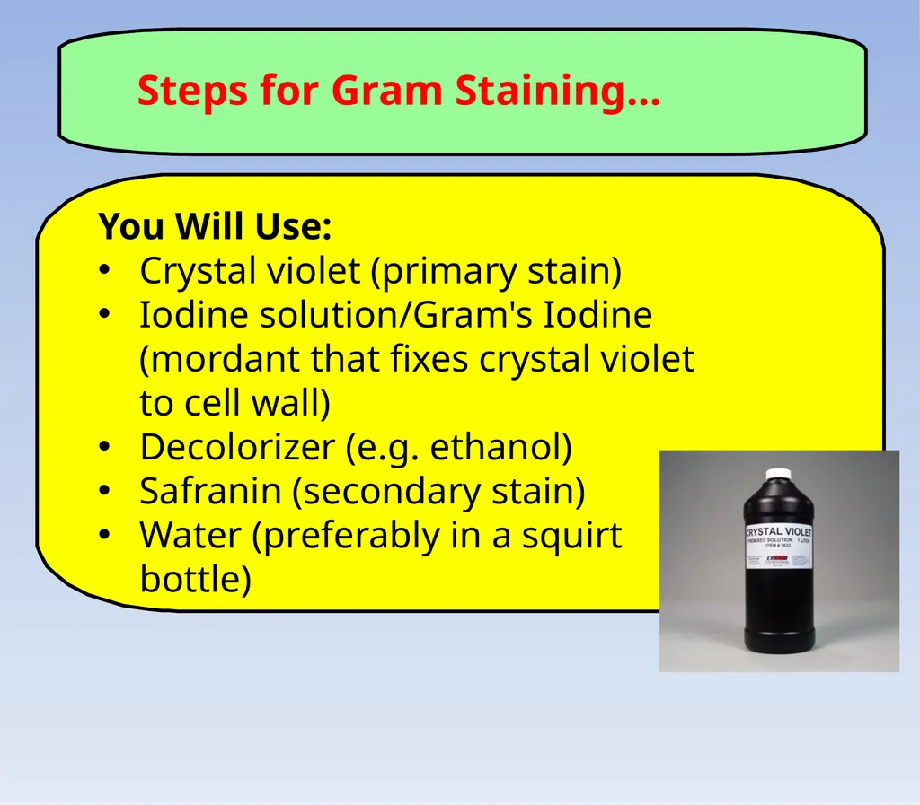
Steps for Gram Staining... You Will Use:
Crystal violet (primary stain)
Iodine solution/Gram's Iodine (mordant that fixes crystal violet to cell wall)
Decolorizer (e.g. ethanol)
Safranin (secondary stain)
Water (preferably in a squirt bottle)<br>
10
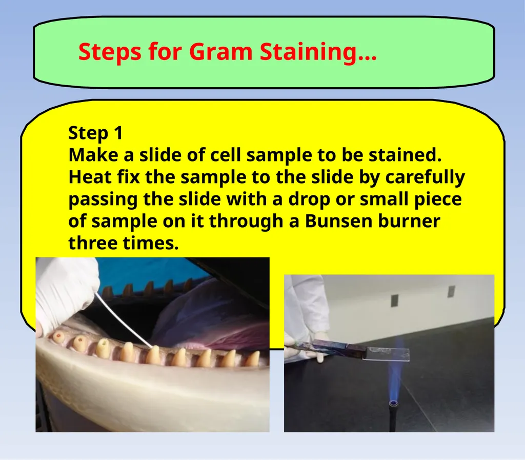
Steps for Gram Staining... Step 1
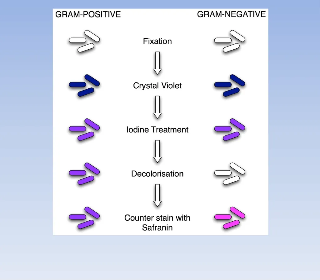
Make a slide of cell sample to be stained. Heat fix the sample to the slide by carefully passing the slide with a drop or small piece of sample on it through a Bunsen burner three times.<br>
11
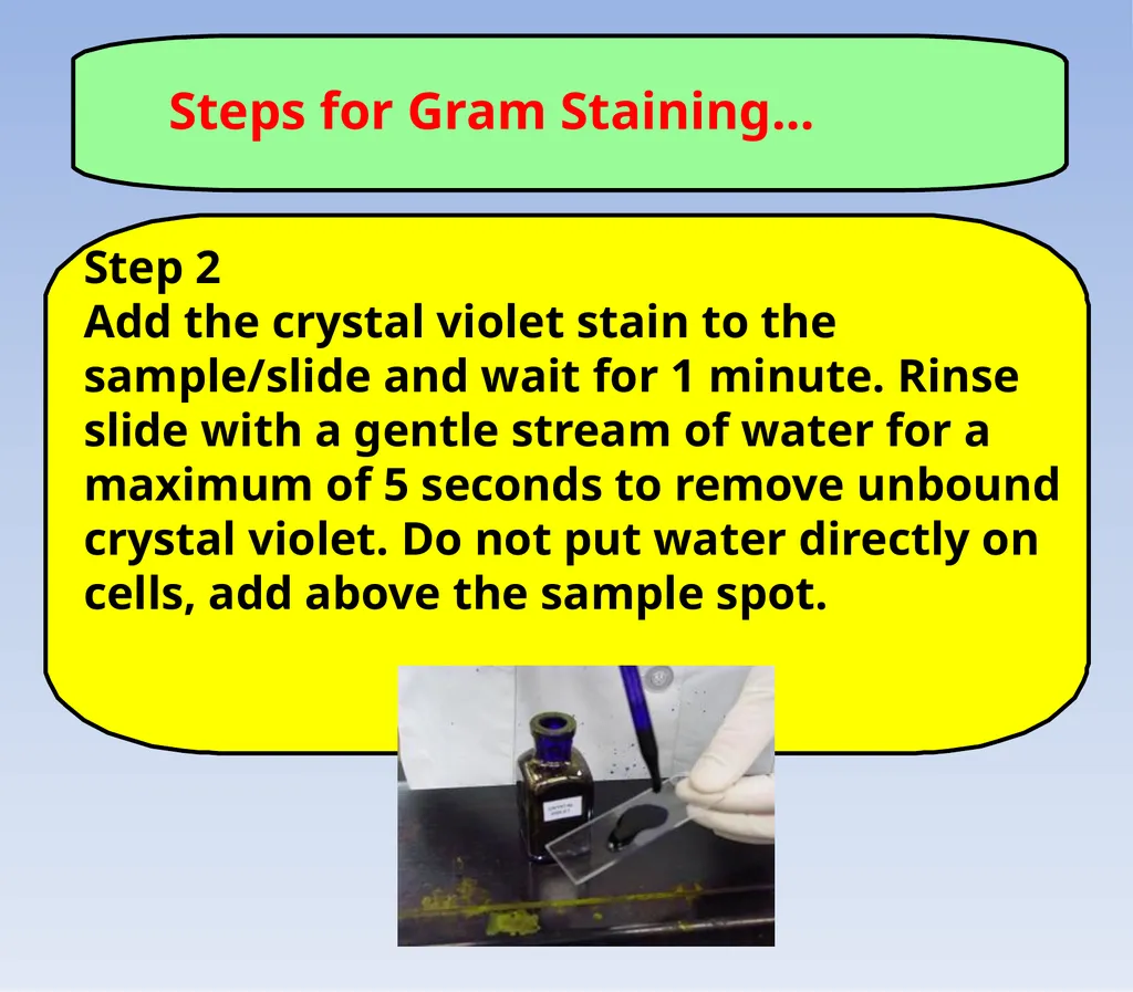
Steps for Gram Staining... Step 2
Add the crystal violet stain to the sample/slide and wait for 1 minute. Rinse slide with a gentle stream of water for a maximum of 5 seconds to remove unbound crystal violet. Do not put water directly on cells, add above the sample spot.<br>
12
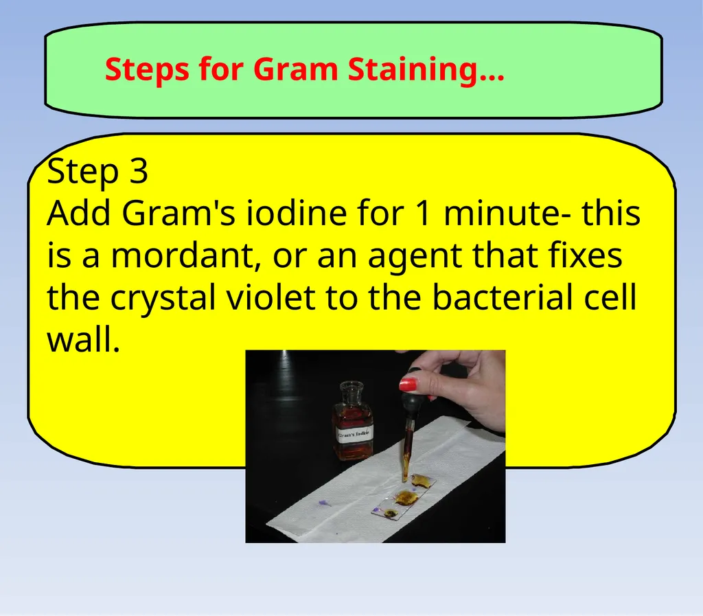
Steps for Gram Staining... Step 3
Add Gram's iodine for 1 minute- this is a mordant, or an agent that fixes the crystal violet to the bacterial cell wall.<br>
13
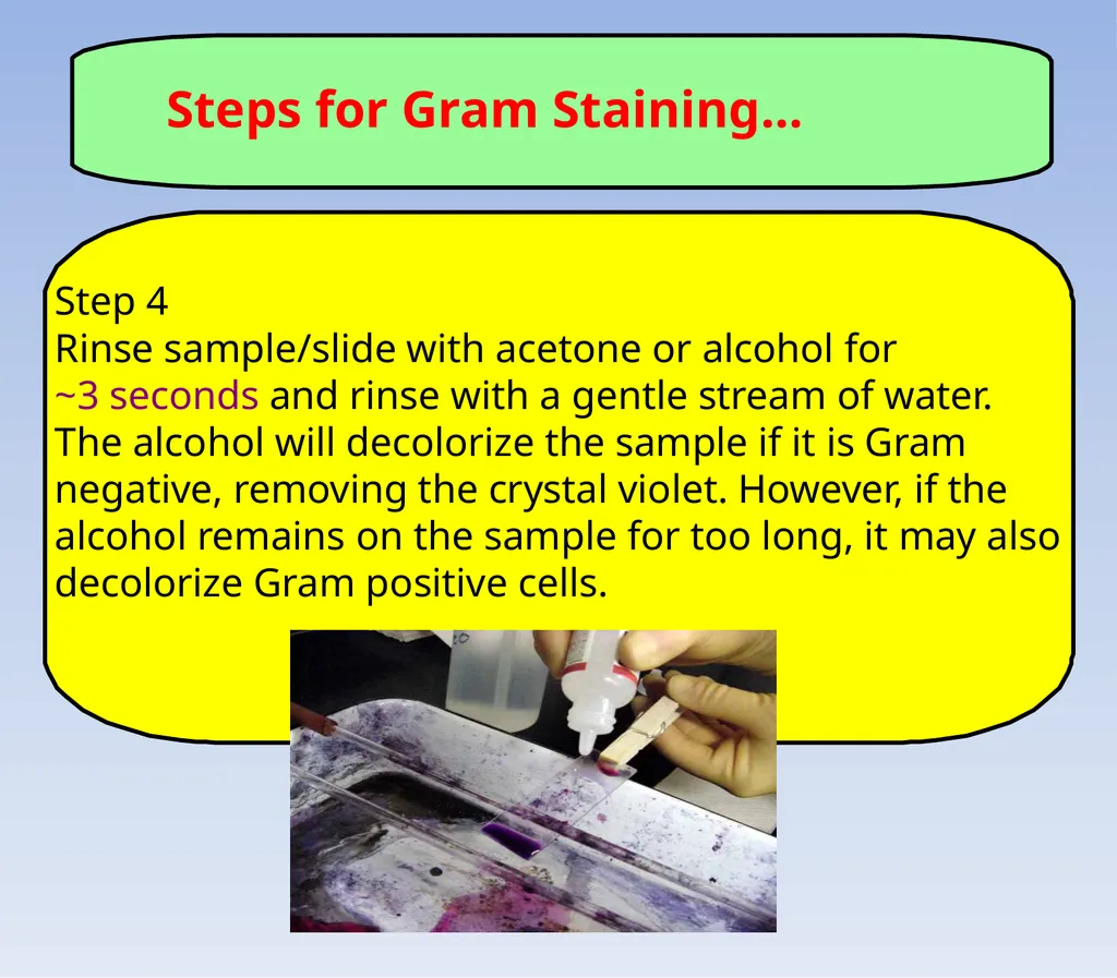
Steps for Gram Staining... Step 4
Rinse sample/slide with acetone or alcohol for
~3 seconds and rinse with a gentle stream of water. The alcohol will decolorize the sample if it is Gram negative, removing the crystal violet. However, if the alcohol remains on the sample for too long, it may also decolorize Gram positive cells.<br>
14
Steps for Gram Staining... Step 5
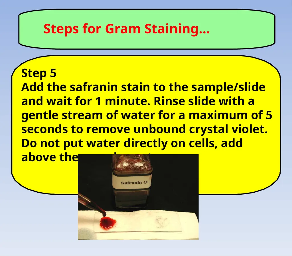
Add the safranin stain to the sample/slide and wait for 1 minute. Rinse slide with a gentle stream of water for a maximum of 5 seconds to remove unbound crystal violet. Do not put water directly on cells, add above the sample spot.<br>
15
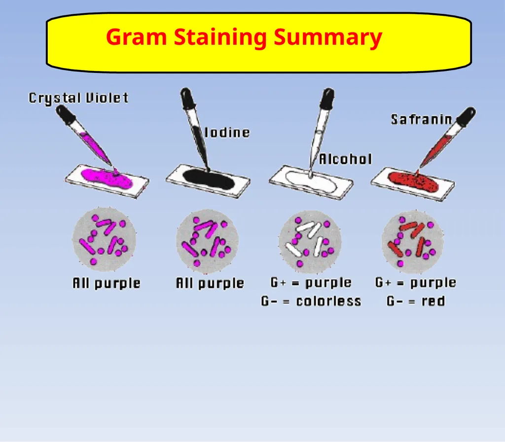
Gram Staining Summary<br>
16
Label these steps as a table team
What are you putting on the sample?
How long does it stay there?<br>
17
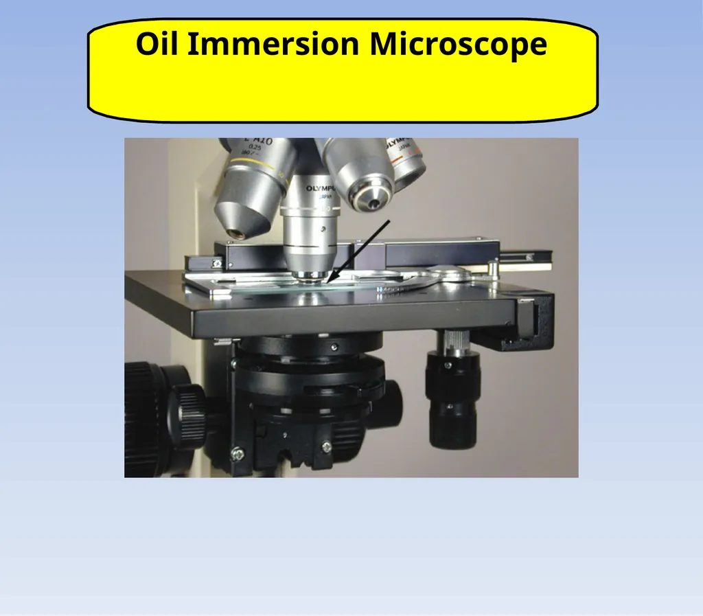
Oil Immersion Microscope<br>
18
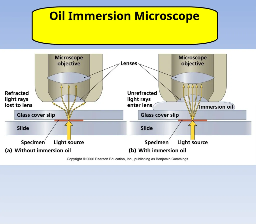
Oil Immersion Microscope<br>
19
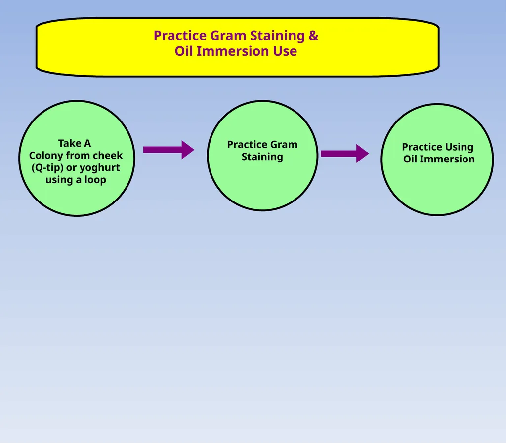
Practice Gram Staining &
Oil Immersion Use Take A
Colony from cheek (Q-tip) or yoghurt using a loop Practice Gram Staining Practice Using
Oil Immersion<br>